Week 2 - EEG Flashcards
Define spatial resolution
- Spatial resolution refers to the accuracy with which one can measure where an event is occurring. How precisely can the origin of a neural signal be located?
- Can spatially separate sources be differentiated?

Define temporal resolution
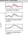
- Temporal resolution refers to the accuracy with which one can measure when an event if occurring. How precisely can changes in the signal be tracked?
- High sample rate, no lag: good temporal resolution
- fMRI has a lag of a second

What is the temporal and spatial resolution for EEG and ERPs?
EEG, MEG and ERP have good temporal resolution but bad spatial resolution

What is EEG?
- Electric potentials in the brain being recorded through electrodes placed on different points on the scalp.
- Raw EEG signal:
- Voltage = difference in electric potential between two sites.
What is the origin of the EEG signal and explain what happens during this
- Neurons communicate with each other through quick pulses of electrical current called “action potentials”. They bring about the release of neurotransmitters, which are absorbed by adjacent neurons. Action potentials occur at a rate of over 200Hz and are highly localised, they don’t create a diploe which makes them impossible to pick up by electrodes placed on the scalp.
- The type of electrical acitivity, that makes up the EEG signal, is the post-synaptic potential. Once a neuron receives an action potential from a neighbouring neuron and chemical transmitters have been released, this generates a current of ions in the cell. The ion current then causes a build up in electrical potential - this is the postsynaptic potential. If the postsynaptic potential reaches a certain level, it will cause an action potential to be released from the neuron. Postsynaptic potentials create a tiny dipole due to change in ion distribution allowing an EEG signal to be measured.

What are the requirements for a measurable EEG signal?
- A large number of simultaneously active neurons are needed to generate a measurable EEG signal
- Neurons must be highly synchronous – if they fire with a delay, there is not enough charge to affect the electricity on the scalp side.
- Currents must have same direction (mostly inhibitory or mostly excitatory neurons) – otherwise they might cancel each other out.
- Neurons must have same orientation (of the cell itself):
- Subcortical areas (e.g. basal ganglia) - neurons are not aligned
- Cortical layers – neurons are aligned
What is the inverse problem?
- Inverse problem – we can’t trace where the original EEG signal comes from.
- EEG systems typically have 32-256 electrodes:
- Each electrode = one observation
- Each dipole = one variable to solve
- Increasing the number of electrodes does not solve the inverse problem because we would still have millions of dipoles.
- Because of the inverse problem, the spatial resolution of EEG is poor

What is the history of the EEG signal?
- 1875 - Richard Caton used galvanometer to observe electrical impulses from surfaces of living rabbit and monkey brains
- 1890 - Adolf Beck found that frequency of electrical activity of dog and rabbit brains is modulated by light intensity (input changes frequency of electrical activity)
- 1924 - Hans Berger measures first human EEG (from scalp sites), finds different frequencies for open and closed eyes
How do you measure an EEG signal?
- Participant with EEG cap – with single active electrodes:
- Active electrodes show impedance (resistance) via LEDs
- Green < 5k
- Yellow <20
- Red > 20
- Active electrodes improves the signal-to-noise ratio by amplifying the signal at the scalp.
- Shielded cabin – to avoid other signals interfering:
- Reduction of electrical noise is crucial
- Faraday Cage – positive and negative ions. Negative ions move to one side which causes a second magnetic field to build up in the opposite direction of the field on the outside. Electrical charges in cage’s material redistributed → cancel out the magnetic field’s effects in the cage’s interior.
- Eye and muscle movements and sweat need to be reduced (drifting electrodes)
- Standardisation of scalp locations
- High viscosity contact gel (necessary for low impedances) – air and hair bad for electrical conductors
How do you decompose a raw EEG signal?
- With a Fourier Transform, the raw EEG signal can be decomposed into frequencies. Similarly, chords can be decomposed into notes.
What are the frequency bands in the EEG signal?
- Neurons tend to fire in temporal synchrony with each other; but at different frequency rates
- Different frequencies are related to different states in the brain

What are the two ways to analyse EEG data?
- Spontaneous EEG:
- Measurement of voltage differences throughout longer periods
- Useful for:
- Activiational state (e.g., sleep research)
- Clinical research (e.g., epilepsy)
- Event-related potential (ERP):
- Voltage difference in time window relative to specific ”event“
- Measure for cognitive, motoric or sensoric process
How is the spontaenous EEG signal used for the sleep-wake cycle?
- Different oscillation frequencies characterise the different phases of sleep-wake cycle
- Awake - beta and alpha bands.
- No-rapid-eye-movement (NREM) sleep:
- Stage 1: light sleep with low-amplitude waveforms. Theta bands.
- Stage 2: sleep spindles (the higher-frequency waves) and K complexes
- Stage 3 and 4: slow-wave sleep (SWS) with high amplitude waves and a deeper level of unconsciousness. Delta bands.
- Rapid eye movement (REM) sleep associated with activity and, in some individuals, with low-amplitude sawtooth waves and a higher level of consciousness. Beta bands.
- Finding out about sleep wake cycle can help with learning

What are ERPs?
- Event-related potentials (ERPs) are very small voltages generated in the brain structures in response to specific events or stimuli
- The positive and negative peaks are labelled with “P” or “N” and their corresponding number. Thus, P1, P2, and P3 refer to the first, second, and third positive peaks, respectively. Alternatively, they can be labelled with “P” or “N” and the approximate timing of the peak. Thus, P300 and N400 refer to a positive peak at 300 ms and a negative peak at 400 ms ( not the 300th positive and 400th negative peak!). ERPs are measured by EEG signals.
How do we get from spontaneous EEG activity to event related potentials?
- Steps involved in pre-processing:
- Segmentation (e.g. -200 – 500ms)
- Sort segments according to condition
- Baselining – necessary because of spontaneous voltage fluctuations in raw EEG:
- Principle - the average baseline activity (e.g. 200 ms epoch before stimulus onset) is subtracted from entire segment activity
- Result - the waveform is shifted towards zero - allows comparison between trials!
- Artefact rejection and correction – necessary because of noise:
- Artefact correction – correcting the signal based on what it would look like without environmental interference.
- Saccades, blinks, drifting voltage, high noise level all need to be removed.
- Averaging – for each condition and electrode separately
- Exporting and grand average:
- Export - calculate mean amplitude for all conditions and all electrodes in time window of interest –> run statistics
- Grand average - average of averages: across trials and subjects → Display purposes (this is what is typically shown in figures in EEG papers)

Why do we need hundreds of trials?
- More trials allow us to identify the real evoked potentials more easily
- Random noise averages out to zero

What are Topo maps?
- Amplitude is depicted in terms of colour
- Spatial representation (scalp distribution) of EEG signal – even though spatial resolution is not good for EEG, its important to look at topo maps to see what cognitive processes are affected.
- Typically for time windows, not single data points
- Allows comparison between conditions or time course

How can the data quality (signal-to-noise ratio) in EEG/ERP experiments be improved?
- Increase number of trials
- Decrease impedance (electrical resistance) – make a good connection between the scalp and electrode
- Avoid noise during recording (Faraday cage, low temperature, no moving, no eye movements, blinks etc.)
- Using active electrodes
These measures are important because the statistical power increases with improved signal-to-noise ratio
What are some examples of ERP components that are associated with cognitive functions?
- P1/N1: Sensory gain, early attentional processes
- N170: Detection and processing of faces
- N2pc: Selective attention
- P3a/b: Evaluation and Categorisiation of stimuli, updating working memory
- N400: Semantic Processing
- LRP (lateralised readiness potential): Preparation of motor responses
Describe the N170 ERP
- N170 reflects processing of faces
- When potentials evoked by images of faces are compared to those elicited by other visual stimuli, the facial images increased negativity 130-200 ms after stimulus presentation.
- Not related to the familiarity of faces, focuses more of perceptual processing of faces – signal decreases if the face is perceptually degraded.
Describe the P300 ERP and how is this related to early vs. late selective attention?
- The P300 (P3) wave is an event related potential (ERP) component elicited in the process of decision making
Early versus late selective attention?
- Early section theories (Broadbent, 1958) assume that after the sensory store, a selective filter picks out small information that goes onto higher level processing
- Late selection theories (Treisman, 1964) assume that a lot of information goes to higher level processing and then we filter out stuff.
- Early and late selection models were based on behavioural data but inconclusive.
Hillyard et al. 1973 – used EEG signals for early vs. late selective attention:
- Task - count number of single tones. Asked to attend left or right ear.
- Found the N1 component was always larger for the attended ear than the unattended ear
- Shows early on that we filter out right stimuli if we listen to the left ear or filter out left stimuli if we listen to the right ear.
- Used the P3 to see when there is a difference between the target and non-target
- P3 only observed for task relevant tones (high tones) (signals), not for standard tones (from 250 ms)
- Differences between attended and non-attended tones (regardless of target versus non-target) suggest filtering at ~80 ms post stimulus.
- Differences between target and non-target tones from 250 ms on. This requires higher level processing.
- Higher level processing happens AFTER filtering
- Evidence for early filter models

Describe the N400 ERP and the study associated with it
- The N400 is part of the normal brain response to words and other meaningful (or potentially meaningful) stimuli, including visual and auditory words, sign language signs, pictures, faces, environmental sounds, and smells
- Detection of semantic incongruity/expectancy
Kutas & Hillyard, 1980:
- Participants saw sentences on the screen
- Divided into three categories:
- No incongruity – sentence is normal
- Semantic incongruity – last word doesn’t fit the sentence
- Physical deviation – last word is bigger
- EEG signal shows:
- Difference between semantic incongruity and no congruity
- Interpreted as a violation of semantic expectancy:
- Congruent to context (classical N400 effect)
- N400 is affected by:
- No relation (classical N400)
- Untypical member (bird – turkey)
- Typical member (bird – sparrow)
- Antonym (opposite)
- N400 tracks effort to retrieve stored conceptual knowledge
- Preceding words serve as cues and make retrieval easier
- Also, N400 independent of sensory modality
- Doesn’t have to be written, can be spoken or visual

What are lateralised and unlateralised potentials?
- Unlateralised potential - single electrodes or pools of electrodes irrespective of laterality on screen
- Lateralised potential - pairs of electrodes in relation to laterality on screen
- Allows us to look at ipsilateral and contralateral activity separately
What is the Lateralised Readiness Potential? and outline the study associated with it
- Lateralised ERP
- Related to response or stimulus – we can look at the response of the participant independent of when the onset was
- LRP reflects preparation of motor activity (e.g. button press)
- Easy to isolate from other ERP components due to ipsilateral
Kato et al. (2009): Go/NoGo task
- Task: Respond to stimulus A, omit response for stimulus B
- Very boring and tiring task
- 1440 trials
- Results:
- Response times increased, accuracy decreased
- LRP decreases with time → response execution decreased
- Mental fatigue impairs response processes
What are the four different advanced EEG analysis methods?
- Independent component analysis:
- Counteracts bad spatial resolution
- Decomposition of electrode activity in activity of statistically independent components (neural ensemble?) – makes it easier to locate the component in the brain
- Analogy: Isolate the sound of a violin from an orchestra
- Classifying methods:
- Support vector machines, forward-encoding models, machine learning algorithms, dipole localisation
- Make use of the complexity of EEG data
- Brain-computer interfaces:
* Control of computers or machines with EEG - Frequency-based analyses:
* can reveal patterns hidden in raw EEG/ERPs
Outline Brain-Computer interfaces
- An algorithm is trained to differentiate between different EEG patterns
- After training, the computer receives input from EEG signal and is thus controlled by person
- Can be used to help the disabled to be more independent
Training:
- Participants have to imagine to move left or right arm
- This elicits different neural patterns that can be measured by EEG electrodes
- This goes into the algorithm
- Linear classifier helps get rid off classification errors
Applying the training:
- They can move a dial by pretending to use their left arm, to stop the dial they can imagine to use their right arm
What is frequency-bases analyses?
- Raw EEG signal or ERPs comprise of many frequencies
- Specific cognitive process can be strongly linked to one frequency band
- Easy way to reveal interesting patterns usually hidden in the EEG signal
- Experimental manipulation or interindividual differences may only be present in one frequency
- Can be hidden in EEG signal
- Fourier-transform allows isolation of single frequency
What are the advantages of ERPs?
- Extremely high time resolution (1000 Hz or higher)
- allows online tracking of cognitive processes (changes over time)
- allows capturing very transient processes
- Identification of cognitive processes
- ERP components are sometimes well-defined markers of a specific cognitive process
- Simultaneous cognitive processes can be examined in isolation
- Non-invasive
* safe, easy, high acceptance, ethics approval, healthy adults or children can be tested - Cheap
- Equipment for full lab starts around 25,000 GBP
- No additional running costs
What are the disadvantages of ERPs?
- Bad spatial resolution
* However: Advanced analysis methods (e.g., Independent Component Analysis) allow reasonably good spatial resolution - Many repetitions required for good signal-to noise ratio
* unsuitable for paradigms that require surprise or that are prone to adaptation - ERP experiments require careful planning to avoid confounds
* Differences in the EEG signal can be affected by many things - Prone to noise
- Careful control of testing environment
- unsuitable or very difficult for experiments that require motor activation (running, talking, eye movements)
What is Magnetoencephalography (MEG)?
MEG is similar to EEG, but:
- Instead of current, magnetic fields are detected
- ERFs (event-related fields) equivalent to ERPs
- Brain‘s magnetic field is tiny: 10-1000 fT (femtotesla) versus 25,000,000,000-65,000,000,000 ft Earth‘s magnetic field
- MEG requires superconducting sensors bathed in liquid helium (-269° C) and shielded chambers for a good signal – this is what makes it expensive and challenging
- Very low impedance allows to detect and amplify magnetic fields
What are the differences between MEG versus EEG?
- Same high temporal resolution
- Both non-invasive
- MEG has excellent spatial resolution (magnetic field passes unaffected through brain tissue and skull, i.e. is less blurred so can identify well where a signal is coming from)
- EEG much cheaper than MEG (purchase and maintenance)
- ERFs less clearly understood than ERPs due to less research for MEGs due to expensiveness
- Depending on origin of neural signal, EEG or MEG more likely picking up signal
What is an application of MEG in medical practice?
- Preoperative mapping of somatosensory cortex – identify areas that you don’t want to cut
- Example: Tumor near sulcus centralis
- Steps:
- tactile stimulation (finger, arm, leg)
- Recording ERFs
- Inverse modelling of dipoles (from overall signal, which is the most likely origin?)
- Result:
- Calculated dipoles for sensory areas (here: toes and fingers in red) can be compared with location of tumor (green)
What are the two types of inhibitory and excitatory postsynaptic potentials?
The brain consists of billions of cells, half of which are neurons, half of which help and facilitate the activity of neurons. These neurons are densely interconnected via synapses, which act as gateways of inhibitory or excitatory activity:
- Na+ flows into cell –> net negativity (Excitatory postsynaptic potential)
- Cl- flows into cell or K+ out of cell –> net positivity (Inhibitory postsynaptic potential). Inhibitory potentials make action potentials at the next neuron less likely.


