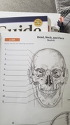
Week 2 bones of skull and movements Flashcards
1
Q
4

A
Frontal
2
Q
2

A
Parietal
3
Q
3

A
Temporal
4
Q
1

A
occipital
5
Q
5

A
sphenoid
6
Q
9

A
Zygomatic
7
Q
11

A
Mandible
8
Q
10

A
Maxilla
9
Q
6

A
Ethmoid
10
Q
1

A
frontal
11
Q
2
A
parietal
12
Q
3

A
Temporal
13
Q
4

A
sphenoid
14
Q
8

A
zygomatic
15
Q
11

A
mandible
16
Q
6

A
ethmoid
17
Q
22

A
Temporal
18
Q
21

A
Occiput
19
Q
23

A
Sphenoid
20
Q
26

A
Palatine
21
Q
24

A
Zygomatic
22
Q
25

A
Maxilla
23
Q
13

A
Parietal
24
Q
15

A
Occiput
25
#20

mandible
26
#19
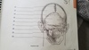
Maxilla
27
cavitys of the skull
Nasal cavity
Orbits
Paranasal sinuses
small cavities in ear for hearing and balance
28
skull has ____ bones
\_\_\_\_\_cranial
\_\_\_\_\_facial
22 total
8 cranial
14 facial
29
What are bony Landmarks?
* Fixed markers on the bones
* Grooves or prominences found on bones, which help us to identify the location of other body structures
30
**THREE GENERAL CLASSIFICATIONS of bony landmarks**
1. Articulations: Where 2 joint surfaces come together
2. Projections: Area of the bone that protrudes above the surface of the bone
3. Hole: An opening or groove in the bone.
31
**Function of Bone Markings**
Hint: (4) Enables, provides, serves, provides
* Enable joints to slide past each other or lock bones in place,
* Provide structural support to muscle and connective tissue
* Serve as a place of attachment for other bones, blood vessels, nerves, and/or muscle
* Provide stabilization and protection to nerves, vessels, and connective tissue.
32
what do Holes & Depressions in bone do?
allow the passage of soft tissue through or along bone and form joints
33
a Cavity is:
: A depression that is associated with a joint articulation
34
a Facet is:
Flat, smooth articular surface
35
a Fissure is
A groove intermediate to two bones
36
a Foramen is
A round or oval opening, or hole in the bone
37
a Fossa is?
A depression in the bone
38
a Groove is:
A slit-like passage
39
Meatus:
A canal, or passageway in the bone
40
Sinus:
Hollow cavity within bone filled with air
41
Notch
An incisure or indentation
42
**Projections:**
*form joints and Serve as attachment points for connective tissues*
43
* **Condyle:**
* **Condyle:** Large, rounded bump – forms a joint with another bone
44
* **Crest:**
* **Crest:** A ridge on a bone; often anterior – usually prominent
45
* **Diaphysis:**
* **Diaphysis:** Long midsection of the long bone (Body of the bone)
46
* **Epicondyle:**
* **Epicondyle:** A space or raised protuberance above or on a condyle of a long bone
47
* **Epiphysis:**
* **Epiphysis:** is the rounded end of a long bone
48
* **Head:**
* **Head:** Rounded structure found at the end of the narrow “neck” of the bone.
49
* **Line:**
* **Line:** A small ridge; often posterior (muscle attachment)
50
* **Notch:**
* **Notch:** Indentation in the bone (curved – to allow for movement of another bone)
51
* **Process:**
* **Process:** A sharp protrusion away from the main body of the bone (much like a finger)
52
* **Ramus:**
* **Ramus:** Arm-like bar of a bone
53
* **Spine:**
* **Spine:** Sharp, slender, often pointed projection
54
Spinous process
: A thin projection
55
* **Trochlea:**
* **Trochlea:** A pulley-like structure
56
* **Trochanter:**
* **Trochanter:** Very large bump (found only on the femur)
57
* **Tubercle:**
* **Tubercle:** Small bump or projection
58
* **Tuberosity**:
* **Tuberosity**: Large rounded projection – Surface may be rough
59
stages of wound healing
Inflammatory phase
proliferative phase
remodeling phase
60
debridement
* removing skin that is too tough to have it regrow into good tissue
61
* burns affect only the epidermis, or outer layer of skin. The burn site is red, painful, dry, and with no blisters. Mild sunburn is an example. Long-term tissue damage is rare and usually consists of an increase or decrease in the skin color.
First-degree (superficial) burns
62
burns involve the epidermis and part of the dermis layer of skin. The burn site appears red, blistered, and may be swollen and painful.
Second-degree (superficial and Deep partial thickness)
63
\_\_\_\_\_\_burns destroy the epidermis and dermis and may go into the subcutaneous tissue. The burn site may appear white or charred
Third-degree (full thickness) burns
64
\_\_\_\_\_\_\_\_\_ burns also damage the underlying bones, muscles, and tendons. There is no sensation in the area since the nerve endings are destroyed
Fourth degree burns
65
Rule of 9's
assesses the percentage of burn
is used to help guide treatment decisions including fluid resuscitation
determines transfer to a burn unit
66
Lund-Browder Chart
* Used with children and infants who sustain burns
* factors in the age of the individual
67
Hypertrophic Scarring
skin is raised, decreased elasticity, and erythematic (red) in appearance
. Scarring can be troublesome, cause contractures, and impair function and lead to deformity
* Surgical intervention may be necessary but typically contraindicated until scar has completely matured
* Therapy plays an important role in minimizing these sequelae
68
69
OT intervention for burns
* Compression Therapy
* Skin Lubrication and scar massage
* Splinting
* Range of Motion
* Strengthening and Conditioning
* Psychological Healing
* Planning for discharge
70
Fitted to the patient to encourage better orientation of collagen fibers
Compression Garments:
71
Approved unscented lotion to prevent cracking and massage to increase pliability and promote desensitization
Lotion and Scar Massage:
72
* Preserve/increase range of motion, prevent scar contracture formation and protect underlying structures
Splinting
73
: Daily stretching and elongation are crucial to improve
Range of Motion:
