Week 2 Flashcards
Scapula label
Where is scapula connected to clavicle?
What does it form with clavicle?
Acromion
Shoulder gridle
What is special about clavicle with respect to the development?
What is the orgin?
First long bone to ossify wk5-6 and last one to fuse 25 yo
membranous origin
Two joints of clavicle
Sternoclavicular joint
Acromioclavicular joint
What is one structure that connects arm to the axial skeleton?
Clavicle
Label ligaments
Where does the shoulder separation occur?
WHere does the shoulder dislocation occur?
Where does the clavicle break most often?
AC joint
glenohumeral joint
Middle/lateral thirds
What is unusal about Strenoclavicular joint
What is the disc made of?
Two bursas separated by articular disc
Fibrocartilage
Movements of the Sternoclavicular Joint
Axis? Movement
Vertical axis —– protraction/retraction
AP axis —– elevation/depression
Oblique axis —– medial/lateral rotation
How much degree of abduction occurs at glenohumeral joint?
How much occurs at sternoclavicular joint?
What is the name of this movement?
120
60
scapulohumeral rhythm
Movements of the Trapezius
Verical axis?
AP axis?
Oblique axis?
Retract
(Superior) elevates (Infectior) depresses
Rotate
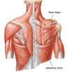
Trapezius Muscle
Nerve supply
Spinal accessory nerve (XI) Supplies motor
Cervical segments C3 & C4 Supply proprioceptive
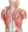
Latissimus Dorsi
Direction of movement?
Nerve supply?
Connects between?
Adduction / Extension
Thoracodorsal nerve (middle subscapular)
Thoracolumbar fascia to humerus
Superficial (extrinsic) Back Muscles
Movement?
Nerve?
Medially rotate scapula
Dorsal Scapular nerve
Levator scapula
Movement?
Nerve?
Elevates and medially rotates scapula
Dorsal Scapular
Pectoralis Minor
Movememnts?
Nerve?
Medial Pectoral Nerve
Depresses, Protracts, Medially Rotates
Serratus Anterior
Movement? Structural importance?
Innervation?
Depression, protraction, lateral rotation
Holding medial border of scapula close to ribs
Lorng Thoracic Nerve
What is winging
Loss of serratus anterior function e.g. loss of the neve
Muscles of the SC-joint
Protraction
Retraction?
Elevation?
Depression?
Medial rotation?
Lateral rotation?
Movements of glenohumeral joint
Horizontal transverse axis —– flexion/extension
Horizontal AP axis —– abduction/adduction
Vertical axis —– rotation
Structures protecting muscle around Glenohumeral joint
Subdeltoid bursa
Subacromial bursa
Biceps brachii tendon
Pectoralis Major
Nerve?
Movement?
Medial and Lateral Pectoral
Flexion, Adduction, medial rotation
Deltoid
Movement?
Innervation?
Axillary
Flexion: Anterior
Extension: Posterior
Abduction: Middle
Adduction: Posterior and Anterior
Medial Rotation: Anterior
Lateral Rotation: Posterior
Teres Major
Innervation?
Middle Subscapular or thoracodorsal (Latissimus)
Lower Subscapular (Teres)
Extension
Adduction
Medial Rotation
SITS
Function?
Supraspinatus
Infraspinatus
Subscapularis
Teres Minor
Stabilization of glenohumeral joint
How medial vs. lateral rotation affects abduction?
External rotation of the humerus during flexion or abduction prevents the greater tubercle from hitting the acromion.
Name the rotator cuff
Glenohumeral joins all movements and muscles
Blood supply and nerves for
Supra/infraspinatus?
Teres minor?
Subscapularis?
Supra and infraspinatus- suprascapular nerve + artery
Teres minor –axillary nerve and post humeral circumflex artery
Subscapularis – upper and lower subscapulars + circumflex scapular art
Collateral Flow of glenohumeral
Dorsal scapular
Suprascapular artery
Posterior circumflex humeral artery
Circumflex scapular from subscapular artery
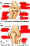
Bones
Bones
Axillary arteries
superior thoracic
thoracoacromial trunk
lateral thoracic
subscapular
circumflex scapular
- thoracodorsal
- posterior humeral circumflex
anterior humeral circumflex
What is axilla?
The space below the shoulder joint, bounded by the pectoralis major anteriorly, the latissimus dorsi posteriorly, the serratus anterior medially, and the humerus laterally.
Posteior wall of Axilla
Members?
Subscapularis
Teres major
Latissimus dorsi
Thoracodorsal artery + nerve (to latissimus dorsi)
Quadrangular space with axillary nerve and posterior circumflex humeral artery
Anterior circumflex humeral artery
Medial Wall of Axilla
what muscle is it?
Serratus anterior
Long thoracic nerve
Lateral thoracic artery
What part of brachial plexus passes through axilla?
Medial cord
Lateral cord
Posterior cord
Brachial plexus
Roots: C5-T1
Turnks: Upper, Medial Pattern
Divisions: Anteior Posterio
Cord: Lateral Medial Posterior
Branches: Ulnar, Radial, Musculocutaneous, Median, Axllary
General patters of innervations to hand
Innervations of anteior compartment of arm?
Innervation of posterior compartment of arm?
Anterior compartment
anterior arm-musculocutaneous
anterior forearm-all median except FCU+2 heads of FDP (ulnar)
anterior hand-all ulnar except thenar comp muscles + lumbricals to the functional midline on radial side (median)
Posterior compartment
shoulder-axillary (deltoid and teres minor)
posterior arm & forearm-radial
What are the branches from each cord?
Lateral=lat pec,
Post=upper, middle and lower subscapulars,
Medial=M. pectoral, M. brachial, M. antebrachial cutaneous
Two compartments of arm and their components
Components for compartment of arm
Biceps brachii
Long head
Short head
Coracobrachialis
Brachialis
Triceps 3 heads
Damage of Terminal Branches affecting the Arm
Axillary nerve?
Musculocutaneous nerve?
Radial nerve?
Axillary nerve-severely weakened abduction, weakened lateral rotation
Musculocutaneous nerve- almost no forearm flexion, weakened arm flexion
Radial nerve-no forearm extension, arm may be slightly flexed
(note also has major effect on forearm and hand, see later)
Three joints on hand
metacarpophalangeal (MP) proximal interphalangeal (PIP) distal interphalangeal (DIP)
What flexor acts on
distal interphalangeal joint?
proximal interphalangeal joint?
Flexor digitorum profundus - distal
Flexor digitorum superficialis - proximal
Structure that surrounds all ligments of the hand?
Retinaculum
Where does most of posterior muscles originate from?
Exceptions?
lateral epicondyle or the supracondylar ridge
The exceptions are the three thumb muscles, the extensor indices and the supinator
Posterior Forearm (Thumb Muscles)
Abductor pollicis longus (APL)
Extensor pollicis brevis (APB)
Extensor pollicis longus (EPL)
Anatomical Snuffbox
Contents
Radial artery
Base of 1st metacarpal
Styloid process of radius
Crossed by dorsal digital branches of radial nerve
Posterior Forearm Muscles
Posterior forearm muscles that don’t cross the wrist
Pronators and Supinators of forearm
Pronator Quadratus
Pronator Teres
Supinator
Biceps
What is innervated by deep Radial Nerve?
Posterior forearm muscles except brachioradialis and the externsor carpi radialis longus ECRL
Which radius branch is motor?
Which radius branch is sensory?
Deep
Sensory
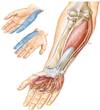
Hand innervations
Compartment Syndrome
Arteries of the arm
Brachial artery
Profunda brachii artery
Radial recurrent artery
Ulnar recurrent artery
Interosseous arteries
Radial
Ulnar
Where does the blood supply come from to thumb/index?
Where does the blood supply the rest finers?
Deep palmar arch
Superficial palmar arch
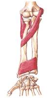
Connective tissue between radius and ulna
Intraosseus membrane
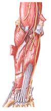
Arteries of the forearm
List five muscles in superficial layer of the anterior forearm
- Pronator teres
- Flexor carpi radialis
- Palmaris longus
- Flexor digitorum superficialis (hidden – intermediate layer)
- Flexor carpi ulnaris
What attaches anterior foream superficial muscles?
Common flexor tendon
Deep muscle the the anterior forearm
What supplies deep anteiror forearm muscle group?
* Except two heads of Flexor digitroum profundus (FDP)
Anterior interosseus branche of the median nerve
Wrist flexors
Flexor carpi radialis
Palmaris longus
Flexor carpi ulnaris