Urological pathology Flashcards
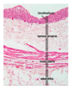
What is the histological structure of the wall of the renal pelvis, ureter, bladder and urethra?
- urothelium
- lamina propria
- muscularis propria (detrusor in bladder)
- adventitia (perivesical fat in bladder)

What are the zones of the prostate gland?
- transition
- central
- peripheral

Prostate gland produces alkaline secretion to neutralise acidic environment of vagina. Gland is made up of numerous acini (glands) + ducts lined by epithelial cells, embedded in a stroma composed of sm muscle cells + fibroblasts.
What do the prostatic acini do?
- secrete prostatic juice
- drain into the prostatic urethra via duct system
What do the stromal cells contain?
- 5 a-reductase
- converts T -> DHT (more potent)
- maintaining suitable levels of androgens in prostate
What are common causes of macroscopic (frank) haematuria?
-
UPPER TRACT:
- kidney cancer (renal cell carcinoma)
- stone in kidney or ureter
- trauma
-
LOWER TRACT:
- bladder cancer
- BPH
- infection (bacterial cystitis)
For macroscopic haematuria, following a full history and examination, what investigations are useful to request?
- MSU for MC+S
- urine cytology
- flexible cystoscopy +/- biopsy
What are LUTS?
- lower urinary tract symptoms
- frequency, urgency, nocturia, hestiancy, poor flow + terminal dribbling
- suggests problem in bladder or prostate
- important to realise that LUTS not specific for any particular pathology
What are some causes of LUTS?
- BPH
- UTI
- urinary tract stones
- bladder cancer
- prostate cancer (LUTS are a late feature of this)
What is the most common malignant tumour of the kidney?
-
renal cell carcinoma (RCC)
- most common type of RCC -> clear renal cell carcinoma
- RCCs are adenocarcinomas, arise from epithelia lining renal tubules
What are the risk factors for development of RCC?
- male gender (4M:1F)
- inc age (most cases occur in over 50)
- smoking
- obesity
-
familial syndromes - Von Hippel Lindau sydndrome
- rare autosomal dominant genetic disease
- predisposes individuals to certain benign + malignant tumours incl RCC + phaeochromocytoma
How is renal cell carcinoma graded?
Fuhrman grading system
- grade 1: tumour cell nuclei closely resembal normal -> less aggressive (better prognosis)
- grade 4: tumour cell nuclei larger + pleomorphic etc -> more aggressive (worse prognosis)
How is renal cell carcinoma staged?
- TNM system
- size of the primary tumour important in determining the T stage
What is the clinical presentation of RCC?
There is a triad, however it is uncommon in clinical practice:
- loin pain
- loin mass
- haematuria
Other common presentations of kidney cancer include:
- incidental finding on scan
- presentation w/ symptoms/signs of metastatic disease (lung or bone) eg. bone pain, SoB
- paraneoplastic syndrome eg. hypercalcaemia, erythrocytosis, amyloidosis
What type are the majority of bladder cancers?
- urothelial carcinomas
- malignant tumour arising from urethelium
- transitional cell carcinoma is an old term
Wht are risk factors for developing urothelial carcinoma?
- cigarette smoking
- industrial exposure to certain industrial dyes + solvents (eg arylamines)
Bladder cancers are staged using the TNM system. What are key features of the TNM system regarding bladder cancer?
- superficial tumours are Ta or T1
- muscle invasive tumours are T2, T3, or T4
- CIS is Tis

How do you clinically classify bladder cancer?
Three main groups:
- low-risk bladder cancer (superficial tumours Ta T1 + low grade)
- high-risk bladder cancer (muscle invasive T2 or worse + high grade)
- carcinoma in situ (CIS) - precancer

Describe low-risk bladder cancers and their extent of invasion
- confined to mucosa or invade into lamina propria
- do not invade into muscularis propria (detrusor) or beyond
- often low grade
- tend to be composed of frond-like papillary growths
- papillary refers to finger-like projection (w/ fibrovascular core)

How are superficial urothelial carcinomas (low-risk bladder cancers) managed?
- removed at cystoscopy by TURBT
- after removal:
- high chance tumour will recur
- low chance tumour will transform into high risk
Therefore, pts diagnosed w/ superficial urothelial carcinoma require regular check cystoscopies
Describe high-risk (muscle-invasive) bladder cancer and how they differ to low-risk cancers
- invade into detrusor muscle or beyond
- tend to be solid rather than papillary
- almost always high grade
- have much worse prognosis than low-risk tumours
- more likely to spread to regional nodes + metastasise to distant sites
- radical treatment required for cure, often cystectomy (removal of bladder) +/- other organs eg. uterus
Describe urothelial carcinoma in situ (CIS)
- flat lesion
- ureothelium contains cells that display nuclear features
- associated w/ malignancy (eg pleomorphism, mitoses etc)
- BUT no invasion through basement membrane
- form of precancer: left untreated, ~40% progress to muscle invasive cancer
- diagnosis of CIS bladder v serious due to chances of prog
What is blue light cystoscopy?
- may be used to identify CIS
- ~1hr before cystoscopy, Hexyl aminolevulinate (HAL) inserted into bladder via a urinary catheter
- urologist performs cystoscopy using special cystoscope + blue light
- CIS cells absorb chemical + then fluoresce red in blue light
- background normal urothelial cells do not absorb chemical so do not fluoresce
- thus, urologist can identify areas of CIS bc they stand out

How is urine cytology helpful in picking up CIS?
- highly atypical cells shed into urine
- abnormal urothelium loses its normal cohesiveness + begins to fall apart
- loss of urothelium -> intense LUTS (dysuria, inc freq)
- important to note that negative urine cytology does not exclude a significant pathology

What is the molecular classification of bladder cancer?
- low risk (superficial) bladder cancers strongly associated w/ mutations in HRAS and FGFR3
- CIS + high-risk (muscle invasive) bladder cancers strongly associated w/ mutations in TP53 and RB1
What is BPH?
- overgrowth of prostatic tissue in transition zone of gland
- overgrowth of prostatic tissue -> formation of large nodules
- nodules form due to hyperplasia of all cellular elements in prostate ie. epithelial cells, sm muscle cells + fibrous tissue
BPH is due to a subtle hormonal imbalance which occurs w/ age. What is this imbalance?
- oestrogen levels in blood increase with age
- vicious cycle set up as more stromal cells produce more potent androgens which induce more growth and so on

What is the most important complication of BPH?
- gradual compression of prostatic urethra
- causing urinary tract obstruction
- two components underlying development of obstruction:
- inc tone of sm muscle in prostate, making organ more contracted around urethra
- mechanical effect due to physical bulk of prostate
Wha timpact does BPH have on the bladder?
- gradually progressive bladder outflow obstruction
- in order to generate higher pressure required to eject all urine from bladder, bladder detrusor muscle undergoes compensatory hypertrophy (visible as trabeculations)
- eventually, detrusor ‘decompensates’ + relaxes
- bladder begins to dilate until it’s converted into a big flabby sac w/ little power over contraction
- pts w/ chronic urinary retention have a palpably enlaregd distended bladder (typically painless)
- in contrast, acute urinary retention is painful
- if bladder outflow obstruction not relieved, hydroureter + hydronephrosis may develop on both sides of urinary tract
What is hydronephrosis?
- literally “water inside kidney”
- refers to distension + dilation of renal pelvis + calyces
- caused by obstruction of free flow of urine from kidney
- untreated -> progressive atrophy of renal parenchyma
- -> progressive loss of renal fxn in both kidney
- development of chronic kidney injury

What is obstructive nephropathy (uropathy)?
- renal dysfunction
- due to obstruction to flow of urine
- BPH common cause of obstructive nephropathy in adults
What are possible complications of obstructed urinary tract?
- infection
- hypertension
How might BPH present?
- LUTS
- acute urinary retention
- urinary tract infection
- renal dysfunction (obstructive nephropathy)
BPH may also give rise to a significantly raised serum PSA level
What is the risk of prostate cancer?
likelihood of…
- developing = 1 in 3
- causing problems = 1 in 10
- dying = 2-3 in 100
What are risk factors for developing prostate cancer?
- increasing age
- afro-caribbean origin
- germline BRCA mutations
- Lynch syndrome
There are two distinct ways in which prostate carcinoma may present: latent prostate cancer or symptomatic prostate cancer.
What is latent prostate cancer?
- inc # of men diagnosed when they have no symptoms
- eg. 50yo man asks GP for PSA test
- PSA is elevated -> pt undergoes prostate biopsy
- -> diagnose prostate cancer
Symptomatic prostate cancer refers to pts presenting w/ symptoms. These may be due to the primary tumour in the prostate (local) or due to effects of metastases.
What are local and metastatic symptoms that present first?
- symptoms due to metastatic disease typically occur first - eg. bony mets common in prostate cancer + may be mode of presentation eg. persistent back pain
- local symptoms eg. bladder outflow obstruction typically occur late - bc prostate cancer occurs in peripheral zone of prostate + so obstruction of urinary flow through prostatic urethra occurs late in natural history of disease
Which cancers most commonly metastasise to bone?
- breast
- lung
- prostate
- kidney
- thyroid
Remember, any cancer has theoretical potential to metastasise to bone. Bone mets are usually osteolytic, the one major exception to this is prostate cancer, in which bone mets are typically osteosclerotic.
What is the pathology of prostate cancer?
- adenocarcinomas arising from epithelial cells lining acini or ducts of prostate gland
- asymptomatic premalignant lesion = prostatic intraepithelial neplasia (PIN)
- progression from PIN to adenocarcinoma is not inevitable
- prostate cancer is often multifocal
- usually arises from peripheral zone of prostate
How is diagnosis of prostate cancer made?
- transrectal US-guided biopsy of prostate (TRUS)
- biopsies taken from 12 areas within gland

How is prostate cancer graded?
- gleason scoring system
- very important in helping determine most appt treatment + prognosis for patients
- tumour given two scores (1-5) according to microscopic appearance
- scores added together to give overall grade (2-10)
- in practice, nowadays only scores 3-5 are used, giving poss total of 6-10
- 6 (3+3) = indolent, good prognosis
- 7 (4+3/3+4) = intermediate prognosis
- 8+ = agressive, worse prognosis
How is prostate cancer staged?
- TNM system
- CT/MRI
- bone scan
What is the problem with treating latent prostate cancers?
- if we treat all prostate cancers we risk over-treating clinically insignificant prostate cancers
- if we don’t treat any prostate cancers we will under-treat potentially lethal cancers
There are a # of treatment options for latent prostate cancer which aim to cure th disease. The ‘active surveillance’ option attempts to minimise the chances of over-treating clinically insigniciant prostate cancers
What is a radical prostatectomy and its side-effects?
- entire prostate removed
- bladder is rejoined to urethra
- may be done as robotic procedure using a da Vinci robot
-
side-effects include:
- urinary incontinence (5-15%)
- erectile dysfunction (30-100%)
- bladder neck obstruction (<10%)
- death (<1%)
What is radical radiotherapy and its side-effects?
- external beam radiotherapy (EBRT) or brachytherapy
- aim of brachytherapy -> deliver high dose of radiation more directly to prostate + reduce radiation damage to normal surrounding structures
- brachytherapy involves placement of permanent radioactive seeds into prostate gland
- seeds slowly release radiation over couple of months
-
side-effects include:
- urinary incontinence (5%)
- erectile dysfunction (40-80%)
- long term bowel + bladder problems (5-10%)
What is involved in active surveillance?
- careful clinical monitoring
- repeat PCA
- repeat digital rectal examinations
What is the rationale behind active surveillance?
- many pts w/ low risk and low volume cancer have clinically insignificant or indolent cancer
- may not necessarily be causing them problems in lifetime
- provided these pts are appropriately monitored for disease progression + then offered more active treatment, the risk of over-treatment can be reduced and the risks of side-effects of active treatment can be deferred in order to maximise the quality of life
Which patients are suitable for active surveillance?
- relatively low PSA (<10)
- Gleason 3+3 cancer
- those w/ only a small proportion of 12 core biopsies containing cancer
If subsequent biopsies show a large volume of cancer or there is Gleason 4 cancer present, radical therapy would be recommended
How is symptomatic prostate cancer treated?
- pts presenting w/ symptoms due to prostate carcinoma will almost always have advanced disease
- disease not usually curable
- so palliative treatments to control disease employed
-
androgen deprivation used to slow tumour growth
- achieved by surgical castration (bilateral orchidectomy)
- or by pharmacological treatment (anti-androgens or LH-RH agonists)
Where do urinary tract stones most commonly arise?
- anywhere in urinary tract
- but most commonly: renal calyces + pelvis
- lifetime risk of stone formation = 10%
What are the most common types of renal calculi?
- calcium oxalate
- or mixture of calcium oxalate + calcium phosphate
Most patients w/ calcium stones have hypercalciuria but do not have hypercalcaemia.
What are the causes of hypercalciuria?
- absorptive hypercalciuria - overabsorption of calcium from gut
- renal hypercalciuria - defect in renal tubules, impairs renal tubular absorption of calcium in prox tubule
- hypercalcaemia - usually due to primary hyperparathyroidism (minority)
How might larger stones present clinically?
- tend to remain confined to kidney
- may form a staghorn calculus
- fills the entire renal pelvis + calyces
- stones may remain clinically silent or come to light when haematuria or recurrent UTIs are investigated

How do smaller stones present clinically?
- pass into ureter + cause ureteric colic
- if stone becomes impacted in ureter -> severe pain
- agonising pain - sudden + colicky, lasts several mins
- originates in loin + radiates towards groin
Remember, most small stones pass through the urinary tract naturally and so a ‘watch and wait’ policy is appropriate (as long as no complication supervenes)
A stone may become impacted in the urinary system causing obstruction.
What are the three most common places that ureteric stones become impacted?
- pelviureteric junction - large diameter of renal pelvis decreases to that of the ureter
- pelvic brim - as ureters arch over iliac vessels
- vesicoureteric junction - ureter narrows, most common place for impaction!

What are the complications of an impacted/blocked stone?
- stone blocks urine flow in one half of urinary tract
- serious immediate complication = UTI -> pyelonephritis -> sepsis
- in medium to long-term, inc in pressure proximal to impacted stone causes hydroureter + hydronephrosis
- loss of kindey fxn on affected side = unilateral obstructive uropathy
- however, since only one kidney affected, contralateral kidney able to maintain sufficient kidney fxn (eg bilateral obstruction secondary to BPH -> both kidneys affected!)
- hypertension is another long term complication of ureteric obstruction
What is the immediate emergency treatment of urinary stone obstruction?
- nephrostomy
- involves insertion of tube through abdominal wall
- into renal pelvis through which urine can drain freely
- usually performed by radiologists under image guidance

What is the general management of an impacted urinary stone?
- uteroscopy
- +/- laser lithotripsy
- lithotripsy breaks up stone into small pieces which can then be removed


