Lymph node pathology Flashcards
(34 cards)
What are the two main components of the lymphatic system?
- a conducting system (lymph vessels) which transport lymph from the interstitium to the circulation
- lymphoid tissue (lymph nodes, MALT, spleen etc)
Lymph is the name given to interstitial fluid when it enters the lymphatic system. What is the role of lymph within the immune system?
- transports antigen-presenting cells (APCs) to lymph nodes + MALT (mucosa-associated lymphoid tissue)
- APCs present antigen to naïve lymphocytes in the lymph nodes + MALT thus stimulating an adaptive immune response
What is lymphadenopathy?
enlarged lymph nodes
What is a useful investigation for lymphadenopathy?
FNA
What are the causes of lymphadenopathy?
- reactive to infection - acute (influenza, infectious mononucleosis) or chronic (TB, HIV)
- malignant tumour - primary (lymphoma) or secondary (metastatic tumour such as carcinoma or melanoma which have spread from elsewhere to involve lymph nodes)
- multisystem disorders - sarcoidosis, SLE, rheumatoid arthritis
Lymphomas are a large group of haematological malignancies. Why do lymphomas occur and thus how do they present?
- lymphomas occur due to mutations in a lymphocyte that has left the bone marrow and taken up residence in a lymph node
- lymphomas therefore tend to present with solid mass lesions, particularly enlarged lymph nodes (rather than abnormal blood counts)
Over 90% of lymphomas are derived from B lymphocytes. Why is this?
- B cells are able to produce specific antibodies to an almost infinite range of antigens
- B cells do this by a process of somatic hypermutation
- in response to antigen stimulation they re-arrange their immunoglobulin genes to produce an infinite range of possible antibodies
- during this re-arrangement phase they are at risk of acquiring mutations in growth-controlling genes which can give rise to malignant behaviour ie. development of lymphoma
- T lymphocytes do not undergo somatic hypermutation and so there is lower risk of malignant transformation in T lymphocytes
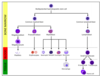
Lymphomas are divided into two broad groups. What are they?

Hodgkin lymphoma represents a special group of lymphomas which account for 1 in 3 lymphomas. What is the distribution of incidence?
- bimodal distribution of incidence
- peak in young adulthood (15-35yo)
- second peak in 55+ age group
The aetiology of Hodgkin lymphoma is poorly understood. What are the established risk factors?
- history of Epstein Barr Virus (EBV) infection
- immunosuppression eg. HIV infection
- family history
How does Hodgkin lymphoma present clinically?
- lymphadenopathy (typically supraclavicular or cervical)
- there may be ‘B symptoms’ (systemic) eg. fever, weight loss, night sweats
Diagnosis of Hodgkin lymphoma requires exicison of a node for histological examination. What is the defining pathological feature of Hodgkin lymphoma?
- presence of Reed-Sternberg cells
- the tumour cells seen in Hodgkin lymphoma
- thought to be derived from B-lymphocytes
- typically large cells w/ 2 nuclei (binucleate)
- prominent pink nucleoli imparting an ‘owls eye’ appearance
Non-Hodgkin lymphomas (NHL) account for about 2/3 lymphomas. Where do most arise?
- most arise from B lymphocytes (~90%)
- minority arise from T lymphocytes
The classification of NHLs is complex; you are not expected to know all the different types of NHL.
How can the two types of NHL be divided?
-
B-cell NHLs can be divided into 2 groups depending on their clinical behaviour:
- indolent
- aggressive
- _T-_cell NHLs usually show aggressive clinical behaviour
Describe characteristics of indolent non-Hodgkin lymphomas
- eg. marginal zone lymphoma
- tend to present w/ widely disseminated disease at diagnosis involving several nodal + extranodal sites
- typically follow a slowly progressive course
- can be controlled w/ treatment but cure rarely achievable
- survival is typically in region of 8-10 years
Describe characteristics of aggressive non-Hodgkin lymphomas and what the most common type of Non-Hodgkin lymphoma is
- eg. diffuse large B cell lymphoma (most common NHL)
- tend to present w/ short Hx of rapidly growing mass at a localised nodal or extranodal site
- typically follow a rapidly progressive course
- usually rapidly fatal if untreated but therapy may lead to complete cure
Describe the lymphoma staging system
Modified Ann-Arbor system:
- stage I - involvement of 1 lymph node region
- stage II - involvement of 2+ lymph node regions on same side of diaphragm
- stage III - involvement of lymph node regions on both sides of diaphragm
- stage IV - involvement of extranodal sites eg. liver, bone marrow, lungs
A - no B-symptoms
B - B-symptoms present (night sweats, fever, weight loss)

What is sarcoidosis and what causes it?
- multisystem disease of unknown aetiology
- characterised by presence of non-caseating granulomas in tissues + organs
- an inflammatory response to some unidentified environmental agent in a susceptible host
- infective cause has long been sought, in particular a mycobaterium due to the granulomatous response
- however, no study has equivocally proven an infective aetiology
- inhaled antigens such as pine pollen + peanut dust have been incriminated
- existence of familial clusters suggests genetic factors may be involved
Remember that a granuloma is an aggregate of activated macrophages. How do the granulomas in sarcoidosis differ from TB?
granulomas in sarcoidosis do not show caseous necrosis
Sarcoidosis may involve virtually any organ but some are involved more than others.
What is the involvement with lymph nodes?
- virtually all cases of sarcoidosis involve lymph nodes
- any lymph node may be involved
- most common are hilar and mediastinal nodes
- enlargement of these nodes usually picked up on CXR
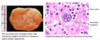
What is the involvement of sarcoidosis within the lungs?
- lungs frequently involved by sarcoidosis
- healing of granulomas may lead to varying degrees of lung fibrosis
- severe sarcoidal lung disease -> cor pulmonale (RHF due to lung disease) + resp failure
What is the involvement of sarcoidosis with the skin?
- skin involvement typically presents as lupus pernio
- an asymptomatic maculopapular rash on face and trunk
- erythema nodosum may be associated w/ sarcoidosis
What is the involvement of sarcoidosis and the eyes?
- typically causes uveitis (=inflammation of uveal tract)
- anterior uveitis -> red painful eye w/ photophobia, often self-limiting
- posterior uveitis -> floaters due to inflammatory cells appearing in vitreous w/ some blurring of vision, more chronic form of the disease
What is the involvement of sarcoidosis and the lacrimal/salivary glands?
- dry eyes
- dry mouth