Unit 4 - General Concepts of Cancer and Protecting the Genome Flashcards
cancer - biology vs molecular level
out of control cellular proliferation (bio)
damage to genetic material (mutations and epigenetic) that affect cellular proliferation
3 types of mutations in cancer cells
ONCOGENIC
TUMOUR SUPPRESSIVE
NEUTRAL
oncogenic mutation
ras
myc
cyclin D
normal growth promoting genes (proto-oncogenes) become either hyperactive or inappropriately active
dominant mutations = mutation of only 1 allele required
tumour suppressive mutation
normal growth restraining genes become lost or down-regulated
recessive mutation = mutation of both alleles required (to lose tumour suppressive function)
BRCA1 and 2
TP523
neutral mutation
cancer cells have 1000s of mutations to genes that have little or no effect on aetiology of cancer
cancer evolves a mutagenic phenotype
protooncogenes when mutated =
driving with foot on the accelerator
normal (stem) cells
low instability
no cells have genetic alteration required to overcome the selection barrier
NO TUMOUR
increased genetic instability
at least 1 cell contains the requisite genetic alteration to overcome the selection barrier (clonal selection)
barrier traversed and population of mutant cells eventually accumulates the new mutations required to cross the next selection barrier
significant step towards tumourigenesis

too much genetic instability
too many mutations accumulate to allow viability
cells die - apoptosis, necrosis
NO TUMOUR
6 selection barriers
reduced requirement for growth factors - autocrine stimulation
insensitivity to inhibitory signals
escape from senescence (cellular immortality)
evasion of apoptosis
stimulated angiogenesis
invasion/metastases
how are these selection barriers overcome
by inactivating tumour suppressors and activating oncogenes
tumours - hostile cellular environments
e.g. periods of anoxia, malnutrition
fluctuating hormonal influences and immune attack
a fertile breeding ground for mutations
similarly, bacteria with higher levels of genomic instability, but not too high, adapt to and eventually dominate new environments
challenge posed by the somatic mutation hypothesis
very low mutation rate - 1 x 10⁻¹⁰ nucleotides/cell/division for human somatic cells
diploid human genome = 6.4 x 10⁹
1016 cell divisions in a human lifetime are insufficient to permit a single cell to obtain the estimated 5-7 advantageous mutations required to produce a cancer
⇒ cancer shouldn’t occur with such a low mutation rate
the mutator (or genetic instability) hypothesis
cancer cells have significantly elevated (just-right) mutation rates
Min tumours
where is there instability
type of karyotype
prevalence
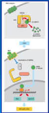
microsatellite instability
instability at the nucleotide level e.g. mutation of MMR results in 10-100 fold increase in mutation
most easily seen at microsatellites
normal karyotype
relatively uncommon e.g. 15% colorectal cancers
Cin tumours
where is there instability
karyotype
prevalence
chromosomal instability
instability at chromosomal level
not clear what mutational events initiate Cin tumours - loss of p53, mitotic checkpoints?
abnormal karyotype
most frequent - 85% of colorectal cancers
similarity between min and cin
NOT FOUND TOGETHER
same oncogenes and tumour suppressors appear to be targeted in both min and cin tumours
what is the lifetime risk of many cancers dependent on
the total number of divisions of adult stem cells in the particular tissue rather than on environmental or inherited mutations
the more cell divisions the more likely that random mutation to key cancer driver genes will occur during cell division - 65% of cancer
what explains the extreme variation in cancer incidence across different tissues
> cell divisions ⇒ the more likely that random mutation to key cancer driver genes will occur during cell division
cancer risk for different tissues
- 9% - lung
- 08% - thyroid
- 6% - brain
- 003% - pelvic bone
- 00072% - laryngeal cartilage
proportion of cancers that have an inherited component
5-10% of cancers
what effect does exposure to mutagens have
mutagens or viruses cannot account for a 24x variation of lifetime risk throughout alimentary canal
LI - 4.82%
stomach - 8.6%
oesophagus - 0.51%
SI - 0.2% - 3x LESS COMMON than brain tumours even though the brain is protected from environmental mutagens by the BBB
lifetime risk vs total stem cell divisions

tumour
an abnormal uncontrolled growth without physiological function, that can be either benign or malignant
benign tumour
confined and not life threatening
malignant tumour
invades surrounding tissue and may spread to other parts of the body
neoplasia
process of forming tumours (benign or malignant)
hyperplasia
small abnormal growth in a part of the body caused by an excessive multiplication of phenotypically normal cells
metaplasia
appearance of invading, microscopally normal cells of a type not normally encountered at that site
most frequent at epithelial transition zone e.g. oesophagus/stomach, cervix/uterus
dysplasia
small abnormal growth in a part of the body caused by an excessive multiplication of cytologically abnormal (variable size and shape, bigger nuclei, increased mitotic) cells
transitional state between benign and pre-malignant growths
dysplasia
adenomas, polyps, papillomas
large benign tumours of epithelial origin
dysplastic but not malignant
usually grow to a certain size and them stop growing
carcinoma
most common (80% of cancer deaths)
malignant tumour derived from epithelial tissue
→ surface layer of an organ/body part
e.g. GI tract
skin
breast
pancreas
lung
liver
ovary
bladder
adenocarcinoma and squamous cell carcinoma
2 main classes of carcinoma that derive from epithelia
secrete substances into cavities/ducts e.g. breast or from simple protective layers e.g. skin
leukemia (dispersed)/lymphoma (solid)
2nd most common (16%) malignant tumour type derived from haematopoietic and lymphatic tissues
sarcoma
rare (1%) malignant tumour derived from CT/muscle
e.g. fibroblast
adipocyte
osteocytes
myocytes
neuroectodermal tumours
rare (2.5%)
malignant tumours derived from central and peripheral nervous system
e.g. gliomas, neuroblastomas
most common tumours
- carcinoma
- leukemia/lymphoma
germ cell tumours (GCT)
tumours derived from germ cells
germ cell tumours can be cancerous or non-cancerous
normally occur inside gonads (ovary and testis)
testicular cancer - curable
teratoma
tumours with tissue or organ components resembling normal derivative of all 3 germ layers
although resembling normal tissues, often dissimilar to surrounding tissues (teeth and hair)
encapsulated and hence usually benign
multiple fluid-filled cysts can form within the capsule
teratoma within a large cyst can sometimes form a structure resembling a foetus
immature teratomas occasionally malignant, rare but slightly more common in males
mature vs immature teratomas
mature
typically benign
rare
more common in females
immature
malignant
rare
more common in males
colorectal cancer - cin occurence
structures in colon
107 crypts in colon
each crypt has 1000s of differentiated cells (fast growing - lot of apoptosis - 1010 cells are lost)
1-10 stem cells (slow growing and self renewing)
mutation can occur in any 1 cell cycle
early studies of tiny adenomas ⇒ that >90% had allelic imbalances of 1+ of 5 chr tested
⇒ supports the idea that CIN occurs early
85% of cases of sporadic colorectal cancer initiated by inactivation of APC (adenomatous polyposis coli)
< 10% by activation of β-catenin as both function in WNT signalling
sporadic colorectal cancer causes
85% of cases of sporadic colorectal cancer initiated by inactivation of APC (adenomatous polyposis coli)
< 10% by activation of β-catenin as both function in WNT signalling
Wnt signalling
regulated by
target
what is included in its complex
proliferation of epithelial cells is regulated by mitogen Wnt
key target = β-catenin - activates transcription of genes involved in proliferation
no Wnt ⇒ no β-catenin - constitutively associated with an inhibitory cytoplasmic complex including APC, Axin and GSK3β (glycogen synthase kinase 3β) which regulates phosphorylation of β-catenin and thus targets it for destruction via the SCF E3 ubiquitin ligase
Wnt present ⇒
dissociation of APC complex and GSK3β inactivated, therefore releasing β-catenin to perform its function

loss of APC or activating mutations of β-catenin itself in colon cancer
constitutive activation of β-catenin signalling
where else does β-catenin function
in cell adhesion
where also does GSK3β function
in glycogenesis
familial adenomatous polyposis (FAP)
associated gene
early tumour initiation
APC gene regulates colorectal cell proliferation via wnt signalling
referred to as gatekeeper

hereditary non-polyposis colorectal cancer (HNPCC)
gene
also known as
rapid tumour progression
MMR mutations cause the MIN class of genome instability
15% of sporadic colorectal carcinomas display MIN phenotype
Lynch syndrome

function of gatekeeper genes
required for net cellular proliferation
maintenance of a constant cell number in renewing populations
mutation of a gatekeeper leads to
a permanent imbalance of cell division over cell death
role of gatekeepers in different tissues
can be expressed ubiquitously
may function as gatekeepers in only 1 or a few tissues
redundant, expendable or perhaps play different roles in other cell types
examples of gatekeeper genes
NF1 in schwann cells
Rb in retinal epithelium
VHL gene in kidney cells
** not yet reported for majority of human malignancies but will be important for our understanding and future treatment of cancer
Rb and gatekeeper concept
how can a retinoblastoma arise
human retinal progenitor (stem) cells give rise to 7 different cell types but only in 1 of these, the cone-precursor, does loss of Rb result in transformation (in others - either no effect or apoptosis)
molecular circuitry of cone-precursor (possibilities identified by authors include - high expression of N-myc, SKP2 and MDM2) allows them to proliferate and become transformed when Rb is lost
specific molecular circuitry likely to be a paradigm for all so-called gatekeeper genes

what is DNA damage response (DDR)
biochemical signalling pathways that respond to structural perturbations in the genetic material e.g. DNA damage
transient “normal” structures generated during cell proliferation also sensed e.g. ongoing DNA replication prevents exit from S phase until replication is complete
DDR regulates co-ordinated cellular responses to DNA damage and replication structures = checkpoint responses
once repair is complete cells re-enter the cell cycle and resume proliferation in a regulated process - recovery
inefficient DDR =
genome instability
easily repairable lesions are converted into mutations
biological response to DNA damage

checkpoint in G1
prevents cells going into replication - damage is repaired
checkpoint in S
detects any damage
checkpoint in G2/M
prevents cells going into mitosis e.g. DNA double strand break
part of the chromosome is not attached to centromere - during mitosis chromosomes would segregate but broken fragment of chromosome would stay in the middle of the cell
BRCA1 and BRCA2
key players in the DDR
recruitment and activation of protein kinases
typical of signal transduction in general
2 TYPE OF PKs
- phosphatidylinositol 3-kinase-like kinases or PIK kinases e.g. ATM and ATR - do not phosphorylate the lipid - phosphatidylinositol, at the 3 position -OH of inositol ring, but instead serine/threonine directed protein kinases
- checkpoint kinases or CHK kinases e.g. CHK1 and CHK2
2 types of PKs - DDR
- phosphatidylinositol 3-kinase-like kinases or PIK kinases e.g. ATM and ATR - do not phosphorylate the lipid - phosphatidylinositol, at the 3 position -OH of inositol ring, but instead serine/threonine directed protein kinases
- checkpoint kinases or CHK kinases e.g. CHK1 and CHK2

ATM mutation names
pattern of inheritance
symptoms
when does it develop
ataxia telangiectasia, BOder-Sedgwick or Louis-Bar syndrome
rare, autosomal recessive disease (non-essential)
progressive loss of purkinje cells in the cerebellum, affecting motor skills (ataxia = poor co-ordination)
telangiectasia = prominent BVs in whites of eyes
symptoms develop in toddler years and post patients die by age 20 from bronchopulmonary infection and/or malignancy
increased incidence of tumours - lymphomas/leukemias
half of patients have immune problems
poor appetite
hypersensitivity to ionising radiation
ATR mutation names
pattern of inheritance
seckel syndrome
microcephalin primordial dwarfism
harper’s syndrome
bird-headed dwarfism
rare autosomal recessive disease (ATR is an essential gene - caused by hypomorphic rather than null mutations - wouldn’t get past early development)
1 of several proportionate dwarfisms of prenatal onset
severe microcephaly with a bird-like head appearance - protrusion of nose, large eyes, low ears, small chin, mental retardation
activation of PIK kinases (ATM/ATR)
ATM - DNA double strand break
ATR - single stranded DNA

activation of CHK kinases (CHK1/CHK2)
role of adaptors/mediators
CHK1 and CHK2 are released from lesion sites

G1 checkpoint
drives expression of p21

hypothesis - proto-oncogene and DNA
early in development of cancer oncogene activation of a proto-oncogene causes increased DNA replication stress resulting in activation of the DDR and cell cycle arrest or apoptosis

evidence to support that DDR is activated at early stages of lung cancer
- NSCLC, as well as for malignant melanoma that the DDR is activated at early (pre-neoplastic) stages of tumorigenesis
- bladder carcinoma, as well as for carcinomas of the breast, colon and lung that the DDR is activated at early stages of tumorigenesis
- DDR is not appreciably activated in any normal proliferating tissues e.g. normal colonic crypts, which have a higher proliferation index than most cancers, or even in tissues experiencing inflammation (hyperplasias)
IMMUNOHISTOCHEMISTRY, EXPERIMENTALLY INDUCED HYPERPLASIA, OVEREXPRESSION OF ONCOGENES
⇒ constitutive activation of the DDR pathway commonly occurs at pre-invasive stages of major types of human tumours
is the DDR activation in the earliest cancer lesions
yes - in hyperplasias as well as earliest cancer lesions

S phase promoting oncogenes and DDR
DDR induced in cultured cells upon expression of S phase promoting oncogenes

what does oncogenic activation result in
replication stress (SSBs/DSBs) which both activate DDR/checkpoint
activation of DDR
checkpoint dependent apoptosis (p53 dependent) and senescence suppress expansion of precancerous lesion i.e. tumour suppressive
subsequent mutations of DDR results in loss of these crucial tumour suppressive mechanisms and further evolution of cancerous phenotype
telomere attrition and hypoxia can also contribute to formation of DSBs

conclusions
