Tour of MCB Block 1 Flashcards
What is an enzyme?
Chemically active proteins (or RNAs)
What is the general name for the enzymes that copy a DNA strand during normal transcription?
RNA polymerase
How many chromosomes are carried by a normal diploid human cell?
46
What is the name for the barrier that separates the eukaryotic nucleus from the cytoplasm of the cell?
E. Nuclear envelope
Roughly how many protein-coding genes are found in humans?
20,000
Which of these processes is part of normal RNA maturation in eukaryotes, but not found in prokaryotes?
RNA splicing
HOw does a fully mature mRNA get into the cytoplasm of a cell?
It is exported through the nuclear pores.
The crossing-over observed betwwen chromosomes during human meiosis is better termed:
Homologous recombination
Approximately what percent of the human genome is repetitive DNA?
60%
Which of these processes is part of normal RNA maturation in eukaryotes, but not found in prokaryotes?
RNA capping
What is produced by meiosis I?
Two haploid cells containing bivalent chromosomes.
Are prokaryotic Genes “polycystronic”
NO
How much of the human genome is unique sequence?
40%
What is a SINE?
Short interspersed nuclear element
What percent of the human genome codes for functional RNAs?
1.6%
What of these is the best desription of the human mitochondrial genome?
Small 16,500 bp DNA circular chromosome
Why do we include coverage of bacterial plasmids in our course?
They are the main mechanism for antibiotic resistance in bacteria.
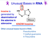
What is the correct name for a purine or pyrimidine base linked covalentlky to a pentose sugar?
Nucleoside
In adenosine monophosphate (AMP) where is the phosphate attached to the sugar?
at the 5’ position
What are the LINEs in the human genome?
Long interspersed nuclear elements.
What is a retrontransponson?
It is a genetic component that can copy and paste itself into different genomic sites-using RNA as an intermediate.
(using RNA as intermediate)
What is the size of the repeat unit in microsatellite DNA?
2-4 bp
What is the simplest name for this molecule?

Deoxyadenosine triphosphate
Which of these is a simpler way of writing pCpApTpGpGpC?
CATGGC
Specific hydrogen bonds normally only form between specific pairs of bases to drive the formation of a DNA double helix. What is the name given to this hydrogen bonding pattern in DNA?
Complementary Base-pairing.
Which of these processes is part of normal RNA maturation in eukaryotes, but not found in prokaryotes?
RNA Capping.
DNA is “incredibly thin.” Just how thin is that really?
2 nanometers wide.
You have notes showing you the shape of a DNA duplex, and you have figures showing you the DNA-binding proteins. Most DNA-binding proteins recognize specific DNA sequences. How do they read the base sequence?
By hydrogen bonding or sticking to the portions of the base-pairs exposed at the side of the double helix.
If the sequence of a DNA duplex contains 22% A, then it also contains —–% of G?
28%
22% A ——> 22% T ==> A/T
28% G —-> 28% ==> G/C
RNA is trnascribed from a DNA sequence that is an inverted repeat (a palindrome). What structure can this single strand of RNA assume?
A hairpin
An investigator is performing a “DNA hybridization assay” using a selected piece of DNA to probe for a target sequence among the millions found in his sample of chromosomal DNA fragments. He first heats the chromosomal DNA fragments in this sample to 96° C? What is he doing?
He is denaturing the sample DNA so that it melts into single-strands.
The term “stringency” is used to describe the rigor of the conditions under which annealing or hybridization of DNA strands occur. What does “high stringency” mean?
Only exact complementary base-pairing is allowed in the assay.
Do Chargoff’s rules apply to a single-stranded nucleic acid?
NO
If you deaminate the adenine component of adenosine, what nucleoside is formed?
Inosine
Chromonsomal DNA is ragmented into pieces by an enzyme. The investigator then loads small amounts of this material ont an agarose slab gel and performs electrophoresis. This procedure separates the pieces of DNA according to their ________.
A. Leght (# of bp)
HIstone proteins are designated with an H. How many basic classes of histone proteins are there?
5
Note: Even though two of the histone proteins are labeled H2A and H2B; they are 2 separate classes—-> H1, H2A, H2B, H3, H4
Techniques exist that allow investigators to track RNA polymerase II molecules. Where will this protein be found in an interphase nucleus?
Interspersed throughout the euchromatin.
Several types of histones are used to compose the core nucleosome. What histone binds to the outside edge of the core nucleosome to serve as a type of “handle”?
H1
The core histone proteins all share an important structural feauture. What is this?
They possess very flexible tails that are enriched in basic, positively charged amino acids.
(like lysine and arginine)
What happens when an enzyme acetylates lysines or arginines within core histone proteins?
Some of the negative charges on the protein are neutralized.
What happens when an enzyme deacetylates lysines or arginines wihtin core histone proteins?
Promotes the formation of heterochromatin.
There are 5 major epigenetic mechanisms known to be important in humans. DNA methylation, histone modification, and expression of variant histone proteins are well known. Which of these is another important epigenetic mechanism operating on our chromosomes?
Movement of nucleosomes by chromatin remodeling complexes.
An important process drives the inactivation of 1 of the 2 X chromosomes within every female cell. What process is this?
Directed large-scale DNA Methylation
Neurons are clearly a different type of cell than a hepatocyte. One mechanism by which tissue differentiation is accomplished involves “permanently” swtching off selected genes. What is the major way in which this is done?
DNA methylation of specific genes.
What does a DNA polymerase need– that an RNA polymerase can do without?
An exposed 3’ OH on a primer to act as a starting place.
WHich of these is true for a DNA polymerase?
Has 5’ to 3 polymerase activity.
Most DNA polymerases have a proofreading activity. What is the term for this activity?
3’ to 5’ exonuclease activity
The bacterial origin of replication is marked with specific proteins, and when a swetch is flipped, enzymes are recruited to begin replication. What is the first enzyme to begin replication?
Helicase.
2nd. Primase
3rd: Polymerase III
4rth: Polymerase I
Seal the nick Ligase.
In the figure below, which direction is the replication for moving– AND– Which single-strand is the lagging strand?

Right-to-left, top
IF you were planning ot make cDNAs against all the mRNAs exposed in liver cells, how would you ensure that only mRNA was copied into DNA during the first step?
Prime the reaction with an oligo-dT primer.
IMPORTANT KNOW PLEASE
What is the common term used to describe an RNA-dependent DNA polymerase?
Reverse transcriptase.
What is the common term used to describe an RNA-dependent DNA polymerase?
Reverse Trascriptase.
What kind of an RNA virsus carries its own reverse transcriptase to convert its genome into double-stranded DNA that can insert itself into eukaryotic chromosomes?
Retroviruses
What Kind of an Enzyme is a telomerase?
It is aribonucleoprotein that functions as an RNA-dependent DNA Polymerase.
In prokaryotes, DNA polymerase I is able to replace the RNA primer at the beginning of every Okazaki Frangment. What happens in eukaryotic Systems?
A DNA polymerase pushes the RNA stretch out to the side as a “flap” and a ribonuclease cuts it off.
Why is it necessary for a telomerase to add on some DNA to the ends of all four chromosomes?
Becuse the RNA primer that used to exit at the 5’ end of one strand in each new DNA dubplex is removed before the end of replication.
(Most Probably will not Test on Telomerases)
You use an electrical field to move things thorugh some sort of a restrictive medium. What do you call this process?
Electrophoresis.
Gel Electrophoresis separates macromolecules according to what properties?
Their size, length, number of bp.
What are the 3 basic steps in a PCR cycle?
Melting, reannealing, and syntheis.
What is the most curcial specific reagent in ‘Alllele-specific PCR”?
One specific primer that corresponds to a known mutated sequence.
Specific primers have been developed to permit an assay for the evolution of a VNTR locus in the members of a family. How will you measure the results of this assay?
The leght of the PCR product observed on an electrophoresis gel.
How does dideoxyATP differ from deoxyATP?
An additional Hydroxyl is missing from the 3; position of the sugar.
Sanger Dideoxy DNA sequencing” is based on what biochemical process?
Primer Extension.
What happens in a Sanger reaction when a small amount of ddCTP is added to the four normal dNTPs?
Some DNA chains are terminated at C.
If you had to start at time=0 with only 1 DNA duplex, how many DNA duplexes would have by the end of the PCR reaction depected below?

64
WHat is one thing that both PCR and Sanger sequencing have in common?
They are both based on primer extension.
What is the sequence shown on this sequencing gel?

GATCTGCA
Need to read the gel from the bottom to top, not the other way around.
If RED=T, Yellow= G, Blue= C, and Green= A, what is the template strand sequence that generated this profile?

CATCAAACAGG
you Read it from 3’ to 5’ and write it from 5’ to 3’
If you are hoping to use PCR to amplyfy up this region of interest, which of these is your pair of primers?
5’ GATGTACC——————CGATATGC 3’
3’ CTACATGG——————GCTATACG 5’
GATCTACC & GCATATCG
WHat is a more complete name for RNA Polymerase?
DNA-Dependent RNA Polymerase
From this duplex DNA example, which sequence below represents a possible RNA transcript?
5’ - ACGGGCATTTTAAATGC- 3’
3’ -TGCCCCAAAATTTACG- 5’
GGGCAUUUUAA
What does the abbreviation Tss mean?
Transcription start site
What DNA binding protein recognizes both the -35 box and the -10 box in a bacterial promoter?
The family of Sigma Factors.
About how large is the region of RNA/DNA duplex formed within the active site of an RNA Polymerase during transcription?
5-7 bp
Apprximately how large is the area of a single-stranded DNA held open by the RNA polymerase (and termed the “transcription bubble”) ?
15-17 bp
What mediates the Rho-independent termination of transcription bacteria?
A Strong GC-rich RNA Hairpin
What mediates the Rho-dependent termination of trnscription on bacteria?
The-Rho Helicase.
What streptomyces-derived antibiotic directly binds to the bacterial RNA polymerases to inhibit thier activity and stop transcription?
RIfampin
What RNA is produced by eukaryotic RNA Polymerase I?
MOst of the ribosomal RNA.
One of the most important eukaryotic basal transcription factors is built aroun da protein that is often simple referred to as a “TBP”. WHat is TBP?
The TATA Box Binding Protein
Of al the basal transcription factos TFIID seems important? What does it do?
Position the RNA Polymerase and the other factors at the Tss.
If you are hoping to use PCR to amplify up this region of interest, which of these is your pair of primers?
5’ CGATATGC———-GATGTACC-3’
3’ GCTATACG———CTACATGG-5’
GGTACATC & CGATATGC
What is an important structural feauture of RNA Polymerase II that is not found in RNA Polymerse I or III?
IT has a Prominent CTD tail
RNA Elongation is tightly coupled to RNA processing. THe enzymes responsible for the processing steps ride on the phosphorylated CTD tail until an RNA sequence is revealed that they recognize. What is the FIRST RNA-processing step?
5’ Capping
Which one of these is an enzyme that is involved in capping a nascent RNA made by RNA polymerase II?
Guanylyltransferase.
Which on of these is an enzyme that is involved in capping a nascent RNA made by RNA polymerase II?
Guanine-7-methyltransferase
The ribonucleoprotein molecules that splice the eukaryotic preRNAs have an interesting abbreviation that reminds everyone of an old animated cartoon show? what are these?
snRNPs
What is the name of the GU site at the 5’ end of an intron that is recognized by the U1 snRNP at the beginning of the RNA-splicing Process?
Splice Donor site
What is the name for the AG site at the 3’ end of an intron that is cut by the snRNPs to release the intron as a lariat structure?
Splice Acceptor Site
Cleavage of the nascent RNA from RNA polymerase II is tightly coupled to recognition of an RNA sequence within the 3’ UTR of the sequence. THe protein recognizing this sequence also perfom the final processing step as soon as the RNA comes free. What enzyme do this?
Polyadenylate Polymerase
Multiple proteins cote the poly(A) tail and a protein complex covers the 5’ cap. What marks the sites where the introns were spliced out?
Exon Junction Complexes
What is used to mark the areas within a mature mRNA where introns have been spliced out?
Poly(A) binding proteins
An important structural protein is encoded by a single gene. Cellular analysis indicates that the protein is not modified in a major way following translation, yet different versions of this protein (long, short, and medium-sized) exist in diferent tissues such as neurons, kidney and stomach. How must these different-sized version of the protein be generated?
Alternative RNA Splicing
What lable indicates the 3’ end of the coding strand?

D
What is one important feauture of an eukaryotic mRNA that determines its half-life?
Lenght of the poly(A) tail
Where would one find most of the RNA processing enzymes wihtin an eukaryotic cell?
In the nucleus, largely concentrated on the CTD tail of the RNA polimerase II.
What is the last step in the process of a transfer RNA?
CCA is added to the 3’ edn
What is one very important DNA element athat helps to position RNA polymerase II and the basal transcription factors in the correct position near Tss?
The TATA bos
What is the most crucial modificaiton of the nascent preRNA coding region that results in a mature mRNA?
Splicing
If the microRNA that brings the RISC complex to an mRNA sequence has 100% homology with that sequence, what will happen?
RISC will bind tightly to that sequence and the Argonaute endonuclease will splice up the mRNA to destroy it.
There is a 5’ UTR and a 3’ UTR in an normal mature human RNA. What does UTR stand for?
Unstranslated Region
A large primary miRNA molecule is processed to procduce a pre-miRNA molecule that is then diced to produced an immature miRNA duplex. What kind of an RNA is a mature single-stranded microRNA?
A guide RNA
WHat is the average half-life of most bacterial mRNAs?
Only a few minutes.
What does RISC stand for?
RNA-induced silencing complex
WHat RNA is produced by eukaryotic RNA polymerase II?
All of the tRNAs + 5s rRNA
If you are hoping to use a PCR to amplyfy up to this region of interest, which of these is your pair of primers?
GCATATCG————GGTACATC
CTACATGG———-CCATGTAG
GCATATCG & GATGTACC
Which kind of an RNA is CRISPR RNA?
A Guide RNA
What is the 3-letter abbreviation for the aminoacid Isoleucine?
Ile
WHich of these is NOT a grouping of amino acids with similar properties?
Alkaloid
What is linked together to form a polypeptide bond?
Amino Acids
Which of these is a peptide bond?

A
Protein structure seems constructed from “secondary structures”. One of the most important is the alpha-helix. This stiff coil is usually at least 10 residues long and is stabilized by hydrogen bonds between the oxygen of the carbonyl group and the hydrogen of the amide group______/
On either side of the two peptide bonds spaced 4 aminoacids apart.
You are deducing the sequence of a protein from reading the series of coding elements (codons) on an mRNA that you have sequenced. You notice that sequence leu-ile-trp-leu. Where would you predict this sequence will be found in a final folded protein?
Tucked down inside a hydrophobic pocket.
Whic of these is an amino acid that has a side chain that will function as an aice in a chemical reaction and bear a negative charg at pH 7.2?
Glutamate
What are the circled reactions in this depiction of a protein complex?

Contact between subunits
Which one of these is amino acids has a side chain that is polar but will not bear a formal charge at pH 7.2
Leucine
Tryptophan
Isoleucine
Asparigine
Proline
ALanine
Asparigine
What is the main force holding together secondary structues as protein folds?
Hydrogen bonds betwwen portions of the polypeptide backbone.
WHa tis the term used to describe a 180 degrees reversal in a protein strand and Which amino acid is usually involved in this structue?
Beta sheets; Proline
What is the single-lettter abbreviation for Lysine?
K
Especially important in extracellular and secreted proteins such as insulin, what is the most common type of covalent bond used to stabilize a protein’s tertiary structure?
Cys-Cys disulfide bond.
What is the most prevalent and important type of interaction that holds togeter the primary sequence of proteins into the known secondary structures?
Hydrogen Bonds
What type of an amino acid is valin considered to be?
Nonpolar, aliphatic
After denaturation an reduction, proteins are separated according to their size (chain leght) in a way very similar to the way in which DNA is sized an agarose gels. What is the common name for this method of sizing proteins?
SDS polyacrylamide gel electrophoresis (PAGE)
Which of these proteins would elute first from a size-exclusion chromatography column?
Myosin, 200,000 daltons.
What is the specific term used to describe proteins from diferent species that have extemely similar structures and idential functions?
Orthologs.
What is the name for the type of electreophoresis that causes proteins to concentrate in a pH where they carry no net charge?
Isoelectrofocusing
Many proteins seem to be constructed from semi-independent modules that fold and function largely independent of the rest of the protein. These 3D modules are termed:
Domains
In this depiction of hemoglobin, what is indicated by the arrow?

Beta reverse turn
Which of these proteins would elute first from a cation-exchange column?
7-methylguanyltransferase, 1 positive and 7 negative surface charges.
The following sequence is predicted from the series of coding elements (codons) on an mRNA: asn-Gly-Thy-Ser-Thr. Where would you predict this sequence will be found in the final folded protein?
Exposed to water in a hydrophilic area.
Which of htese analytical methods would give the result shown below?

Mass Spectroscopy
What is the circled region in this ribbon depiction of a multifunctional vitamin D receptor protein?

An important domain.
ANtibody proteins have the strongest binding properties that we know about. Which of these values would describe the binding of a very high affinity antibody to its specific antigen?
KD = 10-12 M
A Binding prtein is depicted below that shows a KD of 10-9 for its ligand. If the ligand is presented at a 10-9 M concentration, then what percent of it is bound by the protein?
50%
WHich of these proteins would be the first to elute from an anion-exchange column?
Rennase, 6 positive and 2 negative surface charges.
How many tRNAs match up with athe codon UAA?
0
With what protein do the eukaryotic release factors interact to cause normal termination of trasnlation?
PABP
Which factor causes translocation of the ribosomes in eikaryotics?
eEF-2-GTP
In eukaryotes, where is the first initiator tRNA placed by the initiator factor?
P site.
Does f-Met ever show up during protein synthesis in eukaryotic cells?
Only within the mitochondria.
Both chloramphenicol and ricin do similar things to prokaryotic or eukaryotic ribosomes, respectively. WHat do they do?
Inactivate the peptidyl transferase activity .
Which factor causes translocation of the ribosome in prokaryotes?
eEF-2-GTP
What codon Serves as the Start codon?
AUG
If an mRNA codon reasd GAU, then what is the tRNA anticodon?
AUC
Approximately how many high-energy phosphates are spent for each aminoacid that is linked into a protein?
4
Some cells can use as many as 70 different tRNAs during their lifetime. How many aminoacyl-tRNA synthetases do they use?
70
WHat is the size of the larges rRNA found in the eukaryotic ribosomes?
28S
Which prokaryotic antibiotic r= produces an effect on translation very similar to that produced by the eukaryotic diphteria toxin?
Erythromycin
Which factor brings the sencond charged tRNA into the A site of a prokaryotic ribosome?
EF-Tu-GTP
In what way is an aminoacyl-tRNA “ Charged” ?
An amino acid is linked to the 3’ A thorugh an acyl link.
WHat is the name of hte “ ribosome recognition site’ found in many prokaryotic mRNAs?
Shine-Delgarno Site
How does puromycin work?
It tricks the ribosome into attaching the nascent polypeptide to it instead of a new aminoacyl-tRNA
What catalyzes the formation of the peptide bond in eukaryotes?
28S rRNA
What brings the new aminoacyl-tRNA into a site of the eukaryotic ribosome?
eEF-2-GTP
What factor causes translocation of hte ribosome in prokaryotes?
EF-G-GTP
During the general protein folding process, what forms first-right after the promary sequence is laid down?
Secondary structure.
What are the names of the proteins that guide a nascent protein along a maturation pathway and oftern help it forld correctly into an approximation of its final shape?
Protein chaperones
Wha tis one feauture in a partially folded protein that the chaperon proteins complex seem to recoginize?
Exposed hydrophobic patches.
Which of htese bacterial antibiotics binds specifically to the small subunit portiion of the site and blocks anything else from binding there?
Tetracycline.
What factor causes translocation of the ribosome in prokaryotes?
EF-G-GTP
Generally, when a large “ precursor protein” is trimmed down to size or cut up to generate multiple smaller proteins, this is referred to as:
Post-translational proteolytic processing
Fifty to sixty percent of human proteins are directed into the lumen of the rough endoplasmic reticulum while being translated. THese proteins are destined to be associated with membranes for much of their life, and many of these proteins are secreted. What directs these proteins into the rER?
A signal sequence.
Which of these is not a postranslational modification typycally foun din normal proteins?
GLycation
What is the name of an enzyme that attaches a phosphate group to the side chain of an amino acid wihtin Protein Chains?
Kinase
What is the name of an enzyme that removes a phosphate group from the side chain of an amino acid within a protein chain?
Phosphatase.
Which amino acid is know to be hydroxylated in certain proteins to modify how tertiary and quaternary structures can form?
Pro
A nonsense-mediated mRNA decay, the UPF proteins promote and mediate interaction betwwen the eRF’s and an importatn proteins . THis interaction triggers the recruitment of decapping enzymes, exonucleases and endonucleases that destroyu the mRNA. What is the protein complex that interacts with the other proteins?
EJC
Which aminoacid can by carboxylated to form a binding site for calcium ion?
Glu
What is the name given to infectious proteins that procduce mammalian spongiform— such as scrapoie or Creutzfeldt-Jacob Disease?
Prions
Which aminoacid is known to be hydroxylated in certain proteins to modify how tertiary and quaternary structures can form?
Pro
What is the given name to the inactive precursor form of the enzyme chymotrypsin?
Chymotrypsinogen
What is the common name given to a membrane immunoblot- a method wherein proteins are sized by SDS PAGE, transferred to a membrane and probed with a specific antiboy—- THat is used to dected proteins?
Western Blot
What type of post-translational modification is dependent on fat soluble Vitamin K.
Carboxylation
What is the name of the enzyme that attaches ubiquitin(s) onto a phosphorylated lysine in a protein?
Ubiquitin Ligase
Which Amino acid is linked to the specific type of oligosacharide chain that is attached to nascent proteins inside the rough endoplasmic reticulum?
Asparagine
Aside from possible co-translational removal of the signal sequence, the trimming or curring up of the laarge “precursor protein” to produce multiple smaller proteins is referred to as?
Post-translational proteolytic processing.
Inside which cellular compartment is an O-linked glycoprotein made by adding certain types of oligosaccharide to a serin or threonine?
Golgi body
Which of these protein complexeds is essential to the operation of the molecular mechanism that brings the nonsense-mediated mRNA decay in mRNAs that possess a “premature stop codon”?
EJC
Which of these protein modificatarions would provide a signal that leads to the old protein being ground up and recycled by a proteasome?
Attachment of several ubiquitins in a chain to a phospholysine
What is caused by attaching a fatty acid chain to a C-terminal cysteine of a protein?
Protein is tethered to a membrane.
What is this diagram trying to show up?

THe alpha to beta transition typical to the protein misfolding diseases.
What is the name of the compound shown below?

Dimethyl Lysine
Which amino acid can be carboxylated to form a binding site for calcium ion?
Glu
From your study of this topic and your inspection of the recorded lecture, which kindd of secondary structure wil interact with others like it so that they will stackup like a deck of cards?
Beta sheets
What does the “chromosomal mutation” mean?
There are large-scale change to chromosomal sequences-large rearrrangements, deletions, or insertions involving many genes.
Which of these could be considered a “genomic mutation? “
THere is a achange in chromosome number.
What is “polyploidy”?
Number of all chromosomes is increased in a uniform manner; increased set of chromosomes
What is aneuploidy?
Additional or missing of one to several individual chromosomes.
What does the term “gene mutation” mean?
There are extra or missing copoies of specific genes or areas within genes- or there are changes in sequence within individual genes
Where do “genomic mutations” come from?
Mistakes in meiosis
What are the main sources of DNA damage that generates “gene mutations?
Replication error, spontaneous or induced physiochemical changes
What is a cause of increased base deamination?
PResence of nitrosamine and other nitrous acid precursors
If a cytosine base is deaminated– what is produced?
Uracil
If the base in an adenosine nucleside is deaminted–What is produced?
Inosine
Incresed UV irradiation is linked to a large increase in skin cancer over the past 30 years. How does UV light directly damage our DNA?
Produce pyrimidine dimers
SIngle base substitutions are often referred to as point mutations. WHat type of a base substitution is shown below.

Missense
Single point mutations can have big effects. What type of mutation is this?

Frameshift
Single base substitutions are often referred to as point mutations. What type of base substitution is shown below?

Nonsense
Mistakes occur during replication. Many are corrected through polymerase activity know as________?
Proofreading
Suppose an alkylating agent has added a methyl group to a guanine base to produce O-methylguanine. What’s the problem with that?
O-methylguanin will pair up with thymine instead of cytosine.
If a mistake escapes proofreading by an eukaryotic polymerase, what type of repair si still possible immediatly after the polymerase has moved on down the sequence?
Mismatch repair.
If there is a mistake on one strand of a DNA duplex, what general type of repair mechanism will make the needed corrections?
Some version of excision repair.
What is the main cause for the autosomal dominant disease, hereditary nonpolyposis colorectal cancer?
Mutations in MSH2 or MLH1 genes
What is the main cause for the autosomal recessive disese Xeroderma Pigmentosum?
Mutations in the XP gene family.
What is one possible reason for diseases caused by a dysfunctional base-excision repair mechanism?
Mutations in the gene encoding the AP endonucleases.
What is one possible cause for diseses that feauture defects in double-stranded DNA repair pathways?
Mutation in ATM or BRACA genes
What is the first enzyme to go into action during base excision repair?
DNA glycosylase
How does base excision repair differ from nucleotide excision repair?
Repairs specific damage to individual bases– rather than damage to several nucleotides.
A double stranded RNA genome isolated from a virus in the stool of a child with gastroenteritis was found to contain 15% uracil. WHat is the percentage of Guanine in this Genome?
35%
Endonuclease activation and chromatin fragmentation are characteristic feautures of eukaryotic cell death by apoptosis. WHich of the following chromosomes structues is the least dense (loose DNA) and therefore will be degraded first?
A. Barr body
B. 10-nm Fiber
C. Centromere
E. Heterochromatin
B. 10-nm Fiber
Dyskeratosis Congenital (DKC) is a genetically inherited disese in which the proliferative capacity of stem cells is markedly impaired. THe defect has been traced to inadequate production of an enzyme needed for chromosome duplication in the nuclei of rapidly dividing cells. Structural analysis ahs shown that the active site for this progein contains a single stranded RNA that is required for normal catalytic funciton. Which step in DNA replication is most likely deficient in DKC patients?
A. Synthesis of Centromeres
B. Synthesis of Okazaki fragments
C. Synthesis of Telomeres
D. Synthesis of RNA Primers
E. Removal of RNA primers
C. Synthesis of Telomeres
In the human genome there are an estimate of 20, 000 genes that code for proteins. What is the percentage of the genome that is protein-coding?
1.5 %
In huntingtoon disease, the severity of the disese in patients is proportional to the number of CAG repeats in the Huntington gene. THe three base repeat in Huntington disease would be classified as what type of repeat?
A. LINE
B. SINE
C. Minisatellite
D.Satellite
E. Microsatelite
E. Microsatellite
Which of these DNA sequences woud take the most energy to break apart from it complement strand?
A. ATTACGTA
B. CAATTAAG
C.CGATTACG
D. ACGCGCGT
D. ACGCGCGT
Find the Palindrome.
A. ATTAGCGCGA
B. TATAGTATAG
C. CACGTACGTG
D. GCGTAATGCG
C. CACGTACGTG
Which of these levels of DNA organization is considered as Heterochromatin?
A. DNA strand
B. 10-nm Fiber
C. 30- nm Fiber
D. Loop
D. Loop
Which of these DNA polymerases is the only one that has 5’-3’ Exonuclease activity?
A. DNA Polymerase I
B. DNA Polymerase II
C. DNA Polymerase III
D. DNA Polymerase Delta
E. DNA Plymerase Epsilon
DNA Polymerase I
In what stage of the Cell cycle does synthesis of DNA occur?
S.
In an experiment you use RNA polymerase without its sigma factor for transcriotion. WHat will be the result that you observe?
A. More transcription
B. Less transcription
C. More random Transcription
D. More specific transcription
C. More Random Transcription
A cruel PA in a research group has decided to mix up four different antibiotics and expects you to figure out which substance is which antibiotic. You begin examining the first substance on an unsuspectign bacterium and found the substance binds strongly to the double stranded DNA betwwen a GC pair prevention transcription. This antibiotic is?
A. Actinomycin
B. Puromycin
C. Streptomycin
D. Rifampin
Actinomycin
After beating the PA by correctly deducing each antibiotic, the PA decides to play one more prank. THis time you are given a sample with the words alpha-amanitin written on the dish. The PA ask to describe the effects of this substance on a eukaryotic cell. Being somewhat fair, the PA tells you there is a significant decrease in all mRNA of the affected cell?
A. Inhibit RNA polymerase II
B. Inhibit Rna polymerase I
C. Inhibit RNA polymerase III
D. Inhibit RNA Primase
Inhibit RNA Polymerase II
Which factor phosphorylates the serine residues in RNA pol II CTD?
A. TFIIA
B. TFIIB
C. TFIIH
D. TFIID
C. TFIIH
Which nucleotide is present in 5’ cap?
A. ADP
B. GDP
C. CDP
D. UDP
GDP
Splicing consensus sequence is ______ ?
A. Exon/GU-intron-AG/exon
B. Exon/UG-intron-AT/exon
C. Exon/GU-intron-CA/exon
D. Exon/AU-intron-CG/exon
A. Exon/GU-intron-AG/exon
The poly a tail protects the 3’ end from _______?
A. 5’-3’ Exonucleases
B. 3’-5’ Exonucleases
C. Translation
D. Export
B. 3’-5’ Exonuclases
Which snRNP are involved in the lariat formation during splicing?
A. U1 and U2
B. U4 and U6
C. U1, U2, U5
D. U2, U5, U6
D. U2, U5, U6
A 10-year-old boy with severe progressive skin ulceration, decreased resistance to infection, and impaired cognitive ability has been diagnosed with a genetic deficiency of the enzyme Prolidase. Mutation analysis has indentified a single base substitution at the 3’ end of intron 6 of the mutant allele as well as a deletion of a 45-base exon (the entire exon 7) in the Prolidase cDNA. Which type of gene mutation was most likely inherited by this boy?
A. Frameshift mutation
B. In-frame mutation
C. Missense mutation
D. Nonsense Mutation
E. Splice-site mutation
E. Splice-site muation
Eukaryotic ribosomal subunits are_____?
A. 30s and 50s
B. 40s and 60s
C. 40s and 80s
D. 50s and 70 s
B. 40s and 60s
Which of these is transcribed by RNA pol III?
A. 16s
B. 28s
C. 5.8s
D. 5s
D. 5s
The catalytic activity (peptidyl transferase) of the ribosome is located in _____?
A. EF-G
B. 50S ribosome subunit
C. 30s ribosome subunit
D. EF-Ts
B. 50s ribosome subunit.
Puromycin is a ribosomal antibiotic. It binds to ____?
A. E site of the ribosome
B. 23S subunit
C. A site of the ribosome
D. 16S subunite
C. A site of the ribosome
In an experiment you add streptomycin at low concentration first and then gradually increase the concentratrion till it completely inhibits peptide formation. What will be its effect at low concentration?
A. No effect
B. Shorter Peptide
C. Slower formation
D. Wrong aminoacid incorporation ( Misreading)
D. Wrong amino acid incorporation (Misreading)
Which initiation factor prevents association of ribosome when not bound to mRNA?
A. IF1
B. IF2
C. IF3
D. IF4
C. IF3
Accumulation of heme in reticulocytes can regulate globin synthesis by indirectly inactivating elF2. Which of the following steps is most directly afected by this mechanism?
A. Attachement of spliceosomes to pre-mRNA
B. Attachement of the ribosme to the ER
C. Met-tRNAmet binding to the P-site
D. Translocation of mRNA on the ribosome
E. Attachment of RNA polymerase II to the pronmoter
C. Met-tRNAmet binding to the P-site
WHich of the enlongation factor binds to the aminoacyl tRNA?
A. EF-Tu
B. EF-Ts
C. EF-G
D. GDP
A. EF-Tu
What is the eukaryotic homologue for EF-G?
A. eEF1
B. eEF2
C. eEF3
D. eEF4
B. eEF2
What protein modification gives a signal for degradation by the proteasome?
A. Polyubiquination
B. Monoubiquination
C. N-glycosilation
D. Methylation
A. Polyubiquination
The deficiency of an excision endonuclease may produce an exquisite sensitivity to ultraviolet radiation in xeroderma pigmentosum. Which of the following functions wuold be absent in a patient deficient in this endonuclease?
A. Removal of introns
B. Removal of pyrimidine dimers
C. Protection against DNA virus
D. Repair Of mismatched bases during DNA replication
E. Repair of mismatched bases during transcription
B. Removal of pyrimidine dimers.
Cytosine arabinoside is used as an affective chemotherapeutic agent for cancer, although resistance to this drug may be developed. In certain cases, resistance is related to an increase in the enzyme cytidine deaminase (Takes away the amine group from cytosine) in the tumor cells. This enzyme would inactivate inactivate the agent by turning cytosine into?
A. Cytosine
B. Cytidylic acid
C. Thymidine
D. Uracil
E. Adenin
D. Uracil
Hereditary nonpolyposis colorectal cancer (HNPCC) is an Autosomal dominant disease that makes up 2-6% of colon cancer patients. It is mainly characterized by two feautures: The expasion or reduction of microsatellites in DNA and mutation in a gene affecting which type of DNA repair?
A. Base Excision Repair
B. Nucleotide Excision Repair
C. Homologous Recombination
D. Non-Homologous Recombination
E. Mismatch REpair.
E. Mismatch Repair.
Which on of these changes is considered as a transversion?
A. A-> C
B. C-> T
C. G-> A
D. T-> U
A. A->C
Which of these proteins belongs to the Base Excision repair mechanism?
A. BRCA1
B. XPC
C. DNA Glycosylase
D. MSH2
C. DNA Glycosylase
Which Type of mutations occur the most frequently?
A. Aneuploidy
B. Chromosomal Translocation
C. Non-sense Mutation
D. Missense mutation
A. Aneuploidy
Which DNa repair mechanism is used to fix methylation?
A. Base Excision Repair
B. Nuecleotide Excision Repair
C. Homologous Recombination
D. Non-Homologous End Joining
E. Mismatch Repair
A. Base Excision Repair
During Bacterial mismatch repair, how does the bacteria know which strand is the original parental strand?
A. Detecting the shine-delgarno sequence
B. Detecting proximal enhancer elements
C. The original strand is methylated unlike the new one.
D. It just knows
C. The original strand is methylated unlike the new one.
Which of the following mutations would cause an in-frame mutation?
A. A 4-base mutation
B. A 22-base insertion
C. A 28-base deletion and a 4-base insertion
D. A 15-base insertion and a 2-base deletion
C. A 28-base deletion and a 4-base insertion
In Sickle Cell Disease, there is a single-base mutation that changes glutamine into a Valine. THis mutation would be considered as?
A. Silent mutation
B. Conservative Missense mutation
C. Nonconservative Missense mutatioin
D. Nonsense mutation
C. Nonconservative Missense mutation
A genetic element is found to contain several genes and to catalyze its own movement wihtin and between chromosomes. Inverted 35-nucleotide repeats are present at the ends of this genetic element. This genetic element in hepatocytes is incapable of replicating independently. Based on this description, this genetic element is best categorized as which of the following?
A. Complex Transponson
B. Insertion Sequence
C. Plasmid
D. Regulon
Complex Transponson
Transfer RNAs
Function as adaptors that match mRNA codon to aa at the ribosome
Ribosomal RNAs
Participate in protein synthesis
Messenger RNAs
Code for proteins
Describe the properties of the genetic code.
- 3 bases= codon
- 64 codons: 1 start + 3 stop codons
- Start Codon: AUG (Met)
- Stop Codons: UAA, UAG, UGA
- Redundant
- Directional products are always made 5’ -> 3’
Explain the WOBBLE Hypothesis
- Wobble happens between 5’ base of the anticodon and the 3’ base of the mRNA codon
- Redundancy
Describe the classes of mutations that result from changes to a codon or a set of condons.
NONSENSE
- Produces a STOP codon too early in the RNA sequence
- Disrupts termination steps
- UAA, UAG, UGA
Explain why aminoacyl-tRNA synthetases are the molecular determinant of the genetic code.
Aminoacyl-tRNA synthetase carry out 2 coupled rxns:
- Amino acid is activated by rxn with ATP (This Costs 2 phosphatases and forms AMP intermediate)
- Activated amino acid is coupled to the terminal 3’ A in CCA of the tRNA by the same enzyme
They Determine which aa corresponds to a specific anti-codon
- Each enzyme is specific for ONE aa and all the matching tRNAs
- aa-tRNA synthetases can edit to correct incorrect coupling
Describe the protein and rRNA components of both prokaryotic and eukariotic ribosomes.
1. Prokaryotic
- Smaller: 70s
- Subunits: Large: 50S (23S: Catalyzes peptide bond formation) Small: 30S (16S)
2. Eukaryotic
- Larger: 80S
- Subunits: Large: 60S (28S) Small: 40S (18S)
3. Functional Sites:
- A site: Acceptor holds mRNA
- P site: Peptidyl transferase site hold mRNA codon
- E site: Exit
- Component of Shine-Dalgarno sequence found at 4’ end of the 16S rRNA within the small subunit.
- 23S rRNA in the 50S subunit is the peptidyl transferase (ribozyme)
- 28S rRNA has the peptidyl transferase activity within the eukaryotic 60S subunit.
Describe the processes of initiation, elongation and termination of translation in both Prokaryotes
Prokaryotic Translation:
-
Shine-Dalgarno: mRNA recognition sequence
- Pairs with the 3’ end of the 16S rRNA
- Translation is INITIATED on the next downstream AUG
- In the ribosomal P-site
- __There are multiple sequences because prokaryotic mRNA is polycistronic
-
Translation Initiation Factors.
-
IF-2 is the star
- Binds to GPT
- G-protein
- When the large 50S subunit shows up
- IF-2 splits the GTP to GDP and releases all
-
IF-2 is the star
-
Translation Elongation Factors:
-
EF-Tu (temp unstable)
- The GTP bound protein
- EF-Ts (Temperature stable)
- The G-exchange protein
- EF-G
- Translocation via hydrolysis of GTP
-
EF-Tu (temp unstable)
-
Tranlation Termination
- No tRNA for STOP codon
- Release factors
- ~ 4 GTP per aa
KEY POINTS
► EF-Tu is the G-Protein that delivers the aminoacyl-tRNA to ribosome
► IF-1 Prevents the use of A-site for initiation
► IF-3 Prevents 30S and 50S from binding at one time
► IF-2 is the Starter
► The actual signal for IF-2 to comoe in when IF-2 splits GTP to GDP
Describe the processes of initiation, elongation and termination of TRANSLATION in Eukaryotes
Eukaryotic Translation
- Initiation factor:
- elF-1
- elF-2
-
Elongation factors:
- eEF1: brings in new tRNA
- eEF2: Causes ribosome to shift
- Termination:
- Finds STOP codon
- eRF interacts with PABP for dissassembly
KEY POINTS
► Ribosomes displaces all EJCs, reaches STOP codon
►eRF interacts with STOP codon and PABP for disassembly
► mRNA is now ready, FREE of EJC and can be translated.
►elF-2- GTP helps look for AUG Start
►elF-1- GTP brings in the new aminoacyl-tRNA then leaves as eEF-1- GDP
►28S catalyzes peptide bond formation
Explain the function and significance of N-Formyl methionine incorporation into bacterial proteins
- Your body launches an immune response when fMet is detected in extracellular fluids
- Either proteins invading bacteria or proteins released from damaged mitochondria
Give a brief overview and explain the significance of nonsense-mediated decay.
- EJC/UPF recruit decapping enzymes and exo and endonucleases.
- Poly-A tail is removed
- mRNA is degraded
Provide a general estimate of energy consumption during translation.
About 4 GTP per aa
Describe how translation inhibitors, which are used as antibiotics or are toxins, interfere with the process of translation.
1- Inhibition of protein synthesis (Small subuinit-30S)
- Streptomycin
- Binds to 30S subunit
- Distorts shape to cause misreading
- Inhibits initiation of translation
- Tetracycline
- Blocks the A-site
- Nothing can bind
- Puromycin (NOT PROKARYOTIC SPECIFIC)
- Structurally similar to the 3’ end of aminoacyl tRNA
- Binds to the A-site and participates in peptide bonding
- Causes premature termination
- Works in both Eukaryotes and Prokaryotes
2- Inhibition of protein synthesis (large subunit-50S)
-
Chloramphenicol
- Blocks bacterial peptidyl transferase
-
Erythromycin
- Blocks the translocation step
- Resistance is mediated by plasmids
- Sdenosine is methylated
3- Toxins for Eukaryotes
-
Diphtheria: Inhibits translocation by deactivating eEF-2
- ADP-ribosylating a histidine
-
Ricin: inactivates the peptidyl transferase activity
- Depurinating a specific A in the 28S rRNA
- Shigella toxin does the same thing
Central Dogma:
1- Replication: Exact copy (DNA code for DNA)
2- Transcription: rewrite (DNA code for RNA)
3- Tranlation: Change languate (RNA code for Protein) (mRNA is use to make Proteins) Ribosomes
Eukaryotic VS Prokaryotic Genomes
- Eukaryotes
- Protein coding genes ~ 20,000 mRNA only
- Non-Protein coding genes ~23,000+ miRNA, siRNA, tRNA, rRNA, NO mRNA
- Total Genome 6,200 Mbp
- Monosistronic, 1 Gene/mRNA
- 1 gene/ 100, 000 bp
-
Prokaryotes
- Protein coding genes ~ 4,300
- Non-protein coding genes ~4.6 Mbp
- Total genome 4.6 Mbp
- 1 gene/ 1,000 bp
Monocistronic
Each mRNA produced will only carry content from one (1) gene
Polycistronic
Only one promoter but independent ribosome binding sites for each gene, they’re Called
→ Shine-DAlgarno sequences
- NO SPLICING, because no introns in the prokaryotes
- gaps betwwen polycistronic genes are NOT introns- Eukaryotic-Encoding Gene Protection
- 5’ cap = signify mature RNA
- poly-A tail = how long mRNA will last but it’s ( Not Encoded by gene)
Meisosis
- Cell division= 4 haploid cells
- Crossing over only happens in Prophase (I) → (average of 2 crossover every meiosis)
- Meiosis I
- 2 haploid cells
- Bivalent chromosomes = 2 pairs of homologous chromosomes held together by at least one DNA crossover.
- Meiosis II
- 4 Haploid cells
- Monovalent chromosomes = exists as just one chromatid
Content of Human Genome
- Hast 2 components
- Nuclear
- Linear DNA (46 Chromosomes Diploid)
- Mitochondrial
- Circular DNA
- Nuclear
- Unique Sequence DNA (40%)
- 1.5% Codes for protein-coding Exons
- 1.6% COdes for Functional RNA
- Repetitive Sequence (60%)
-
Human Mitochondrial Genome
- Circular DNA
- 16,500 bp/ genome
- Densely packed with genes
- 37 genes encoded
- Most proteins involved in oxidative phosphorylation and ETC
- NO HISTONE invoved.
Bacterial Plasmids
- 4,000-24,000 bp in size (Circular Chromosome)
- Self- replicating
- Response to stimulus (antibiotic exposure)
- Used recombinant DNA technology
Repetitive Sequences
- Tandem repeats
- Satallite
- 68 to 171 bp extended to millions of repeats
- In centromeres where spindles attach
- Very Structural
- Minisatellite
- 6 to 64 bp
- Telomeres (process of aging)
- Polymorphic= vary from person to person
- Hayflick limit: mechanism detects and prevents mitosis
- Used in DNA markers in DNA fingerprinting and allele tracking
- Microsatellite
- AKA Short Tandem Repeats (STRs) or simple sequence repeats (SSRs)
- 2,3,or 4 bp
- Polymorphic
- Used in DNA fingerprinting and allele tracking
- Satallite
- Interspersed Repeates
-
SINE- Short Intersperse Nuclear Elements
- Alu elements= most abundant sequence ingenome, Restriction Enzyme →role in unequal crossing over resulting in chromosomal deletions/duplications/ inversions
-
LINE- Long Interspersed Nuclear Elements
- ~6,000 bp
- 21% of genome
- Transponsons & Retrotransponsons (they can cause mutations)
- aka: jumping genes: when they jump, they generate diversity of sequence.
Retrotransponsons
Class I Retrotransponsons
- 18% of Human Genome
- Copy and paste mechanism
- Use reverse transcriptase to turn RNA back to DNA (CDNA)
Transponsons
Class II Transponsons
- 3% genome
- Cut and paste mechanism ⇒ Require Transposase (enzyme)
Human Genomic sequences vary by:
~ 0.1 %
~ 3 million base-pairs differ out of
3 million base-pairs
Individuals carry ~10 unique….
‘Sequence Variants’
Variations in sequence arise by mutation and then…..
spread thorughout the population during evolution causing genetic POLYMORPHISM
NATURAL SELECTION: (Per Darwin, 1959)
- Selected for variants that conferred a survival advantage
- Selected agianst variants that conferred a survival disadvantage (ie. , disease- causing mutations)
- Variants that converred neither advantage or disadvatage spread more randomly
Describe POLYMORPHISM
Simply means having things that exist in different forms. Polymorphism, as used here, describes the condition of having variation in sequence for known genes = having different versions of a known gene. Most polymorphisms represent small changes in sequence.
Alleles:
Different versions of an identified gene normally differ by only a small number of bps
Wild-type allele
The most comon allele for a gene within a given population
A Polymorphism:
An allele that is less common than the wild-type allele, but occurs more than 1% of the time
Variant
Generally just refers to a change in DNA sequence (that may or may not produce changes in observed feautures)
Rare Variant:
Found at a frequency of < 1%
Nucleotides made of:
- Nitrogenous bases
- Pentose sugar
- 1-3 phosphates
►Nitrogenous bases + Pentose sugar= NUCLEOSIDE
Pentonse Ring
- Glycosidic bond to base
- RNA OH → Ribose; H → deoxyribose DNA
- Phophodiester bond in nucleic acids= bond to phosphate group
► Mnemonic
Pure As Gold → Purines = Adenine and Guanine
Pyrimidines → Cytocine, Thymine (DNA), Uracil (RNA)

Unusual Bases in RNA
Deamination of Adenosine
Deamination of Cytosine
Deamination of 5-methyl-cytosine
Adenosine ⇒ Inosine
Cytosine ⇒ Uracil
5-methyl-cytosine ⇒ Thymine

DNA vs. RNA
- RNA more chemically active
- RNA less stable
- RNA= single stranded; DNA = Double stranded
- RNA more flexible strucurally
- RNA can be found inside the nucleus, mitochondria and cytoplasm
- DNA found in the nucleus and mitochondria only
3D RNA Structure
- Formed by H-bonds and base stacking
Chargaff’s Rule
-
For DNA only
- %T= % A
- %G= % C= 100%
- Doesn’t work in RNA
Inverted Repeats = Palindromes
- Read the same 5’ → 3’ but on complementary strands
- Recognition sequences for restriction endonucleases and many transcription factor proteins
- Palindromes can be interrupted in the middle
- Single stranded product = hairpin loop
- Double stranded= Double hairpin/ cruciform
DNA Denaturation (Melting)
- H-bond between bases separate
- Tm is the temperature at which 50% denatured
- Higher GC = Higter temperature required of DNA
DNA Reannealing
Reforming H bonds = reassociation of DNA
DNA Hybridization
- Annealing single stranded DNA to complementary strand of other DNA molecule
-
HighStringency
- High specificity = No mismatch tolerated
- Important for fingerprinting, specific point mutations.
-
Low Stringency
- Used in lab/diagnosis when exact sequence doesn’t matter
Gel Electrophoresis
- Separation of nucleic acids
- Either agarose (big pieces) or polyacrylamide (small pieces)
- Shorter fragments travel faster
DNA Packing (Prokaryotes)
- Supercoiling into twisted looks like to RNA core
- Coating with positively charged polyamines/spermine/spermidine
DNA Packing (Eukaryotes)
- Wrapping around small, positively charged proteins= Histones → Form Nucleosomes.
Heterochromatin
- Condensed closed structrue
- Major form of chromatin during mitosis and meiosis
- Found at nuclear periphery during interphase of cell cycle
- Genes UN-available for transcription
Euchromatin
- Dispersed open structure
- Major form during interphase
- Genes AVAILABLE for transcription
Necleosome
- Basic Repeating unit of chromatin
- 2x (H2A, H2B, H3, and H4) = Octamer of Histone protein→ wrapped with 1.8 turns of DNA= 146 bp
- H1 attached at edge and facilitates higher level packing (of 30nm fiber)→ rings of nucleosome (Acts as a Handle)
- Nucleosomes= beads on a string→ linker “string” = 50 bp
- Histones rich in Lys and Arg = positively charged amino acids that bind strongly to negatively charged DNA (from phosphates)
- DNAase → deoxyribonuclease= endonuclease
- Digests linker DNA (string connecting beads) with high salt condition
- Treatment of intact chromatin results in 146bp fragments (each core nucleosome)
Epigenetic Mechanisms:
Epigenetics
Changes in gene function that can be inheerited but does not affect DNA base sequence
Dynamic Epigenetic Mechanism:
Cell response to regulatory signal
Stable Epigenetic Mechanism
Passed down from acestral cells
Methylation
1. Methylated DNA bases= silecing
- Only C’s followed by G’s can be Methylated→ CpG islands
►Importance of Methylation:
-
Bacteria
- Control initiation of replication
- Discrimination of self (methylated) DNA from foreign
- Regulation of Gene expression
-
Eukaryotes
- Normal regulation of chromatin structure and gene expression
- Gene silencing
- Imprinting (loss of parental identity) and X chromosome inactivation) (balances gene expression between males and females)
Histone Acetylation
- Histone Acetylation
- Lysine and Arginine acetylation= neutralized + charge= weakens histone binding (doesn’t bind well with - Charged DNA) → open chromatin structures → euchromatin = Active gene expression
- Deacetylation = heterochromatin= gne silencing
- HATs = histone acetyltransferase → add acetyl group (euchromatin)
- HDACs = histone deacetylases → remove acetyl group (heterochromatin)
SWI/SNF Remodeling Complexes
- Large SWI/SNP protein complex binds DNA wrapped around histone core octamers and binds ATP → Loosening of chromatin structure→ remodeling (octamer transfers and octamer sliding)
Histone Variants
-H2A, H2A.X, H2a.X
Non-coding, Functional Regulator RNAs
- microRNA (miRNA) ⇒ Interferes with protein-coding mRNAs
- Long Noncoding RNA (lncRNA) ⇒chromatin remodeling, DNA binding proteins, transcription complexes, methylation, and demethylation processes
- Guide/scanning/scaffolding/enhancer RNA
DNA Replication in DNA Metabolism
Define Semiconservative
Strands of parent double helix are separated and each used as a template for synthesis of new daughter complex.
DNA Polymerases
-
DNA Polymerases
- DNA-dependent DNA Polymerase
- Synthesize new daughter complementary single-stranded DNA in 5’→3’ direction
- Phosphodiester bond formed
- 5’ end= phosphate
- 3’ end= Open -OH
- dNTPs (deoxyribonucleotide) added 1 at a time.
DNA Chain Elongation
- Only 3’- OH can be added to
- 3’- OH of primer attacks incoming dNTP
- Template DNA read 3’ →5’ ; Synthesized 5’ → 3’
- DNA polymerase= highly processive
- Processivity= ability of enzyme to catalyze multiple rxns without releasing its substrate.
Primase
- DNA-dependent RNA polymerase
- Synthesizes a short RNA primer (~10nt)
- WHY? To provide 3’-OH to which dNTPs can be added by DNA polymerase.
- PRIMASE = Activity of DNA polymerase ALPHA.
► NOTE
RNA Polymerases DON’T need primers, unlike DNA Polymerase
Origin of Replication
- Specific Squences where DNA replication can begin
- From every origin → 2 replication forks migrate in opposite directions
Replication Fork
-
Leading Strand = Continous Synthesis
- All synthesis happens in 5’ → 3’
- Same direction of fork (reading 3’ → 5’)
-
Lagging Strand = Discontinous Synthesis
- Opposite direction of fork’s movement→ Synthesized in Okazaki fragments Proofeading
- DNA Polymerases make mistakes every 10,000-100,000 bp
- 3’-5’ endonuclease activity (In most DNA Polymerases)
- Proofreading improves fidelity (ability to accurately replicate template) by 100-1000 fold.

Prokaryotic Replication
- One Origine of Replication
- Initiator proteins bind to Ori C- origin of replication
- Helicase binds to the Ori C.
- Helicase opens the helix using ATP recruits primase. Induces supercoi.
-
SSBs bind to the open strands, prevent nuclease attach and reanniling.
- Single strand binding proteins
-
Primase makes a short RNA primer
- 8-10 bases of RNA primer
- DNA Polymerase III begins synthesizing the leading and lagging strand. Has 3’ → 5’ exonuclease activity
-
Topoisomerase introcudes negative supercoils. THink of uncoiling the positive supercoils introduced by helicase.
- DNA Polymerase I replaces the RNA primer with DNA and displaces the nick forward. Has both 3’ → 5’ activity as well as 5’ → 3’ exonuclease activity found in DNA Polymerase I.
- Ligase seals the nick. It is just a Phosphodiester Bond
Topoisomerases
- Relax DNA structure
- Removes supercoil (made by helicase) -
- 2 Families of enzymes:
- Type I ⇒ Cut one strand: No energy required
- Type II ⇒ Cut boths strands: Requires ATP
BACTERIAL TOPOISOMERASES
- Two Type I
-
Two Type 2 (often used in chemotherapy)
- →One know as Gyrase: (Most famous type 2 topisomerases)= Introduce negative supercoils using energy from ATP hydrolysis to relax DNA.
♦ Topoisomerase Inhibitors (Type 2)
- Inhibitors of DNA replication
- Ex: CIPROFLOXACIN
- It can be used in humans for Chemotherapy
Eukaryotic REPLICATION
- Allowed in S phase of the cell cycle. Checkpoints to find any DNA damage or errors to prevent the mutations from passing to daughter cells.
- Linear chromosome. Multiple origin of Replication.
- ORC complex
- MCM 2-10 is a hexameric Helicase. Regulated by CDK (only active in S Phase)
- DNA Pol alpha → Contains both Primase and Pol activities only on Lagging strand. No Proofreading.
- DNA Pol delta → Primary, highly processive, synthesizes Both Leading and Lagging Strands
- Proliferating Cell Nuclear Antigen (PCNA) - clamp for DNA Polymerase to slide.
- DNA Pol epsilon → alternative processive polymerase. Involved in DNA repair also.
- Primers removed as flap → DNA Pol delta or epsilon displaces the primer. Fen-1 or Rnase H degrades the flap.
- DNA ligase seals the nick

Reverse TRANSCRIPTASE
- Carried by retroviruses (LINE type I)
- RNA dependent DNA Polymerase
- cDNA= entire coding sequence form protein with no introns ( a DNA that’s made of mRNA using Reverse Transcriptase)
- No Proofreading ability
Telomerases
- Contains its own RNA template to replace shortened telomeric repeats at end of chromosomes.
- RNA dependent DNA Polymerase
- Solves chromosome shortening ⇒ added to the 3’ end of chromosomes
- Telomeric repeats (TTAGGG) ⇒ act as buffers ⇒ telomeres shorten instead of genetic info
- Found in stem cells and cancer cells, (Not normally active in Somatic Adult Cells)
Heat Stable Polymerases
- DNA dependent DNA polymerases
- Function at High Temps ⇒ Ex: Taq Polymerase (used in PCR)
DNA Sequencing
Sanger Sequencing
- Starting with tons of DNA
- DNA polymerase will extend and add nucleotide bases to growing strand
- Randonm incorporations of ddNTPs= dideoxynucleoties will terminate chain
- Because there is no 3’ OH for additional lenghtening
-
Separation by gel electrophoresis gives sequence
- Separates fragments based on size
DNA Sequencing
Dye-Terminator Dideoxy Sequencing
- Gives DNA syntheis determined from random terminations at specific bases
- Uses ddNTPs labelled with different fluorescent dye → combine everything in one reaction and one gel.
- DNA fragments separated by size + fluorescence detector using chromatography → generates peaks of color in order of the sequence.
Dideoxynucleotides
- Replace the -OH on the dNTPs ribose → no 3’ OH that is required for elongation
PCR Reactions
- Amplify certain regions of DNA
- Exponential in-vitro amplification of specific DNA sequences.
- High sensibility = only need little DNA
- High sensitivity = susceptible to combination- Need info to select primers
► What you need:
- Template DNA
- Large amount of 2 primers
- All 4 dNTPs
- Buffer, Mg 2+
- Taq DNa Polymerase
- DNA doubles with each cycle of PCR
► PCR Cycle
- Denaturation at 95° C
- Break H-bonds, strands come apart
-
Extension at 73° C
- Taq Polymerase extends
-
Annealing at 55° C
- Primers attach to DNA Strands
PCR PRIMERS
- Forward
- Identical to the 5’ end of the region to be amplified of the given strand
- Reverse
- Complementary to the 3’ of the region of interest of the given strand

Reverse Transcriptase- PCR (RT-PCR)
- Amplification of RNA by PCR
- Used to detect RNA viruses
- Extract viral RNA from sample → Use reverse transcriptase to turn RNA to cDNA → Use gel elecrophoresis and a control to study the virus
Quantitative PCR (qPCR)
- Measures production of fluorescently lablled PCR product in real-time as cycles proccessing.
RNA Metabolism
- RNA polymerase transcribes DNA (from Coding strand) in 3’ → 5’ direction = Template strand
(IF TEMPLATE STRAND IS GIVEN TO YOU IN 5’→ 3’ DIRECTION, FLIP IT TO 3’→5’ THEN FIND COMPLEMENT BASES TO MAKE YOUR RNA TRANSCRIPT THAT IS SYNTHESIZED.)
- RNA transcript synthesized 5’ → 3’ = mRNA
RNA Polymerase in Bacteria
- DNA dependent RNA Polymerase
- No proofreading → error 1/10,000 bases
- No Primers needed → Use helper proteins
Prokaryotic Promoters & Initiation of Transcription
- First nucleotide in coding strand = + 1 and the second is + 2, etc.
- Nucleotide upstream (5’) of start site = -1 numbering refers to the coding strand
- Two regions for most bacterial promoters:
- -35 to -25 region= Hogness box
- -7 to -10 region = Pribnow box
- These regions are recognized by helper proteins → Help RNA poly, start up
- Sigma Factor
- Detachable subunit of RNA polymerase that guides it to specific promoter
- When sigma factor bind promoter → bring core polymerase to specific area that must be transcribed.
- Core polymerase + sigma factor= holoenzyme that initiates transcription
Prokaryotic Transcription
- During transcription only a small region of the transcribed gene is ever open
- 15 - 17 bp of DNA remain unwound during transcription = Transcription bubble
- RNA/DNA hybrid duplex is only about 5-7 bp
- One past the promoter → RNA Polymerase changes conformation → cannot go backwards
- Multiple RNA polymerases can simultaneously transcribe the same gene in Prokaryotes
- And ribosomes hop on and start translaating mRNA immediately
Prokaryotic Termination of Transcription
- Rho = Helicase that binds RNA sequence band clibs up to transcription bubble where it breaks hybrid duplex.
-
Rho-Dependent Termination (uses ATP_
- Binds CA-rich region in nascent RNA chain upstream of termination site
- Climbs up RNA strand until it catches up with RNA Polymerase
- Moves in and unwinds the mRNA-DNA duplex to free RNA
-
Rho-Independent Termination
- RNA G-C region forms a strong hairpin structure (palindrome)
- RNA Polymerases pauses
- Combined effects of Weak U-A bonding in RNA/DNA hybrid immediately adjacent to Strong GC RNA hairpin
- RNA Pulled off the DNA template
Antibiotics inhibiting Transcription
Streptomyces-derived
-
RIFAMPICIN
- Binds to RNA Polymerase → alter its confirmation so that it cannot initiate transcritpion. ** Eukaryotic poly, are not affected**
-
ACTINOMYCIN
- Binds strongly to duplex DNA → Prevent DNA from acting as a template (High concentratrions inhibit DNA replication)
- Intercalates in between two adjacent GC base pairs
Eukaryotic RNA Polymerase I
- Location: Nucleolus
- Cellular Transcripts: 18S, 5.8S, 28S rRNA
Eukaryotic RNA Polymerase II
- Location: Nucleoplasm
- Cellular Trancripts: mRNA Precursor, snRNA and miRNA
Eukaryotic RNA Polymerase III
- Location: Nucleoplasm
- Cellular Transcripts: tRNA and 5S rRNA
►KEY POINT
- Structure of RNA Polymerase I, II, III = Similar → Except their C-terminal part:
- Substantially longer for RNA Polymerase II.
RNA Polymerase II (Eukaryotic)
- Core promoter elements: TATA/INR (Initiator element) = binding sites for Basal Transcription Factors:
- TBP= The TATA box binding protein (it bends the DNA)
- TFIID = Polymerase II transcription Factor D ** important basal TF**
- TSS (transcription start site) = some promoters have it, some don’t.
CTD= Carboxyl-terminal Domain
Eukaryotes RNA Polymerase
- Tail of RNA Polymerase II that helps control the polymerase
- 52 repeasts made of 12 subunits
- RNA Polymerases (I and III) do not have the tail.
Basal Transcription Initiation Complex
- ~ 30 Different interacting proteins needed for basic initiation.
- Basal/general factors surrounding/riding RNA Polymerase II to get it positioned and ready for start up.
Initiation of Transcription
- RNA Polymerase needs basal transcription factors to start = basal transcription initiation complex (no Sigma Factor)
- Helicase activity and CTD phosphorilation (using UTP, ATP, CTP, GTP) → disassembly of most general transcription factors.
RNA Elongation
- After 60-70 nt of RNA → Most initial transcription factors are released
- Elongation factors trhen jump onto RNA Polymerase so it can read through nucleosomes
- Displace nucleosomes (Attracting & formting ATP-dependent Chromatin remodeling complexes)
- Topoisomerase activity usually type II also removes (+) supercoils in from of RNA polymerase.
- When transcription finishes → RNA Pol (II) is turned off and recycled by desphosphorylation of its CTD tail (Which works as a switch)
- ** patterns of phosphorilation control RNA polymerase II and the cooperative enzymes it recruits.
RNA Processing (Eukaryotes)
-
5’ RNA capping
- Enzymes bind 5’ppp (triphosphate) of nascent RNA → Capping by nuclear enzyme: Guanyllyltransferase. ⇒ Methylation of N7 by Guanine-7-methyltransferase
- Methylated cap immediately bound by cap binding complex (CBC)
- Protects mRNA from degradation
- Needed to export out to cytoplasm and recognized before translation
-
Splicing
- Splice out
- EJC = Exon Juction Complex = Proteins added after splicing (snurp) + associated proteins to splice-out the introns.
- snRNA + associated proteins= snRNP= ribonuclear protein RNA to form a spliceosome.
- U2 snRNP= catalytic RNA= most important!! Which causes the RxN to happen.
- U1 snRNP also get splicing started.
- Binds the 5’ donor site
- Splice donor site = GU
- Branch A= Lariat formation; introns form lariat
- Splice acceptor site = AG (end of intron)
-
Alternative Splicing
- Regulated mechanisms to splice RNAs differently in different tissues
- Exons can be skipped/added/exluded → Altered Protein
- 95% of human genes produce multiple proteins through alternative RNA splicing
-
Polyadenylation of capped and spliced RNA
- Enzyme that adds poly-A tail
- Polyadenylation Signal= within 3’ Untranslated Region (UTR) of the nascent RNA— Followed after a space by termination signals.
- Polyadenylate Polymerase directly adds 50-250 A’s to 3’ end of nascent RNA as soon as it is cut loose.
- Poly-A tail
- NOt encoded within DNA gene sequence
- Binds tail and stabilized mRNA
- Help with nuclear export, guide assembly of trasnlation complex, protect RNA
- No translation withouth Poly-A tail
- In cytosol poly-A tail is gradually shortened
- PABP= Poly-A binding protein
- 5’ & 3’ UTR have regulatory sequences important to stability, fate and translatability
► I REMEMBER THE 3 IMPORTANT mRNA-BINDING PROTEINS
- CBC
- EJC
- PABP UTR
RNA Degradation (Eukaryotes)
- Survival of mRNA regulated largely by: RNA Binding proteins and Poly-A tail.
- Nonsense-mediated mRNA decay (mRNA degradation)
- Triggered by RNA-binding proteins that sense the precense of a stop codon in wrong place.
Biogenesis of rRNAs
- 4 different rRNAs in eukaryotes and 3 in Bacteria
- S= sedimentation coefficient → corresponds to size
- 28S = ~ 5000 nt
- 18S = ~ 1900 nt
- 5.8S= ~ 120 nt
- Processing of Eukaryotic pre-rRNA—– Chemical modification:
- Frequent methylation of RNA bases and/or ribose and isomerization of uridine to pseudouridine
- Cleavage
- Degrads regions of nucleotide sequence
- Result: 18S rRNA, 5.8S rRNA, 28S rRNA
- rRNA genes organized in clusters in both prok and euk ⇒ Except 5 rRNA
- Transcription of eukaryuotic rRNA gnes and processing of pre-rRNA takes place in the nucleous
Transfer RNA (72-95nt)
- Both bacterial and eukaryotic tRNAs have CCA sequence at the 3-end
- In eukaryotes the CCA sequence is added post-transcriptionally
- Modified bases fromed in tRNA’s
- Allows for better Protein-RNA recognition-especially in loops
- Inosine put into anticodon → Recognition of multiple codons
Processing of tRNAs in Eukaryotes

- Primary transcript cut by Rnase P → base modification = 5’ cleavage, 3’ cleavage, CCA addition
- Noncoding RNAs are studied:
- Guide/scanning RNA: Guide: Crisper (Prokaryotic Guide RNA). Scanning RNA: target RNA scaffold.
- Long noncoding RNA (lncRNA): Telomerase RNA
- Micro RNAs: are the best studied so far.
BIOGENESIS OF microRNAs
General idea behind it:
If you want to stop the gene from being transcribed, you can turn off the message that’s the gene makin, mi-RNA comes into place to knock-out the (TF) thats making the gene being transcribed.)
microRNAs (22-28 nt)
- Cytoplasmic pre-miRNA = Large piece RNA → Folded to form double stranded hairpin
- Nuclear pore transport to the cytoplasm by Exportin 5
- Dicer = cuts end off hairpin ** makes “ double stranded’ RNA* = immature miRNA
- Release immature duplex miRNA (22-26bp)
- Immature miRNA unwound
- One of the single strande miRNA loaded into RISC (RNA-Induced Silencing Complex)
- RISC assembly⇒ inhibits stability and translation of related families of mRNA
- __RISC loads the mature miRNA to guide it to an mRNA sequence
- Extensive match to target mRNA
- Argonaute (endonuclease component of RISC) → slicing using ATP = very rapid mRNA degradation; no protein made
- Less extensive match
- Mature miRNA inhibit translation because mRNA less stable; less protein made
- In double stranded RNA means that there is usually a virus in there.
- Transcription factor (p53): it’s a Tumor SUPRESSOR
Protein Structure
Primary
- Primary= Peptide bonds
- Amino acid residues
- Covalent amide bond between alpha-carboxyl group of one amino acid & alpha-amino group of next aa Peptide bonds
- Rigid, planar, has dipoles= no rotation

Protein Structure Secondary
-
Secondary = Hydrogen bonds
- H-bonds are hydrophilic, which interact with water.
- Hydrogen bonding betrween the carbonyl and amide groups linked by peptide bonds= hydrogen bonds between parts of the “ Polypeptide backbone”
- R Groups Not Involved
- Linear chain fold into localized Structures.
-
Alpha Helix
- Regid rod-like helix (3.6 amino acids per turn)
- Most energetically stable secondary structure
- H bonding between groups surrounding the peptide bond, but 4 residues apart
- Proline = Helix breaker
- Glycine = Too much flexibility
-
Beta Sheets:
- 2 or more peptide chains arranged in parallel or antiparallel to each other (forming Pleated Sheets)
- Hydrogen bonds between adjacent peptide chains
- R groups alternate above and below that plane of the Sheet
-
Beta Turns:
- 180° turn in peptide chain
- Helps form globular shapes
- 4 amino acids usually incluidng Proline and Glycine
- Proline + Glycine = Shapr turn/kind in backbone ribbon
- Hydrogen bond between peptide bonds 2 residues apart
-
Alpha Helix

Bonds in TERTIARY and QUATERNARY Structures
- Structural Alteration, can abolih and alter the shape function
- Stability in structures by noncovalent interactions ⇒ backbone and side chain involved
- H-bonds, hydrophobic, ionic, van der Waals.
- Also further stabilized by covalent bonds.
- Disulfide brdiges/bonds: –Cys-Cys

Nonpolar, Aliphatic R groups: Hydrophobic
Amino Acids
- Glycine: (Gly, G) (Special AA, due to an H in the R-group)
- Alanine: (Ala, A)
- Proline (Pro, P) ⇒ Disrupter and Breaker of proteins
- Valine (Val,V)
- Cysteine (Cys,C) ⇒ Can be both Polar and Non-Polar (Due to Sulfur-R group)
- Leucine: (Leu, L)
- Isoleucine: (Ile, I)
- Methionine: (Met, M)
Aromatic Amino Acids: Ring R-group. All Hydrophobic
- Phenylalanine (Phe, F)
- Tyrosine: (Tyr, Y) Can be hydrophilic sometimes.
- Tryptophan (Trp, W)
Polar, Uncharged: (Hydrophilic Amino Acids) (Basic AA)
- Serine (Ser, S)
- Threonine (Thr, T)
- Cysteine: (Cys, C)
- Asparagine: (Asn, N)
- Glutamine: (Gln, Q)
Positively charged Amino Acids (Basic AA)
- Lysine: (Lys, K)
- Arginine: (Arg, R)
- Histidine: (His, H) Very weak Base act as a buffer
Negatively charged Amino Acids: ACIDIC
- Aspartate: (Asp, D)
- Glutamate (Glu, E)
Domain Structures in Proteins
- Units of 3D organization that act like functional modules
- Usually separate folding units, often each having its own hydrophobic core
- 3D stable arrangements, of 2D and 3D = Semi-independent structure.
- Small proteins- one domain = few functions
- Large Proteins- Multiple domains= multiple function
Orthologs
- Genes/proteins in different species, evolved from a common ancestor gene/protein
- Generally the same function in the different species – Slightly different structures
Paralogs
- Imperfect copies of genes produce a similar but non-identical protein wihtin the same species (members of gene/protein families)
- Generally different but likely to have related functions
Protein Analytical Methods:
Column Chromatography
- Separates based on some property of th protein
- Surface charge, size, bindin, properties, hydrophobicity, etc.
- Small proteins pass first.
Protein Analytical Methods
Size-Exclusion Chromatography
- Separates based on size ⇒ Largest proteins pass more freely = First to elute.
- Large molecules come out first. Large molecules less likely to enter the pore so it goes around beads (Migrate more quickly).
Protein Analytical Methods
Ion Exchange Chromatography
- Beads with resin carrying charge
- Cation exchange = beads negative = cation (+) elute last
- Anion exchange = bead positive = anions (-) elute last
- The greater of the overall net charge of the protein, the greater the migration.
Affinity Chromatography
- Bind Ligand with high affinity
- Proteins that bind ligand stay in the column
Gel Electrophoresis
- Separation of molecules driven by an electric field through a porous sieving medium Polyacrylamide →Proteins
- Agarose → DNA/RNA
- Migration towards electrode of opposite charge
Denaturing SDS-page
- Basis of size
- Proteins unfold
- Denaturation of proteins by SDS
- Uniform negative charge
- Small proteins migrate quicker ( in contrast to Size Exclusion Chromatography = used to estimate the size/weight of protein)
Isoelectric Focusing of Proteins
- Separation of proteins on the basis of inherent isoelectric point (pl)
- pl = no net charge = stop migrating in either direction
- Ampholytes in gel → Establish pH gradient in electric field
2D Gel electrophoresis
- Proteins separated through polyacrylamide in 2 perpendicular directions
- 2 methods:
- Isoelectric focusing
- SDS page
- Proteomics = Large scale study of cell’s protein composition
- Thousands of proteins separated → Identify each by mass spectrostomy
Immunological Methods
- Used purified protien to elicit the production of an Antibody
- Monoclonal antibody bind to target very tightly
- The bases for many modern precision sensitive tests/assays
- Producing a monoclonal antibody
Protein Binding and Binding Affinity
- Proteins always bind other molecules
- High Affinity means that it binds really tightly
- Antibodies = Best Binders
- Binding and lettng go = change shape
Receptor Ligand Interactions
- Selective binding of protein to other molecules
- Weak noncovalent bonds
- Ligand = small molecule being bound by the protein
- Receptor = Protein that binds
- More H-bonds, the tighter the binding
Equilibrium Constants:
- The stronger the binding b/w ligand and receptor, the Higher Ka and Lower Kd
- Kd equals the concentration of a ligand at receptor binding site which is 50% full
- Most Prefer to use Kd since it’s used in Molarity, and its easier to relate how much ligand needed for 50% saturation
- Usually called the association constant (Ka) (Units M-1 )
- The inverse of it is the dissociation constant (Kd) (units)
- Used as measure of strength of binding affinity
- One is the inverse of the other
- Kd, since it’s in molarity, easier to relate how much ligand is needed for 50% sat.
- Kd=1/Ka
STOP CODONS
UAA, UAG, UGA
Start CODON
AUG (Methionine)
Codon is on
mRNA
Anticodon is on
- tRNA (3’ to 5’)
-
3’ base of a codon is a wobble
- G & U wobbles gives 2 pairings
- (I) Inosine wobble gives 3 pairings.
- Wobble happens b/w 5’ base of the anticodon & 3’ base of the mRNA codon

Aminoacyl-tRNA Synthetases
- Carry out two couples RxN’s
- AA activated by ATP (to AMP, not ADP) and forms aa-AMP “activated aminoacid” (that costs 2 phosphates instead of 1)
- The Aminoacyl-AMP is coupled to 3’ A of the CCA (can carry AA) on the tRNA (same enzyme does both these steps).
- Aminoacyl-tRNA Synthetases are very precise: they determine which amino acid corresponds to a specific anticodon.
Prokaryotic Translation
- Shine-Dalgarno mRNA sequence pairs with 3’ end of 16S rRNA of the 30S subunit. Translation initiates on the next downstream AUG 6 bases down, which goes into the P site
► Polycistronic mRNA has multiple internal Shine-Dalgarno sequences
- Initiation condon pairs with fMet-tRNA
- IF2-GTP (IF= Initiation Factor) assists fMet-tRNA in binding P-site (so it helps it to bind at the right place) → 30S subunit scans and pairs with start AUG.
- IF1 blocks A site from use.
- IF3 keeps 50S subunit from binding small subunit. (you don’t allow to larger subunits to join yet)
- Once everything is in place, 50S subunit arrives (is the signal); GTP dephosphorylated and IF2 releases the tRNA, and IF1 and IF3 are released at the same time. Ribosome assembles into its functional form: 70S
- EF-Tu-GTP (elongation factor Tu: (temperature unstable) loads aa-tRNA #2 into the A site.
- EF-Ts (elongation Factor Ts: Temperature Stable) just recharges EF-Tu with GTP
- 23S rRNA catalyzes Peptide bond formation; adds peptide from P-site tRNA onto the aa of the A-site tRNA
- EF-G-GTP catalyzes translocation, elongation, translocation ect… until STOP codon.
-
Release Factor (tRNA shape, but it’s NOT tRNA)
- There is no tRNA for a STOP codon. Terminations of translation by release factor (proteins with tRNA shapes) This process happens by hydrolyzing a H2O molecule.
- SO, Release factor binds STOP Codon. Ribosome peptidyl transferase adds water to the Peptidyl tRNA in P site, causes a release. THIS REQUIRES GTP.
- For Prokaryotes, 4 GTP required per every aa added to chain (FOUR high energy phosphate bonds are consumed for each AA)
- The additional GTP for initiation also brings first aa-tRNA to P site; the GTP to terminate is compensated for by. not needing a first translocation step.
Eukaryotic Translation
- M7G cap (5’ end of mRNA) binds an elF which binds to the PABPs of the 3’ end to form an “mRNA loop.” CBC is replaced by 2 or more elFs.
- Brings elF complex to the 40S subunit.
- There are 12 elFs.
► eEF1 ortholog of EF-Tu
► eEF2 ortholog of EF-G
- Initiation codon pairs with Met-tRNA
- elF2-GTP (or partner) has helicase activity to unwind 5’ UTR of mRNA using 1-2 ATP.
- elF2-GTP assists Met-tRNA in binding P-Site.
- 40S Subunit scans and pairs with START Codon AUG.
- elF2 and other elFs dissociate, then 60S subunit binds to complete ribosome assembly: 80S
- eEF1-GTP loads aa-tRNA #2 into the A site (There is no equivalent EF-Ts; no helper protein needed to recharge eEF1/eEF2 with GTP)
- 28S rRNA catalyzes PEPTIDE BOND FORMATION; adds peptide from P-site tRNA onto the aa of the A-site tRNA
- eEF2-GTP Catalyzes ribosome shift forward one codon
- Elongation, translocation, elongation, translocation, etc. Until STOP Codon.
- Release Factors (Several) use at leaset 1 GTP; another ATP spent to separate large and small subunits.
- The eRFs interact with the STOP codon and the PABPs; interaction allows ribosome to dissemble.
- For Eukaryotes, also 4 GTP per every AA, then add 1 for initiation and 1 for terminatio, plus 1 ATP to separate ribosomal subunits.
Why do we care about fMet?
- Body launches an immuno-response, whenever fMet is detected in extracellular fluid.
-
Immuno-Response Come from:
- Proteins invading Bacteria
- Proteins released from Damaged Mitochrondria.
► If there is abundance of (formal Methionine) fMet, it could be an indication that there is something wrong either a lot of bacteria present, or it could be damage to the mitochondria.
► fMet: Normally is not that abundant. But as mentioned before if it’s abundant could damage things up.
Streptomycin
- Inhibit Translocation step protein synthesis in Prokaryotes
-
Binds to 30S subunit
- Small subunit of Prokaryotic Ribosome, it distorts the shape causing misreading of genetic code.
- Inhibits Initiation of translation at Higher Concentrations
Tetracycline
- Inhibit translocation step in prothein synthesis in Prokaryotes
- Inhibit Small subunit 30S
- Blocks the A site
Puromycin
- Inhibit Translation step protein Synthesis in Prokaryotes and Eukaryotes
- Inhibit 30S small subunit
- Structurally similar to 3’ end of aa-tRNA; binds to A site, attaches to Peptide as peptidyl-puromycin, causes premature termination.
- Causes Disruption of Elongation
Chloramphenicol
- Inhibits (Translation Step) of Protein Synthesis in Prokaryotes
- Binds the Large subunit 50S
- Blocks Translocation STEP
Erythromycin
- Inhibit Translation Step of Protein Synthesis in Prokaryotes
- Bind to Large Subunit 50S
- Blocks Translocation STEP
► Staph resistance to Erythromycin: Resistance plasmid encodes protective enzyme ( an RNA methylase) that methylates a single adenosine in 23S rRNA so that Erythromycin can no longer bind.
The anticodon of Yeast serinyl-tRNA is 5’-AGC-3’ Which of the following is most likely to be a serine codon?
A. 5’ ACG 3’
B. 5’ CCG 3’
C. 5’ GCU 3’
D. 5’ UAG 3’
E 5’ UCG 3’
C. 5’ GCU 3’
Diphtheria Toxin
- Potent TOXIN (antibiotic) for Eukaryotes
- Inhibits translocaation by inactivating eEF2
Ricin
- Potent Toxin (antibiotic) for EUKARYOTES
- Inactivates peptidyl transferase activity by dpurinating a specific A in the 28S rRNA
► Shigella toxin does the same thing as RICIN.
Nonsense-mediated mRNA decay
- Premature stop codon stalls ribosome in vicinity of an EJC (stop codon is in an intron that didn’t get spliced out…) but far away from PABP
- The further away from the 3’ end, the more likely that NMRD will kick in.
- eRFs associate but now recruit “UPF proteins “ that bind to EJC.
- EJC/UPFs recruit decapping enzymes and exo-endonucleases:
- Poly-A tail removed
- mRNA degraded rapidly from 5’ and 3’ ends.
- Ribosome subunits liberated once mRNA decays.
Co-translational Modification
- Co-translational protein folding occurs.
- Hsp70/ Hsp90 chaperones can act co-translationally.
- Needs ATP to work
- HSP = Heat Shock Protein
Post-Translational Modifications:
- N-terminal Met is removed.
- Frequently, additional N-terminal aa’s also removed
- 50% of Eukaryotic Proteins Acetylate their N-terminal residue (Prlongs half life)
- C- Terminus can be trimmed and modified in various ways.
- C-Terminal residue amidation greatly prolongs half life.
- 50-60% of proteins have RER Signal Sequence—Near N-terminus
Phosphyrilation/De-Phosphorylation
Done by KINASE & PHOSPHATASE
Amino Acid Carboxylation

- Glutamate GLU (important)
- Vitamin K dependent enzymatic mechanism; important in “gla region” of certain blood clotting factors
- Vitamin K antagonists slow clotting
- “Gla regions” bind Calcium ion.
► KEY POINT
They prevent gamma carboxy glutamic acid from being produced, that’s why it slows down clotting, because these factors can’t bind the Ca+ and attach it to the membrane.
Hydroxylated Proline:
- In Collagen triple helix formation: Vitamin C dependent
- Vitamin C deficiency = Scurvy
Glycosylation
- N-linked (Asn), done in rER, Mature in Golgi…. (More Common)
- O-Linked (Ser, Thr), Done in Golgi
- Many extracellular proteins such as plasma proteins and Lubricating Proteoglycans
Lysine Acetylation and Methylation
- Acetylation= histone unbinding, regulating interactions of histones with DNA
- Methylation= Histone interactions with transcription factors.
► KEY POINT
Only Acetylation removes the + charge. So, we’ll have more (negative) so that’s how we get regulation from the gene expression, from THAT!
Protein Degradation
- Ubiquitin ligase, a 76- aa protein, adds polyubiquitin chain to old proteins
- Polyubiquitin chain on a lysine residue directs protein to pretoeasome for very rapid degradation
- Proteasome: large cylindrical ATP-dependent proteasome
- Adding as ingle ubiquitin into a protein, It’s NOT going to get degraded, you have to have a polyubiquitin (at least 4) so you can degrade the protein
Protein FOlding Diseases
- Piled up beta sheets which are amyloid are known
- Sickle Cell Disease: RBCs sickle due to accumulation of rod-like globin-S aggregates in the corpuscle.
- Liver Damage from alpha-1-antitrypsin deficiency, Pro-Enzymes: Zymogens.
- Dysfunction of pancrease can result in Amyloid plaques, due to damage of cells of the pancreas.
- Huntington’s Disease
- Alzheimer’s Disease
PRIONS
- 2 Alpha helices in the prion protein convert into four beta strands and the resulting beta sheets stack.
► CJD in humans
► Bovine spongiform encephalopathy (BSE)
► Scrapie in sheep
♦ Western Blot: Track and measure proteins using antibodies
Mutation
- Any transmittable change to the nucleotides of the DNA base sequence is called a mutation
Gene Mutations
- Mistake in individual genes.
- Mutations result from DNA damage which is caused by Replication Errors
Spontaneous DNA Damage:
Deamination
- Deamination
- 5-Methylcytosine → Thymine
- Usually Cytosine: Uracil
- CG sequences tend to mutation to TG= Mutation Hotpots
Spontaneous DNA Damage
Depurination
-
Depurination
- Loss of a base from a nucleotide → usually purine = apurinic site AP site
Induced DNA Damage
Mutagens:
- Physical, chemical, or biological agents that increase rates of mutations, may also be Carcinogens and teratogens.
From: Induced DNA Damage:
Alkylating Agents
- Alkylating agents
- Add Methyl or ethyl to bases
- Mispairing: Ex: O-Methylguanine pairs with T instead of C
From: Induced DNA Damage:
Addition of Bulky groups
-
Addition of bulky groups
- Benzo (a) prene: Chimney Sweep Carcinoma
- Oxidiezed in cells and binds covalently to guanine, destroying double helix
From: Induced DNA Damage:
Intercalating Agents
-
Intercalating Agents
-
Slide between stacked bases of double helix = destroy helix and increase separation of bases
- Ex: Insertions, deletions, frameshifts mutations
- E.g. ethidium bromide, proflavin, acridine orange
-
Slide between stacked bases of double helix = destroy helix and increase separation of bases
From: Induced DNA Damage
Oxidative Damage:
-
Oxidative damage
- Reactive Oxygen Species (ROS) - oxidative phosphorylation of bases by mitochondria= single or double strand breaks OR Oxidation Species.
From Types of small Gene Mutations:
Base substitution (point Mutations)
Silent Mutation
Mutations that change the codon sequence but NOT athe amino acid. (No impact on the sequence)
From Types of small Gene Mutations:
Base substitution (point Mutations)
Nonsense Mutations
- Mutations that convert the amino-acid codons in nonsense STOP codons (STOP the nonsense). (Nonsense Mediated Decay) mRNA degradation
From Types of small Gene Mutations:
Base substitution (point Mutations)
Missesense Mutation
- Mutations that convert AA codons into a different AA codons
♦ Conservative: New AA has similar properties to the original
♦ Nonconservative: New AA has DIFFERENT properties such as AA CHARGE from the Original.
Telomerases Solves:
END of Replication problem. TAGGG
From Types of small Gene Mutations:
Base substitution (point Mutations)
Nonstop Mutations
- Mutations that convert STOP codons for AA.
Splice Site Mutations
- Mutations to splice donors or acceptor sequences
- Generating new splice donors or acceptors: Abnormal splicing-AA insertions or deletions often FRAMESHIFTS
Regulatory Site Mutations
- Mutation in transcription factor binding sites (factors that initiate the transcription of a gene)
- Abnormal silencing or activation of genes.
Small Insertion and Deletions
-
Small Insertions and Deletions (Frameshift Mutations)
- Results in change in the reading frame
- Only the number of bases deletions/ inserted is NOT multiple of 3.
Large Insertions and Deletions
- Entire gene(S) or a portion is deleted, inverted, duplicated, or translocated.
- Continous Gene Syndromes may occur if more than one gene is affected.
- Ex: Prader-Willi Syndrome
-
Can be caused by transposable elements and retrovirus.
- Ex: LINE insertions have associated with Hemophilia A
Dynamic Gene Mutations
- Tandem Repeat Expansions: It’s prone to expansion during gemetogenesis, due to SLIPPAGE
- Can lead to tandem Repeat Disorders; Important trinucleotide repeat disorders
- Ex: Exons: (Huntington’s Disease) (CAG) repeats.
- Regulatory regions or UTR’s: (happens at the 3’ UTR of the gene) Fragile X, Myotonic Dystrophy (CGG) repeats from 200/2000 repeats
- Introns: (Friederichs Ataxia)
DNA Mutations affecting Translation

- Missense Mutations
- Nonsense Mutations
- Insertions/Deletions
- Frameshifts Mutations (from NON-3N CHANGE)
- 3N Insertions/Deletions
DNA Excision Repair
-
If damage is only on 1 strand
- Missmatch Repair (MMR)
- Nucleotide excision Repair
- Base Excision Repair
- A segment of the damaged strand is excised → DNA polymerase fills in gap using the interactive strand → DNA Ligase seals the nick
Bacterial Mistmatch Repair:
- Differentiation of strands based of differential methylation
-
Following replication ⇒ template strand is methylated and new strand is now MutH: Binds Hemimethylated sites
- “Marker complex” interacts with MutH to direct it onto non-methylated strand
- MutH cleaves the non-methylated (new) strand
- An Exonuclease excises a single strand
- DNA Polymerase III replaces the missing area
- Mismatch repair increases Fidelity of the DNA synthesis to 1/1 billion bases in Prokaryotes.
- In Humans → Replication error rate with proofreading ~1 in 10 billion bases (Error frequency of 10-10)
- After Mismatch repair eukaryotic fidelity clims to
-
Following replication ⇒ template strand is methylated and new strand is now MutH: Binds Hemimethylated sites
~1 error in 1010
Eukaryotic Mismatch Repair
*For Eukaryotic Mismatch repair → use MSH & MLH as main repair proteins that recognize mistmatch and make the repair (NOT done by differential Methylation)
-
Hereditary Nonpolyposis Colorectal Cancer (HNPCC) = Mutations in MSH2 or MLH1 gene
-
Ex: Lynch Syndrome (AD) Disease.
- Increased risk of cancer: (Colorectal Cancer)
-
Ex: Lynch Syndrome (AD) Disease.
-
CpG Dinucleotides Underrepresented in genomes
- Cytosine is CpG frequently methylated
- Deamination of 5’- methyl-cytosine produces THYMIDINE (Results in T-G mismatch)
- If a mismatch repair occurs “outside the window of discrimination” a mistake occur in 50% of the time.
- Most species have a higher A-T content than C-G content which could be protective
Nucleotide Excision Repair (NER)
- Repairs bulky lesions to a single strand
- Pyrimidine dimers, bulky side groups.
-
2 ways to do NER= Global Genomic NER and TRanscription-Coupled NER
-
Global Genomic NER
- Repair either strand
- XPC protein acts as damage sensor (recruits other proteins from XP family)
-
Transcription-coupled NER
- Preferential repair of template strand
- Detected when RNA Polymerase stalls at damage site
- Recruits XP familly Proteins
-
Global Genomic NER
- “Exi-endonuclease” makes 2 cuts on one strand
- XP proteins remove the damaged single-stranded piece
- Gap is filled by a polymerase
- Final nick is sealed by DNA Ligase
Xeroderma Pigmentosum (XP)
-
Xeroderma Pigmentosum (XP)
- Caused by mutation in seven genes. XPA thorugh XPG
- Autosomal recessive (AR)
- XPC are most common
- Unable to repair pyrimidine dimers. Radiation by UV
Base Excision Repair (BER)
- Repairs specific damage to bases (methylation, deamination, oxidation)
- DNA Glycosylases 1st enzyme to go in action during (BER) recognize the damaged base and cut it off the deoxyribose (AP site generated)
- AP endonuclease nicks the DNA
- Exonuclease Removes a stretch of DNA from Damaged Strand.
- DNA polymerase fills in the gap
- DNA Ligase fills in final nick
Double Stranded break Repair
- They are very Mutagenic
- Non-homologous end joining NHEJ
- Recruits DNA-Dependent protein Kinase (DNA-PK)
- Very dangerous, you don’t know how is the DNA broken, but we’re trying to glue it back together.
- Mechanism involved in making antibodies.
- Homologous combination. (preferred mechanism)
- Error FREE but cannot always be done.
► Examples:
Ataxia Telangiectasia (mutated)
Breast and ovarian Cancer (Mutated BRCA1, BRCA2, ATM)