The Skull and Cranial Fossae Practice Questions Flashcards
A nine-year-old boy was playing soccer when he took a blow to the side of the head. He was immediately taken to the hospital where CT scans reveal a lens shaped hematoma around the area where the frontal, parietal, temporal, and greater wing of the sphenoid join. The artery likely damaged, passes through which foramen to gain access to the cranial vault?
A. Foramen magnum
B. Foramen spinosum
C. Foramen ovale
D. Superior orbital fissure
E. Stylomastoid foramen
F. Internal acoustic meatus
G. Jugular foramen
B
The point where the frontal, parietal, temporal, and greater wing of the sphenoid join is known as the pterion. Passing behind the pterion is the middle meningeal artery, which passes through foramen spinosum to access the cranial vault.
The three branches of the trigeminal nerve (CN V) exit the skull through which of the following, respectively?
A. Superior orbital fissure, internal acoustic meatus, jugular foramen.
B. Superior orbital fissure, foramen rotundum, foramen ovale.
C. Foramen rotundum, stylomastoid foramen, internal acoustic meatus.
D. Internal acoustic meatus, foramen ovale, superior orbital fissure.
E. Foramen rotundum, foramen ovale, jugular foramen.
B
The ophthalmic division of the trigeminal nerve (CN V1) passes through the superior orbital fissure, the maxillary division (CN V2) through the foramen rotundum, and the mandibular division (CN V3) through foramen ovale.
You are doing rounds with your attending when you get a patient with a suspected skull fracture. After taking a patient history, you suspect that the fracture is somewhere within the viscerocranium. Which of the following bones is NOT a part of the viscerocranium?
A. Palatine
B. Frontal
C. Lacrimal
D. Ethmoid
E. Zygomatic
B
The viscerocranium can be thought of as the bones that form the face, while the neurocranium is comprised of bones that house the brain.
A patient was diagnosed with a tumor of the vestibulocochlear nerve (CN VIII) where it enters the petrous temporal bone. Which other nerve could potentially be affected as well since it enters the bone through the same foramen?
A. Mandibular branch of the trigeminal nerve
B. Spinal accessory nerve
C. Facial nerve
D. Hypoglossal nerve
E. Vagus nerve
C
Both CN VII and CN VIII enter the temporal bone through the internal acoustic meatus.
A 4-month-old girl presents to the Emergency Department with what the physician suspects to be a form of meningitis. During the physical exam he palpates a clinically relevant area on top of the skull known as the anterior fontanelle to examine for an increase in intracranial pressure. This fontanelle closes at approximately what age, post-partum?
A. 18 months
B. 2 years
C. 2 -3 months
D. 16 months
E. 6- 9 months
A.
At around 18 months, the coronal, sagittal, and frontal sutures fuse to form what is known as the bregma. The anterior fontanelle is no longer palpable.
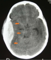
Following a car accident, a patient presents with signs of increased intracranial pressure. Radiologic studies reveal a crescent-shaped hematoma near the junction of the parietal and frontal lobes, which does not cross the midline. Which structure impedes the collection of blood from crossing the midline?
A. Falx cerebelli
B. Superior sagittal sinus
C. Tentorium cerebelli
D. Falx cerebri
E. Cavernous sinus
D
This is an example of a subdural hematoma, which is usually caused by ruptured veins that bleed into the space between the dura and arachnoid mater. The falx cerebri is an extension of the dura mater and since this hematoma is deep to the dura, it is blocked from crossing the midline by this structure. The superior sagittal sinus is contained within the falx cerebri but is not the structure directly preventing the hematoma from spreading.
The cranial fossa is divided into three sections: anterior, middle, and posterior. Within each section there are various foramina for the cranial nerves to exit the skull. In the middle cranial fossa, there is an important foramen that provides the exit for CN III, CN IV, and CN V1, and CN VI. This aperture is also known as:
A. Foramen spinosum
B. Foramen rotundum
C. Foramen ovale
D. Superior orbital fissure
E. Foramen lacerum
D
CN III, CN IV, CNV1 and CN VI all exit the cranial vault through the superior orbital fissure.
The posterior 1⁄4 of the roof of the mouth is formed by which bone(s) of the skull?
A. Maxilla
B. Ethmoid
C. Mandible
D. Palatine
E. Sphenoid
F. Temporal
D
The anterior roof of the mouth is formed by the maxilla, while the posterior 1⁄4 is formed by the palatine bones.
At the level of the internal occipital protuberance, lies the confluence of sinuses. Which dural venous sinuses make up the confluence of sinuses?
A. Superior sagittal, straight, inferior sagittal
B. Sigmoid, superior sagittal, inferior petrosal
C. Transverse, superior sagittal, straight
D. Transverse, occipital, superior petrosal
C
Transverse, superior sagittal, and straight sinuses form the confluence with contributions from the occipital sinus.
The optic nerve (CN II) exits the cranial cavity by passing through which foramen and in which cranial fossa?
A. Cribriform plate; anterior cranial fossa
B. Optic canal; anterior cranial fossa
C. Optic canal; middle cranial fossa
D. Optic canal; posterior cranial fossa
E. Superior orbital fissure; middle cranial fossa
C
CN II exits the skull through the optic canal, which is located in the middle cranial fossa.
Structures that pass thru the
Superior Orbital Fissure
A fissure between the greater and lesser sphenoid wings, connecting the middlecranial fossa and orbit.
Structures passing through include:
- Trochlear nerve (CN IV)
- Ophthalmic division of the trigeminal nerve (CN V1): lacrimal, frontal and nasociliary branches
- Abducent nerve (CN VI)
- Superior and inferior ophthalmic veins
Structures that pass thru the
Inferior Orbital Fissure
A fissure between the sphenoid bone (greater wing) and the maxilla, connecting the infratemporal fossa and orbit.
Connects the infratemporal fossa and orbit.
Structures passing through include:
• Inferior ophthalmic vein
Structures that pass thru the
Optic Canal
Canal that runs through the lesser wing of the sphenoid bone, connecting the anterior cranial fossa and orbit.
Structures:
Optic Nerve [CN II]
Ophthalmic artery
Pterion
where the sphenoid, temporal, frontal, and parietal bones meet
an extremely thin region of the skull.
Weakest part of the skull. The anterior division of the middle meningeal artery runs underneath the pterion. Consequently, a traumatic blow to the pterion may rupture the middle meningeal artery causing an epidural haematoma.

The temporomandibular joint (TMJ) is formed by the articulation of _____________
the mandible with the temporal bone of the cranium.
Clivus
the region of the occipital bone anterior to the foramen magnum
Bones that form the nasal cavity include ______.
frontal, ethmoid, sphenoid, palatine, lacrimal, vomer, nasal*, inferior nasal concha*, and maxilla.
Structures going thru the
Incisive canal
nasopalatine nerve and sphenopalatine artery
Enclosures and bones of
The neurocranium
Encloses: the brain, meninges, vessels, proximal parts of cranial nerves, and internal ear.
Composed of 8 bones: frontal, ethmoid, sphenoid, occipital, temporal*, and parietal*.
Enclosures and bones of
The viscerocranium
Includes: the inferior orbit, nasal cavity, and oral cavity.
Composed of 15 bones: mandible, ethmoid, vomer, maxilla*, inferior nasal concha*, zygomatic*, palatine*, nasal*, and lacrimal*.
Two outer surface of the calvaria has a number of important landmarks:
Bregma: where the parietal bones meet the frontal bone.
Lambda: where the parietal bones meet the occipital bone.
Closures of 4 Fontanelles
(membrane covered areas in immature skulls between two bones, where a suture will form later.)
Mastoid fontanelle: closes ~12 months.
Anterior fontanelle: closes ~18 months.
Posterior fontanelle: closes ~2-3 months.
Sphenoid fontanelle: closes ~2-3 months.
Structures passing thru
Superior orbital fissure
Ophthalmic veins
Optic nerve (CN V1)
CN III
CN IV
CN VI
Sympathetic fibres.
Structures passing thru
Foramen rotundum
Maxillary nerve (CN V2)