The Heart and Mediastinum Flashcards
What is the mediastinum?
Where does it extend from/to?
Broad central region that separates the two laterally placed pleural cavities
Extends from the thoracic inlet (aperture) at rib 1 to the diaphragm (T12) and from the sternal manubrium to the vertebral bodies
Is the patient on this x-ray in inspiration or expiration?

Inspiration:
- Flattening out of costodiaphragmatic recess
- Large lung fields
Label the features of the x-ray

Top arrow: arch of aorta
Bottom arrow: Pulmonary trunk
Line: hilum
What is the mediastinum divided by?
Sternal plane (T4/T5) divides the mediastinum into inferior and superior parts.
Inferior part divided into:
- Anterior
- Middle
- Posterior
- Divided by the pericardial sac

What can collect in the costodiaphragmatic recess?
Effusion fluid
What does the anterior mediastinum contain?
What are its borders?
- Anterior border: sternum
- Posterior border: pericardial sac
- Superior border: sternal plane
- Inferior border: diaphragm
Contains:
- Thymus (part of)
- Internal thoracic arteries
- Fat
- Connective tissue
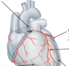
What can the internal thoracic arteries be used for?
Which arteries branch off them?
Which arteries do these meet?
Coronary Artery Bypass Graft (CABG)
Anterior intercostal arteries branch off them
- Form anastamotic relationship with posterior intercostal arteries in each intercostal space.
Label the structures on the diagram


Where is the thymus normally located?
What is the thymic sail sign?
What happens to the thymus with age?
In the anterior and/or superior mediastinum
The thymus is relatively large in children therefore can be seen on x-ray
Thymus shrinks with age

What does the superior mediastinum contain?
Label them on the diagram

Arch of aorta
Great vessels
Trachea
Oesophagus
Thoracic duct
Phrenic and Vagus nerves

Where does the trachea normally bifurcate?
Around T4
Where do the great veins sit in relation to the arteries?
Label the vessels on the diagram

Anteriorly

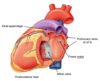
What is the ligamentum arteriosis?
What happens if it stays open?
Was previously the ductus arteriosis vessel, fuses soon after birth. Open in utero to help blood bypass the lungs which are not fully developed.
If it stayed open, it would create a mix of high O2 and low O2 blood from the arch of aorta and pulmonary trunk
Where do the phrenic and vagus nerves travel in lungs?
Which important branch comes off the vagus? Where does it branch?
Phrenic nerve passes anterior to the hilum of the lung and the great vessels
Vagus nerve passes posterior to the hilum of the lung, recurrent laryngeal artery.
- Right recurrent laryngeal branches off around the right subclavian artery in the superior mediastinum
- Left recurrent laryngeal branches off around the arch of the aorta behind the ligamentum arteriosum.
What do the recurrent laryngeal nerves do?
Sensory below vocal chords
Motor to all extrinsic and intrinsic muscles of the larynx except cricothyroid (external laryngeal nerve)
On which side would a hilar lymph node cause a hoarse voice? Why?
On which side could a pancoast tumour cause a hoarse voice? Why?
Hilar lymph node enlargement on the left side
- Could compress the left laryngeal nerve
Pancoast apical tumour could on the right side could compress the right recurrent laryngeal nerve against the right subclavian artery
Describe the route of the vagus nerve in the mediastinum
Vagus nerves form a plexus around the trachea and oesophagus and give off anterior and posterior vagal plexuses on the surface of the oesophagus:
- Left vagus: anterior plexus
- Right vagus: posterior plexus
Where does the left atrium of the heart sit against?
What can this anatomical relationship be used for?
Sits against the oesophagus
Can be used for trans-oesophageal ultrasound of the heart
What does the posterior mediastinum contain?
What are its borders?
- Superior border: sternal plane
- Posterior border: vertebral column
- Anterior border: pericardial sac
- Inferior border: diaphragm
Contains:
- Oesophagus and vagal plexus
- Descending aorta
- Thoracic duct
- Sympathetic chain
- Azygous system
What type of muscle is the oesophagus made up of?
What type of motor supply do they have?
Upper 1/3 = skeletal muscle
- Somatic control
Lower 2/3 = smooth muscle
- Autonomic control
What does the middle mediastinum contain?
What are its borders?
- Superior border: sternal plane
- Lateral borders: parietal pleura of the lungs
- Inferior borders: pericardial sac against diaphragm
- Anterior and posterior: paricardial sac
Contains:
- Heart and paricardium
- Lower superior vena cava
- Lower ascending aorta
- Lower pulmonary trunk
- Phrenic nerves (run along edges of fibrous pericardium)
- Cardiac plexus
What attaches the heart to the diaphragm?
Fibrous pericardium via the central diaphragmatic tendon
Describe the surfaces of the heart
Posterior surface: base
Inferior surface: diaphragmatic surface

Which chambers of the heart can be viewed anteriorly?
Why is this?
- Right ventricle
- Right atrium
- Small amount of left ventricle
Due to the left rotation of the heart during embryological development

What are the atrial appendages?
What is their clinical significance?
Small pouches in the atria
During atrial fibrillation, blood flow becomes turbulent and in atrial appendages can become static where clots can form.
What is the pericardium composed of?
Where is the pericardial cavity?
Fibrous outer pericardium: tough, fibrous layer surrounding entire heart and anchors to diaphragm.
Serous pericardium: 2 layers:
- Parietal (outer): lines internal surface of pericardium
- Visceral (inner): lines external surface of heart, forms the epicardium
Pericardial cavity between the parietal and visceral paricardium
- Filled with pericardial fluid for lubrication

What is cardiac tamponade?
What can it be caused by?
What is inflammation of the pericardium?
Pericarditis: inflammation of the pericardium
- Can result in pain if it affects the parietal layer
Cardiac tamponade: caused by increased pressure in the pericardial cavity caused by air, fluid e.t.c. Compresses heart allowing less room for contraction. This impairs contractility and prevents adequate refilling of the heart.
- Can be caused by haemopericardium (blood in pericardial space)
How is the pericardium formed embryologically?
What pockets are formed from this process?
During development heart tube becomes surrounded by pericardial sac with reflections at great vessels.
Folding of heart tube creates a passageway (transverse pericardial sinus) between arterial outflows & venous input
Reflection of serous layer creates blind ended oblique pericardial sinus posterior to heart

What can the transverse pericardial sinus be used for surgically?
To clamp off the arterial outflow from the heart

What are the atrioventricular valves?
What are they formed from?
What is the role of the papillary muscles?
Lie between atria and ventricles and allow for unidirectional flow of blood.
Formed from flap-like cusps that are anchored to the ventricle wall by tendons (chordae tendinae) which are anchored to papillary muscles.
Papillary muscles contract to prevent the cusps from being blown back into the atria during ventricular systole.
- Note they do not contract to open valves, only contract to prevent vales being forced backwards during systole
What may be the result of MI that has caused damage to the papillary muscles?
What would this sound like on auscultation?
They are unable to contract during ventricular systole to hold the cusps in place.
Can result in AV valve incompetence causing blood to regurgitate back into the atrium during ventricular systole.
Can be heard as a murmur on auscultation
What is the fibrous cardiac skeleton?
What is its role?
Dense fibrous connective tissue of the heart
- Provides mechanical stability
- Provides an anchor point for heart valves
- Prevents free conduction of electrical signal from atria to ventricle.
Label the structures on the superior view of the heart


Describe the structure of the aortic and pulmonary valves
How do they move during ventricular systole and diastole?
Semi-lunar, each have 3 cusps with sinuses located before each one.
During ventricular systole they are pushed against the lateral walls of the vessel (sinuses are flattened) allowing blood to pass
During diastole the sinuses are filled with reverse flowing blood which closes the valve (and allows blood to flow into the coronary arteries)

How are the cusps of the aortic valve named in embryology and adult hearts?
(use adult naming of valves)

How are the cusps of the pulmonary valve named in embryological and adult hearts?

Where do the coronary arteries arise from?
Right coronary artery arises from the anterior cusp of the aortic valve
Left coronary artery arises from the left posterior cusp of the aortic valve

What is meant by right or left coronary artery dominance?
Right dominance = RCA gives rise to the PIVA
Left dominance = LCA gives rise to the PIVA
Label the coronary arteries on the diagram
What do they supply?
Is this heart right or left coronary artery dominant?

Right dominant

Label the coronary arteries on the diagram
What do they supply?
Is this heart left or right dominant?

Left dominant

What do the right and left coronary arteries normally supply?
Right:
- Right atrium and ventricle
- AV and SV nodes
- Posterior IV septum (minority of septum)
Left:
- Left atrium and ventricle
- AV bundle
- Right and left bundle branches
- Anterior IV septum (majority of septum)
*
Which is the most common coronary artery to become blocked?
Left anterior descending artery (LAD)
(anterior interventricular artery
Label the coronary arteries on the image
What type of image is this?

Angiogram

Label the arteries on the coronary angiogram


Label the cardiac veins on the diagram
What does the coronary sinus drain into?

Coronary sinus drains directly into the right atrium

How can the surface anatomy of the heart be mapped out?
Right atrium and SVC: 3rd right costal cartilage 1cm from sternum.
Right atrium and IVC: 6th right costal cartilage 1cm from sternum
Auricle of left atrium and pulmonary trunk: 2nd left costal cartilage 1cm lateral to sternum.
Apex and left ventricle: 5th costal cartilage just medial to mid clavicular line

Where are the auscultation points for the 4 cardiac valves?
Aortic: 2nd right intercostal space just lateral to sternum
Pulmonary: 2nd left intercostal space just lateral to sternum
Tricuspid: 4th/5th left intercostal space just lateran to sternum
Mitral: 5th left intercostal space in mid clavicular line

Label the structures on the diagram
Where does the right atrium of the heart receive venous blood from?
What is the fossa ovalis?

Receives blood from:
- IVC and SVC
- Coronary sinus
Fossa ovalis: remnant of the embryological foramen ovale:
- Allowed blood to bypass the lungs in the embryo and be shunted from right to left.
- Seals shut when newborn takes first breath- increase in pressure pushes it closed and it begins to fuse.

Label the structures on the diagram
Where does the left atrium receive blood from?

Receives blood from the 4 pulmonary veins

Label the structures on the diagram
Is this a right or left ventricle?
What is the moderator band?

Right ventricle
Moderator band: muscle that contains the right bundle branches. Spans between the interventricular steptum and the anterior right ventricular wall.

Label the structures on the diagram
Is this a right or left ventricle?
What are trabeculae carnae?

Left ventricle
Trabeculae carnae: irregular muscular elevations in the ventricle wall. Embryological remnants.

Describe the electrical conduction system of the heart
Where does the electrical impulse usually originate from?
Label the structures on the diagram
Which part is responsible for the coordinated contraction of the ventricles?

Usually originates from SA node
- SA node
- AV node
- AV bundle (bundle of His)
- Right and left bundle branches
- Purkinje fibres
Purkinje fibres are responsible for coordinated ventricular contraction.

What may be a consequence of an interventricular septal defect of infarct?
Necrosis can disrupt electrical signal causing a loss of coordianted contraction (and reduced strength of contraction) of the right and/or left ventricles.
What are the impacts of sympathetic and parasympathetic innervation to the heart?
Where does the innervation arise from?
Where do cardiac visceral sensory fibres travel?
Sympathetic (from T1-T4 sympathetic chain):
- Increases heart rate
- Increases strength of contraction
Parasympathetic (from ganglia on heart):
- Reduces heart rate
- Decreases strength of contraction
Cardiac visceral sensory fibres travel back to the CNS via sympathetic nerves entering the spinal cord at T1-T4 (along with somatic sensory fibres from T1-T4 dermatomes).