Superficial Triangles/Cervical Viscera Flashcards
Submandibular Triangle
- borders
- contents
bound by inferior edge of mandible, and anterior/ posterior digastric, hyoid forms the apex

what innervates the platysma, relationship to fascia
cervical branch of the facial nerve (7), superficial to investing fascia
Muscles of submandibular triangle, their innervation, action

Zones of penetrating Trauma
-boundaries, contents

Branchial Arch Derivatives: Nerve, Muscles, Skeletal structures, Ligaments
- First
- Second
- Third
- Fourth and Sixth

Thyroid gland:
- lobes connected by what
- 50% of people have what lobe?
- Thyroglossal cysts?

Contents of the submandibular triangle
suprahyoid muscles (digastric, stylohyoid, mylohyoid, geniohyoid, and hyoglossus)
-hyoid bone, submandibular and sublingual glands, CN12, lingual nerve, submandibular ganglion, fascial A/V, marginal mandibular branch of CN 7, lingular artery, glossopharyngeal N
Carotid triangle contents, borders
Superior – posterior belly of the digastric muscle.
Lateral – medial border of the sternocleidomastoid muscle.
Inferior – superior belly of the omohyoid muscle.

contents of the root of the neck in posterior lateral triangle

contents of carotid sheath
internal jugular vein
vag nerve 10
common carotid
Branchial fistula
-what is it
branchial fistulas are persistent remnants of branchial pouches

Innervation of the SCM
CN 11, small cervical contributions from C2-C3
Posterior (lateral triangle) borders

Sixth cervical vertebra:
importance as a landmark
arch of the cricoid cartilage
the point where the vertebral artery enters the transverse foramina
External Jugular vein formed by?
- relationship to SCM, posterior triangle
- where does it drain?

Relationships of the thyroid gland:
- RLN location?
- origin of superior thyroid artery
- inferior thyroid artery origin
- lowest thyroid artery origin
know that berrys ligament is usually only 3mm anterior to RLN

Parasaggital Zone:
- Muscular triangle:
- boundaries, contents, innervation

innervation of the trapezius, what type of fascia surrounds it
surrounded by investing fascia, innervated by spinal accessory nerve (11) w/ small cervical contribution from C3-C4
lymphatic drainage of the thyroid
-Deep cer

relationship of inferior thyroid artery to recurrent laryngeal

Superficial Branches of the Cervical Plexus, what they innervate
(there are four)

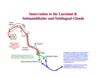
Innervation to the Lacrimal and Submandibular and Sublingual glands
-
both parasympathetic and sympathetic.
preganglionic fibers from the superior salvatory nucleus exit brainstem with the facial nerve and travel through the chorda tympani and join with the lingual nerve before terminating in the submandibular ganglion.
-postganglionic parasympathetic fibers from the submandibular ganglion distribute as numerous short branches to the parenchyma of the gland
postganglionic sympathetic fibers come from the superior cervical ganglion and reach the gland by coursing in the external carotid and facial plexuses in the adventitia of the respective arteries.
what is deep to the CCA and embedded in prevertebral fascia
cervical sympathetic chain
Anterior scalene relationships:
-whats in front, in back
-in front is transverse cervical artery, suprascapular artery, phrenic nerve, subclavian V, inferior thyroid a
posterior you have subclavian A and brachial plex


