Oral Cavity and Pharynx Flashcards
Relatiohships of sublingual region:
- lingual nerve spirals around what in which direction
- hypoglossal nerve
- sublingual salivary gland and its artery

Innervation of the sublingual salivary gland
-parsympathetic, sympathetic

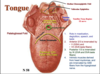
Tongue:
- folds
- what divides it
- valleculae epiglottica
- whats in tonsilar fossa region
- Anterior 2/3 motor and taste
- posterior 1/3 motor and taste
Tongue muscles:
- name them
- innervation
paralysis characteristics
- Hyoglossus muscle arises from the hyoid; retracts tongue
- Styloglossus muscle arises from the styloid process; retracts & elevates the
tongue - Genioglossus muscle arises from the genial
tubercle; protrudes tongue - Palatoglossus muscle arises from the
posterolateral hard palate. It is innervated by
the vagus via the pharyngeal plexus. This
muscle forms the overlying palatoglossal fold,
arch or anterior pillar. It elevates tongue and
closes faucial isthmus during deglutition. - Intrinsic muscles of the tongue comprise longitudinal, transverse, and oblique
fibers which insert onto the median lingual septum

Tongue oral portion vs pharyngeal portion
- type of tissue origin?
- innervation

Tongue:
blood supply
innervation
lymphatic drainage

palate hard vs soft
-incisive foramen
The palate is embryologically and morphologically divided into an anterior hard (bony) and posterior soft palate. The hard or bony palate comprises the anterior two-thirds of the palate and is formed by the palatine processes of the maxillae and the horizontal plates of the palatine bones. The incisive foramen conveys the nasopalatine nerve and vessels. The paired greater and lesser palatine foramina transmit corresponding nerves and vessels.
The soft palate is a movable fibromuscular partition which forms the posterior one-third of the palate. During deglutition, the soft palate closes the pharyngeal isthmus, and prevents reflux of material into the nasopharynx.
muscles of the palate: naso and oropharynx
-muscle names, innervations, actions
A. Muscles of the Palate
1. Muscularis uvulae muscle
This intrinsic muscle of the soft palate and forms part of the midline uvula. It is innervated by the vagus (X) nerve through the pharyngeal plexus.
2. Tensor Veli Palatini muscle
The tensor palati is located anterolateral to the levator palati muscle and auditory tube. After arising from the scaphoid fossa, its tendon loops around the pterygoid hamulus and inserts into the soft palate. The tensor palati is innervated by a small branch of the mandibular nerve (V). The tensor palati tenses the soft palate and opens the auditory tube.
3. Levator Veli Palatini muscle The levator palati is located inferior to the auditory tube on the lateral wall of the nasopharynx. It originates from the inferior surface of petrous temporal bone and part of auditory tube, and inserts in the soft palate. In contrast to the tensor palati, the levator is innervated by the vagus via the pharyngeal plexus. As the name implies, it is an elevator of the soft palate
Paralysis of the tensor or levator palate allows the muscles on the non-paralyzed side to pull
or deviate the uvula toward the normal side.

Unilateral paralysis of tensor or levator palati

Palatine Tonsil:
- location
- artery
- innervation
- venous drainage
This oval-shaped lymphatic gland is located between the palatoglossal and palatopharyngeal folds in the tonsillar fossa. The palatine tonsil is supplied by the tonsillar artery of the facial, and the palatine branch of the ascending pharyngeal. The glossopharyngeal nerve is closely related to the floor of the tonsillar fossa. The venous drainage of the palatine tonsil is principally the tonsillar (paratonsillar) vein drains into the pharyngeal venous plexus and the facial vein. Clinically, the tonsillar vein is a frequent bleeder during tonsillectomy. Lymphatic drainage from this tonsil is directly into the jugulodigastric (tonsillar) nodes

innervation of the pharynx:
- which plexus embedded in what fascia
- GVA from?
- SVE from?
- GVA convey what special reflex

Boundaries of the Nasal cavity:
- roof characteristic
- floor characteristic, made of
- what makes medial wall/nasal septum
- fracture usually occurs where
The roof of the nasal cavity is curved in an antero-posterior direction and is narrower than the floor. However, the floor of the nasal cavity is a relatively flat horizontal shelf, which is curved slightly on the lateral edge. It is formed by the palatine process of the maxilla and horizontal plate of the palatine bones. The floor of the nasal cavity is also the roof of the mouth.
The medial wall or nasal septum is formed by the alar and septal cartilages anteriorly; the perpendicular plate of the ethmoid posterosuperiorly; and the vomer posteroinferiorly. An impact on the external nose often fractures the nasal septum at the junction between the septal cartilage and bone. These fragments may dislocate posteriorly
Lateral nasal wall
- components from which 7 bones
- 3 shelves called what
- superior middle from where? where is inferior from?
Portions of seven (7) bones comprise the lateral nasal wall. These include the maxilla, nasal, lacrimal, inferior concha, ethmoid, sphenoid, and palatine. The lateral nasal wall is characterized by an irregular surface with three overhanging, scroll-like projections called conchae (turbinates), which form underlying gutters called meatuses. In anatomy the terms concha and turbinate are used interchangeably; however, ENT physicians usually call them turbinates

- position of the sphenoethmoid recess?
- what drains into it?
Sphenoethmoidal Recess is located postero-superior to superior concha. The sphenoidal sinus drains into the sphenoethmoidal recess. A highest concha and meatus may be present in this recess.

- Ethmoidal bulla
- opening for maxillary sinus
- frontal recess
- nasolacrimal opening?
- Superior Concha (Turbinate) and Meatus
The superior meatus forms a short oblique passageway over the posterior ½ of the middle
concha. It contains openings for the posterior ethmoidal air cells. The sphenopalatine foramen
is located in posterior to the superior concha in the submucosa. - Middle Concha (Turbinate) and Meatus
The middle meatus is long and continuous with the atrium. It contains the ethmoidal bulla,
hiatus semilunaris and openings for the maxillary, ethmoidal and frontal sinuses.
The ethmoidal bulla forms a bony eminence overlying the middle ethmoidal air cells, which have multiple openings onto it. The hiatus semilunaris is a crescent-shaped trough located anterior and inferior to the ethmoidal bulla. The opening for the maxillary sinus is in the posterior 1/3 of the hiatus semilunaris.
The frontal and anterior ethmoidal sinuses drain into the ethmoidal infundibulum, which is located in the anterosuperior portion of the hiatus semilunaris. If these sinuses drain through a separate opening located anterior to the hiatus, then the opening is called the frontal recess. - Inferior Concha (Turbinate) and Meatus
The opening for the nasolacrimal duct is in the inferior meatus, 1 cm posterior to the anterior edge of the concha.

Blood supply to nasal cavity:
- anterior 2/3 vs posterior 1/3, what are these vessels branches of?
- most common spot for epistaxis
The sphenopalatine artery, the terminal branch of the maxillary artery, supplies the septum and lateral wall of the posterior one-half of the nasal cavity. The sphenopalatine artery branches into the posterior lateral and posterior septal arteries. Branches of the anterior and posterior ethmoidal arteries, which are branches of the ophthalmic artery, supply the anterior one-half of the walls/septum.
A small region of near the vestibule is supplied by septal branches of the superior labial artery. Most nasal hemorrhages or (epistaxis) occurs at the junction of the septal branches of the superior labial and sphenopalatine arteries. Clinically, this region is referred to as Kiesselbach’s area.

nerves of the nasal cavity:
- anterior 1/3 vs posterior 2/3
- what are these branches of
Olfactory Neurons. These bipolar neurons are embedded in the olfactory epithelium. The anterior ethmoidal nerve, a branch of the nasociliary nerve (V1), innervates the mucosa of the anterior one-third of the nasal cavity. Branches of the pterygopalatine (same as sphenopalatine) ganglion supply the posterior two-thirds. These are GVA and some autonomic fibers. Coursing diagonally beneath the septal mucosa is the nasopalatine nerve, which is also a branch of the pterygopalatine ganglion. It innervates the mucosa of the gingiva and hard palate adjacent the upper incisors.

Vidian Nerve carries?, course?
-greater, lesser palatine nerves, what do they carry what is their course
- Vidian nerve
The Vidian nerve (nerve of the pterygoid canal) is formed by the merging of the deep petrosal and great petrosal nerves. It conveys postganglionic sympathetic, GVE parasympathetic, and GVA fibers to the pterygopalatine ganglion where it ends.
- Lesser and Greater Palatine nerves
The greater and lesser palatine nerves are the largest branches of the pterygopalatine ganglion. They convey GSA (V2), GVA (VII), GVE parasympathetic, postganglionic sympathetic fibers to the mucosa of the inferior surface of the soft and hard palate, respectively
- Nasopalatine nerve
The nasopalatine nerve distributes the same components as the palatine nerves to the mucosa of the posterior nasal septum and lateral nasal cavity. Pharyngeal and orbital nerves are small twigs supply the same components as above to the mucosa near the pharyngeal opening of the auditory tube, and a small area of the orbit.
Z-scheme

Maxillary Sinus:
- superior relationship
- inferior
- posterior
- floor of orbit there is what?
- surgical approach
A. Maxillary Sinus
is a large (3.5 x 2.5 x 3.0 cm) pyramidal cavity with its base directly medially and the apex is directed towards the zygomatic process of the maxilla. The medial wall forms the lateral wall of the nasal cavity, and its roof forms the floor of the orbit. The maxillary floor is part of the alveolar portion of the maxilla. It is 0.5-1.0 cm below the level of the nasal cavity, and has elevations formed by the first and second molars. The posterior maxillary wall forms the anterior wall of the pterygopalatine fossa and infratemporal fossa. The maxillary sinus drains by ciliary action, gravity and negative pressure into one or more openings into the hiatus semilunaris.
Maxillary sinusitis may be associated with a toothache of the first or second molars due their close anatomical relationship with the sinus. Because of their intimate relationships with each other, maxillary infections may spread among the frontal, anterior ethmoidal cells, nasal cavity, teeth and maxillary sinus. Transmaxillary surgery. The maxillary sinus is frequently used as a surgical approach to its surrounding structures (transmaxillary approach).
**infraorbital neurovascular bundle can herniate into maxillary sinus if there is bad fracture
Ethmoid sinus:
- air cells open into? A/M/P
- Sphenoidal sinus and its relationships**
The ethmoid sinus is comprised of numerous (4-7) small cavities or cells located in the ethmoidal labyrinth which is between the orbit and the nasal cavity. If the number of ethmoidal cells decreases, they are larger in size. There are three groups of cells according to their openings into the lateral wall of the nasal cavity. Anterior ethmoidal cells open into the anterior part of the hiatus semilunaris. The middle Ethmoidal cells open onto the surface of the bulla ethmoidalis. Posterior Ethmoidal cells open into the superior meatus
C. Frontal Sinus
The frontal sinus is regarded as displaced anterior ethmoidal air cells, which has invaded the frontal bone. This explains the common relationships of the frontal and ethmoidal sinuses. It has a paramedian location behind the supraciliary ridge, and is rarely symmetrical. The frontonasal duct drains into either the ethmoidal infundibulum or the frontal recess of the middle meatus.
D. Sphenoidal Sinus****
This cube-shaped sinus (1.0 cm3) is in the body of the sphenoid with extensions into the greater wings or the pterygoid processes. It drains into the sphenoethmoidal recess. Relationships of the sphenoidal sinus to adjacent structures:
posterior: pons, basilar artery
superior: pituitary
anterior: nasal cavity
Inferior: nasopharynx
lateral: internal carotid, V1, cavernous sinus Sphenoidal sinusitis
Transsphenoidal surgery

KNOW THIS

KNOW THIS
KNOW THIS

Pterygopalatine Fossa boundaries:
Anterior:
Posterior:
Medial
lateral
roof
floor


