Skin Cancer (non-melanoma) Flashcards
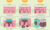
What are the layers of the Epidermis?

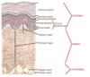
What are the layers of the dermis?

What are the layers of the hypodermis?

Where should melanocytes be located?
Basal Layer

Diagnosis?
• key features?

Basal Cell Carcinoma
• Pearly appearance
• Teleangectasias
• Ulcerated
What cancer is shown here?
• what are the key histologic features?

Basal Cell Carcinoma
KEYS:
• This is a blue tumor on H and E (because these are progenator cells we expect them to have big active nuclei)
• Nodular appearance is very important (especially when differentiating from melanoma)
• Pallisades of blue nuclei that CLEFT AWAY from the surrouding tissue
What pathway and gene is often altered in this cancer?

Basal Cell Carcinoma often has mutations in the PTCH gene in the sonic hedgehog path.
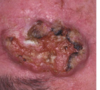
What cancer is this?
• what feature puts patients at the greatest risk of metastasis?

• Squamous Cell Carcinoma (take note of keratin pearls)- if this is found in the EAR or LIP there is a high risk of metastasis.

What group has a rising incidence of Skin Cancer of all types?
Young Females due to tanning bed use
What is the most common neoplasm in the US?
Basal Cell Carcinoma
What is the role of the altered gene that is frequently implicated in this cancer?

PTCH gene is commonly involved in Basal Cell Carcinoma development (thats what makes vismodegib and sonetigib good in BCC tx). This is a TUMOR SUPPRESSOR GENE because it prevents constant activation of the smoothened (SMO) receptor
What risk factors put patients at the greatest risk for Basal Cell Carcinoma?
RISK:
• Chronic but Intermittent UV exposure
• fair complexion
• hx of Sunburns (blistering)
• Family Hx
• Immunosuppression
What is the most common risk factor implicated in the development of Basal Cell Carcinoma?
• Role of immunosuppression in BCC vs. SCC?
- Chonic Intermittent UV exposure like that encounter by people that go on vacations and get sunburned
- Immunosuppression increased incidence 10x in BCC but still immunosuppressed pts. are more likely to get SCC
What is this?
• what would you expect to see on histology?

BCC
Histology will show blue, nodular, pallisading basal cells that cleft away from surrounding tissue

What are the key features of the Skin cancer shown here?

Basal Cell Carcinoma

Note: Clefting is aka Retraction Artifact
Also there are several subtypes of BCC:
• Nodular
• Superficial
• Pigmented
• Morpheaform
• Micronodular
• Cystic
• Infiltrative
What is seen on the left and right in these pictures?

The are BOTH Basal Cell Carcinomas
Left: Superficial Basal Cell Carcinoma
Right: Nodular Basal Cell Carcinoma
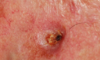
What is shown here?

Basal Cell Carcinoma (nodular subtype)
• well circumscibed with pearly rolled border and central erosion with telangiectasias
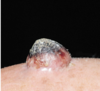
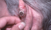
What is this?

BCC - with prominent telangiectasias
What is this?

SUPERFICIAL BCC - note its more patchy, flat and broad, but still has telangectasias and a semi-pearly quality
What is this?

Superfical BCC
What is this?
• what subset of patients is this most likely to happen in?
BCC - note that this is abnormal in that it’s hyperpigmented. This is more likely to happen with a darker skin tone.
Only 20% of people get basal cell carcinomas before age 50. Under what age should you really start to worry about something genetic?
• what genetic defect would you be worried about?
• PTCH tumor suppressor gene in the SHH pathway can be mutated via an inherited defect that predisposes people to Basal Cell Carcinomas
What is is called when people have inherited mutations in their PTCH1 gene?
• How is this inherited?
• what manifestation do we see?
• What are these patients at risk for other than BCC?
Gorlin Sydrome aka Basal Cell Nevus Syndrome is associated with mutations in the PTCH1 gene. These patients often get BCC’s in their twenties with musculoskeletal defects like JAW CYSTS.
RISKS:
• Medulloblastoma
• Fibrosarcoma
• Basal Cell Carcinoma
How common is Gorlin Syndrome?

1:56,000
What is your risk of metastasis in BCC?
VERY low 0.0028% to 0.55% of cases metastasize.
How is BCC treated?
• why do we treat it at all if it doesn’t metastasize?
BCC is locally agressive so we need to treat it so it doesn’t damage too much surrounding tissue
Tx:
• Excision
• Electrodessication and curretage
• Cryosurgery
• Radiation
• Topical Tx
What are some side effects of Vismodegib?
• when is it used?
Vismodegib is only used in severe cases of BCC
• It can cause loss of taste and severe muscle cramps
If a patient presents with a neoplastic lesion on their lower lip. What is it likely to be?
Squamous Cell carcinoma is typically found on the lower lip
**Note: board review books say that SCC is more common on lower face (place that gets chronic exposure) and BCC is more common on upper face (place that gets intermittent exposure) but is variable from person to person**
What is this?

Untreated BCC
What is this?
• Key features?
Squamous Cell Carcinoma
• Crusty and Keratin Topped (also note a lack of telangectasias)

What is the progression of Squamous Cell Carcinoma?
- Actinic Keratosis => minimal atypia
- SCC in situ => full thickness epidermal atypia confined above the basement membrane
- Invasive SCC => invasion through Basment membrane

What is wrong here?

Nothing, this is normal skin histology.
Describe the lesions on this lady’s face.
• what are some key features?
• what would it look like histologically?

ACTINIC KERATOSIS
GROSS:
• **Thin, non-indurated (not hard) lesions
HISTOLOGY:
• Parakeratosis
• Large pleomorphic cells**

What has caused these lesions?
• what are they?

Actinic Keratosis (non-indurated) have been caused by UV exposure

Note: these actinic keratosis may be a bit more concerning because they look more nodular and possible indurated

What is it?

AK

You Biopsy somone’s hyperkeratotic nodule on their lower lip and see this. What is it?

SCC in situ
• Notice FULL THICKNESS ATYPIA you see cells with hyperchromic nuclei in many layers above the basal layer and also continued presence of the nucleus into the corneum (parakeritosis)
What is this?

INVASIVE SCC (not in situ b/c you can see keritin pearls below the basement membrane)
In-situ is shown below

Note: the front of this guy’s lesion is Actinic Keratosis, Middle is In Situ, and the back is invasive

SCC

SCC that mimics basal cell

What is the only gene mutation that has consistently been linked to Squamous Cell Carcinoma?
• p53 mutations
What factors increase your risk of Developing this?

Note: CHRONIC UV exposure is more likely to give you SCC, pts; leukoplakia (white plaque) is associated with chronic tobacco use; SCC developmen after breast ca. radiation is a problem

What determines your risk of Metastasis in Cutaneous Squamous Cell Carcinoma?
- Size
- Depth of Invasion into Dermis
- Anatomic Site (lip and ear)
Which is more likely to Metastasize SCC or BCC?
Both are pretty low risk but BCC is 0.0028 - 0.55% whereas SCC is 5% so SCC is more likely to metastasize.
Which type of SCC is most likely to metastasize?
• Where will it go?
SCC most likely to go to LYMPH NODES and LUNG
High Met Risk:
• Actinic (UV) induced on lip*** (2-16%)
• HPV induced (penile, vulvar, perineal) (30%)
• Leukoplakia
• Marjolin’s (10-30%)
What is it?

SCC on forehead
What makes this a high risk area for SCC?

Sun Exposure is high
Smoking
What is it?
*is this high or low risk of met?
SCC on the tongue - high met risk (as with lip and HPV induced, Marjoins)
What is this?
• What is the course of this disease?

This PAINFUL lesion pops up rapidly in 2-6 weeks then our IMMUNE SYSTEM recognizes it and it spontaneously regresses
Keratocanthomas are dome shaped with a keritin plug
• what does this look like? ddx?

Looks a lot like an epidermal cyts but this one is PAINFUL
What is this?
• cause?

Marjolin’s Ulcer => high risk for SCC development
Ulcerated invasive SCC arising from chronic:
• Inflammation (OSTEOMYELITIS)
• Scarring
• Radiation
• Trauma
