Benign Skin Lesions Flashcards
These bumps have been persistent on this lady’s face for weeks and have not changed. They are not painful.
• What are they?
• Where are they typically found?
• Who gets them?
• What would you do to remove it?
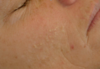
This is a MILIA (small EPIDERMAL cyst) that is painless, persistent, and does not grow. These are common in infants and adults.
LOCATION => SUN EXPOSED AREAS
• Cheeks, eyelids, forehead, genitals
Simple Excision can typically get rid of these
You got stuck with a safety pin in your arm a couple of weeks ago when getting dressed and have since noted the development of this lesion.
• What is it?
• Where is it typically found?
• What aspect of the history is important?
• What happens if you squeeze it?

Dermatolfibromas are common and frequently arise in the extremities as a result of minor trauma (like bug bites). If you pinch this lesion it will likely be positive for “dimple sign”. Note these may be painful and pigmented.
What are some important features to notice in this dermatofibroma on gross inspection?
• what if it continues to grow

NODULE with Central Pallor area and pigmentation at edges is commmon in these
• get a consult if it continues to grow. These things usually max out at 10mm.
What are the key physical features to look for in this seborrheic keratosis?
• typical location?

Typically these are located on the trunk (but can be anywhere) and present as well defined plaques with a hyperkeratotic surface ranging from **millimeters to centimeters. STUCK ON APPEARANCE
****
Often Oval Shaped with long axis following tension lines of the skin
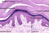
What layers of skin are involved in the formation of this lesion?
• Technical Terms for what has happened histologically?

ONLY the Epidermis is involved in this lesion
Histologically:
• HYPERKERATOSIS and ACANTHOSIS (aka thick stratum corneum due to hyperplasia of keratinocytes)
Define Hyperkeratosis
Thickening of the Stratum Corneum
Define Orthokeratosis.
Normal, basket-woven stratum corneum
Define Acanthosis.
Increased epidermal thickness due the HYPERPLASIA of keratinocytes
Define Parakeratosis.
Retention of nuclei in the stratum corneum
Differentiate the cells involved in Seborrheic Keratosis from those in melanomas and nevi.
Seborrheic Keratosis involves hyperplastic squamous cells (that just happen to get melanin deposited in them). Nevi and Melanomas are caused by hyperplasia of melanocytes
Is this lesion benign or malignant?

This is a benign seborrheic keratosis that has resulted from thickening of the epithelium
What should be your next step in management of this patient who said these lesions popped up in the last couple of months?

SCREEN FOR CANCER -> sudden eruption of multiple Seborrheic Keratoses may be a sign of underlying cancer
What types of cancers are associated with the Leser-Trelat sign?
Cancers associated with Leser-Trelat
• Colon
• Breast
• Stomach
• Lung
When does this lesion typically appear?
• who does it appear in?
• inheritance?

Seborrheic Keratosis
• Typically males are more often and extensitvely affected than females. Usually people are over 30 before these start popping up.
• Autosomal Dominant inheritance
What is the typical description of these lesions?

Seborrheic Keratosis
• Early these are small and light tan but progress to get darker and darker. They often are raised with a stuck on appearance/
**remember SK believed to be autosomal dominant***

Seborrheic Keratosis
What are these?
• Describe them.
• Typical Location?

Cherry Hemangiomas
• These are flat, erythematous, macules
• Typicall on trunk
What is this?
• where is it most likely to be located?
• Who does it present in?

Cherry hemangiomas are neoplasms typically found on the trunk of people in their 40s or older (but may start as early as 30s).
Would you expect this lesion to blanch if you pushed on it?

Yes, cherry hemangiomas are blanching
What features differentiate cherry hemangiomas and petechiae?
*shown here are petechiae

Hemangiomas will Blanch, Petechiae are non-blanching lesion and they are typically more purple.
Shown below is Cherry Hemangioma

What differentiates this lesion from a hypertrophic scar?

- Keloids grow far outside the boundaries of the original injury. Hypertrophic scars do not.
- Keloids also have a much lower threshold for formation than hypertrophic scars
What is this?
• is it painful?
keloid from ear piercing
May itch but is not painful
Who typically gets these?
• genetic predispositions? Gender preference?
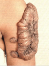
Anyone can get keloids but its typically African Americans in their 30s (no preference for either sex)
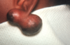
What is an epidermal (epidermoid) cyst?
• Mobile Dermal nodule with Central punctum that is created by the shedding of soft keratin into the center of the cyst. Debris collects here and becomes trapped to for a rancid smelling cheesy material
What is this?
• what should be in your differential?

Epidermal Cyst
• if you see a lump like this you probably want to first rule out metastasis
What is this?
- where does it arise from?
- complications?

Epidermal Cyst
• Arises from theinfundibulum of the hair follicle
• may become traumatized and rupture or become very inflammed or abscessed
most common place for this to occur?

Most commonly these Epidermal Cysts are found on the central trunk and face
What is a pilar cyst?
• where are they found?
• where do they come from?
Cysts that commonly arise on the scap and arise from the hair follicle (similar to epidermal cysts)
What is this?
• Differential?
• what are the chances your kid will have it?

this is a pilar cyst -
• Differentiate from epidermal cyst by lack of a central punctum
• typically present on the head and have AUTOSOMAL DOMINANT inheritance.
