Session 3: Acute Sepsis Flashcards

1
Q

Apply the infection model to a patient presenting with an acute Infection

A

The case: a 19-year old female student presents acutely unwell to the ED. Acute sepsis is part of the differential diagnosis. ED has a ‘sepsis bundle’ for urgent management of acute sepsis.

The history:

  • fit and well until last then non-specifically unwell, temperature and chills
  • This morning felt acutely unwell with fever, severe headache, nausea, weakness, general muscle aches, abdominal pain and eye pain on exposure to light (photophobia).

On examination:

  • Pale, cool extremities
  • Temp 39.5oC, pulse 110/min, BP 80/50mmHg
  • Respiratory rate 35/min
  • Widespread purpuric (non-blanching – not changing under tumbler) rash noted
  • Neck stiffness is noted
  • Mentally alert, GCS 15
  • Immediately recognized as severe sepsis + probable meningitis

Meningococcal meningitis: the likely diagnosis

2
Q

Define Bacteraemia, Septicaemia, Sepsis, Severe Sepsis and Septic Shock

A
  • Bacteraemia is the presence of bacteria in the blood (+/- clinical features)
  • Septicaemia is a clinical term meaning generalized sepsis, suggestive of a bloodstream infection
  • Sepsis is the systemic response to infection and can be defined as: SIRS + Documented or Presumed infection
  • Severe Sepsis = SIRS + organ dysfunction or organ hypoperfusion
  • Hypotension, decreased urine output (due to impaired renal function)
  • Septic shock = severe sepsis + persistently low blood pressure despite administration of intravenous fluids (significant medical emergency)
3
Q

What urgent investigations need to be done if sepsis is suspected?

A
  • Full blood count, Urea and electrolytes (looking at metabolic activity in terms of ions and urea). Look for signs of sepsis with raised white cell count and raised neutrophils.
  • EDTA bottle for PCR
  • Blood sugar
  • Liver function Tests
  • C-reactive protein (CRP): an acute phase protein raised in acute infections. The protein is produced by the liver. NON-SPECIFIC indicator of inflammation and infection.
  • Clotting studies
  • Blood gases
4
Q

What is SIRS?

A

Systemic Inflammatory Response Syndrome (SIRS): a response to a non-specific insult e.g. ischaemia, trauma, infection etc
Two or more of:

  • Temperature <36oC or >38oC
  • Heart rate >90/min
  • Respiratory rate: >20/min (or PACO2 <4.3kPa)
  • WBC <4x10⁹/L or >12x109/L

NB: Temperature, heart rate and respiratory are bedside examinations

5
Q

Describe how microorganisms trigger the inflammatory cascade

A

Endotoxin binds to macrophages => Cytokines: Tissue Necrosis Factors and Interleukins e.g. TNF-alpha and IL-1 [LOCAL] {to stimulate inflammatory response to promote wound repair and recruit reticuloendothelial system) => Cytokines released into circulation [SYSTEMIC] {stimulating Growth Factor, macrophages and platelets. Goal is homeostasis} => Homeostasis NOT Restored {Cytokines lead to activation of humoral cascades and reticuloendothelial system – CIRCULATORY INSULT}

6
Q

Explain about Sepsis and Coagulation

A
  • Cytokines initiate production of thrombin and thus promote coagulation
  • Cytokines also inhibit fibrinolysis
  • Coagulation cascade leads to microvascular thrombosis and hence organ ischaemia, dysfunction and failure.
  • Microvascular injury is the major cause of shock and multiorgan failure.
7
Q

What is meant by phagocytosis?

A

In the blood stream or at the inflammatory site, the neutrophil comes into contact with invading microbes. The encounter initiates phagocytosis of the target. The neutrophil sends out extensions of its plasma membrane called pseudopodia which progressively enclose the particle into a phagosome inside the cell and an arsenal of cytotoxic chemicals and enzymes destroy it. This is known as phagocytosis.

8
Q

What is the Sepsis 6?

A
  1. Deliver high flow oxygen
  2. Take blood cultures and other cultures, consider source control
  3. Administer empirical (best guess) IV antibiotics
  4. Measure serum lactate
  5. Start IV fluid resuscitation
  6. Commence accurate urine output measurement
9
Q

Describe antibiotic treatment for sepsis

A
  • an agent is likely to be active against the pathogens that cause meningitis in this age group (different in neonates and the elderly).
  • An agent that penetrates into the CSF

Empiric choice is CEFTRIAXONE (3rd generation cephalosporin with broad spectrum activity + reaches blood in good concentration)

10
Q

What are possible life-threatening complications of sepsis?

A
  • Irreversible hypotension (life threatening particularly to blood, brain and kidney)
  • Respiratory failure
  • Acute kidney injury (renal failure)
  • Raised intracranial pressure -> may lead to a long term impairment of CNS function
  • Ischaemic necrosis of digits/hands/feet
11
Q

How would you confirm the diagnosis of sepsis?

A
Blood culture (growing the bacteria to confirm and identify the bacteria causing the infection – takes at least 24 to 48 hours)
PCR
Lumbar puncture (if safe):

[*] Culture of cerebrospinal fluid (CSF)

[*] PCR of CSF

12
Q

Describe examination of the CSF sample

A
  • Lumbar puncture only performed after checking contraindications
  • Urgent transport of CSF to laboratory (Glucose and protein estimation in biochemistry as different changes in response to different infections, microscopy and culture in microbiology)
  • Appearance: turbidity and colour
  • Microscopy WBCs, RBCs
  • Gram stain
  • Referral for PCR
13
Q

Describe features of Neisseria Meningitidis

A
  • Bacterial pathogen Neisseria meningitides is gram negative diplococcus and spread by direct contact with respiratory secretions (with either someone who is infected or someone who is colonized)
  • Most people are harmlessly colonized (asymptomatic)
  • In the unlucky few, rapidly progressive and potentially fatal disease if not recognized and treated promptly
14
Q

Describe the mechanism of action of Neisseria Meningitidis

A

In this situation it attaches to the mucosa in the nasopharynx

  • Endotoxins of lipopolysaccharides triggers inflammation
  • Pilus enhances attachment
  • Polysaccharide capsule promotes adherence to host and prevents phagocytosis from phagocytes within the host. Capsule is slippery

Numerous serogroups (e.g. A, B, C, W-135) based on the polysaccharide capsule antigen (evades immune response by preventing phagocytosis).

Outer membrane acts as an endotoxin

15
Q

Describe the features of Meningococcal Disease

A

Up to 25% young adults may be carriers
Spread by aerosols and nasopharyngeal secretions
Acquisitions -> clearance, carriage or invasion
In England 1000 cases/ yr mainly Group B
Fatality rate ~10%
Elsewhere Group A predominates ‘meningitis belt’ across Africa

16
Q

Describe the two types of prevention of Meningococcal Disease

A

Prevention 1. Vaccination

  • Meningococcal C conjugate vaccine (introduced in the UK in 1999 and lead to dramatic decrease in Group C disease)
  • ACWY vaccines available for immunocompromised patients and travel protection
  • Serogroup B vaccine not yet in routine because the capsule is poorly immunogenic and similar to neural tissue (very difficult to introduce as a vaccine) however a vaccine has been developed after screening candidate proteins from genome studies and could be introduced into the UK within the next few years

Prevention 2: antibiotic prophylaxis

  • Meningitis is a notifiable disease
  • Cases reported to the local Health Protection Unit of Public Health England
  • Close contacts (household, student accommodation) can be given antibiotic prophylaxis and considered for vaccination, all to try and clear any carriage of the organism to prevent invasion.
17
Q

What are the factors determining the outcome of the host-pathogen relationship?

A
  • Number of organisms present
  • Virulence
  • Host’s immune response
18
Q

Define Immune System and Infectious Disease

A

Immune System = cells and organs (particularly the spleen) that contribute to immune defences against infectious and non-infectious conditions (harmless substances e.g. dead cells, pollen)

Infectious disease = when the pathogen succeeds in evading and/or overwhelming the host’s immune defences

19
Q

What are the roles of the immune system?

A
  • Pathogen recognition (cell surface and soluble receptors; recognising self from non-self, locating microbes)
  • Containing/eliminating the infection = killing and clearance mechanisms
  • Regulating itself = minimum damage to host (resolution)
  • Remembering pathogens = preventing the disease from recurring
20
Q

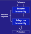
Compare Innate Immunity vs Adaptive Immunity

A

Innate Immunity: immediate protection

  • Fast (within seconds)
  • Recognise group pathogens
  • Lack of memory
  • No change in intensity

Adaptive Immunity: long lasting protection

  • Slow start (takes 3-4 days to start)
  • Specific to one pathogen (can recognise different serotypes)
  • Immunologic memory

Increases in intensity

21
Q

What is the first line of defence (of innate immunity)?

A

First line of defence (of Innate Immunity): preventing pathogen entering and invading

Physical Barriers
Physiological Barriers
Chemical Barriers
Biological Barriers

They all lead to factors that limit entry and growth of pathogens

22
Q

Explain more about physical barriers

A
Skin (surface area 1-2 m2) [any breach would allow pathogens in]
Mucous membranes (secrete an array of different antimicrobial substances): mouth, respiratory tract, GI tract, urinary tract
Bronchial cilia (beat trapped pathogens and dirt particles in mucus out of airways)
23
Q

Explain more about physiological barriers

A

(also seen in cases of non-infections such as allergies, asthma)

Diarrhoea (seen in food poisoning, allergies)
Vomiting (food poisoning, hepatitis, meningitis)
Coughing (pneumonia)
Sneezing (sinusitis)

24
Q

Explain more about chemical barriers

A
  • Low pH (skin 5.5, stomach 1-3, vagina 4.4)
  • Antimicrobial molecules (IgA secreted by mucosal membranes also in tears and saliva; lysozyme in sebum, perspiration and urine; mucus from mucous membranes; Beta-defensins (antiviral peptides) from lung epithelium, gastric acid + pepsin)
  • NB: IgA binds to microorganisms and prevents them from binding to the mucous membrane.
25
Q

Explain more about biological barriers and give examples

A
  • Normal flora: non-pathogenic microbes in strategic locations (nasopharynx, mouth/throat, skin, GI tract, vagina (lactobacillus spp)), absent in internal organs
  • Benefits: compete with pathogens (fierce competition) for attachment sites and resources. Also produce antimicrobial chemicals and synthesise vitamins (K, B12, other B vitamins)

Examples of normal flora that inhabit the skin

  • Staphylococcus aureus (Week 1, Case 1)
  • Staphylococcus epidermidis
  • Streptococcus pyogenes
  • Candida albicans
  • Clostridium perfringens

Examples of normal flora that inhabit the nasopharynx:

  • Streptococcus pneumonia (Week 3, Case 2)
  • Neisseria Meningitidis (Week 3, lecture case)
  • Haemophilus species

NB THEY COULD BECOME PATHOGENIC

26
Q

What happens when normal flora is displaced from its normal location?

A

When normal flora is displaced from its normal location to sterile location, clinical problems start. This can occur in situations such as:

  • Breaching the skin integrity (skin loss – burns, surgery, injection drug users, IV lines)
  • Faecal-oral route (ingestion – foodborne infection)
  • Faecal-perineal-urethral route (UTIs – especially in women)
  • Poor dental hygiene/dental work – commonest cause of harmless bacteraemia (dental extraction, gingivitis – gum disease, flossing)

This can lead to serious infections in high-risk patients

  • Asplenic (no spleen) and hyposplenic (reduced spleen function) patients
  • Patients with damaged or prosthetic valves
  • Patients with previous infective endocarditis

Treatment: may need to give antibiotic prophylaxis

27
Q

In what other situations apart from displacement can clinical problems start in?

A
  • Normal flora overgrows and becomes pathogenic when host becomes immune-compromised (e.g. in Diabetes, AIDS, Malignant diseases, Chemotherapy (causes loss of neutrophils))
  • When normal flora is depleted by antibiotics: e.g. intestine => severe colitis (Clostridium difficile), vagina => thrush (Candida albicans)
28
Q

What is the second line of defence?

A
  • Phagocytes and chemicals lead to INFLAMMATION
  • They are factors that will contain and clear the infection
29
Q

Explain about phagocyte-microbial interaction

A

Recognition process
Killing process of infectious microbes

30
Q

Describe the functions of macrophages, monocytes, neutrophils, basophils/mast cells, eosinophils, natural killer cels, dendritic cells

A
  • Macrophages: present in all organs. Ingest and detroy microbes (phagocytosis). Present microbial antigens to T cells (adaptive immunity). Produce cytokines/chemokines
  • Monocytes: present in the blood (5-7%). Recruited at infection site by cytokines and differentiate into macrophages.
  • Neutrophils (pus): present in the blood (60% of blood leukocytes). Increased during infection. Recruited by chemokines to the site of infection. Ingest and destroy pyogenic bacteria: Staph. Aureus and Strep. Pyogenes
  • Basophils/Mast cells: early actors of inflammation (vasomodulation), important in allergic responses
  • Eosinophils: defence against multi-cellular parasites
  • Natural Killer cells: kill all abnormal host cells (virus infected or malignant)
  • Dendritic cells: present microbial antigens to T cells (acquired immunity)
31
Q

What are PAMPs and PRRS and give examples

A

Pathogen Recognition:

  • Microbial Structures: pathogen-associated molecular patterns (PAMPs) made up of carbohydrates, lipids, proteins and nucleic acids
  • Phagocytes: Pathogen Recognition Receptors (PRRs): best known family is Toll Like Receptors

NB: note how some PRRs like TLR4 can recognize both gram negative and gram-positive bacteria.

32
Q

Explain about opsonisation and give examples of opsonins. Why are they essential?

A
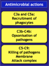
  • Pathogen recognition: opsonisation of microbes (coating proteins called opsonins that bind to the microbial surfaces lead to further recognition and enhanced attachment of phagocytes and clearance of microbes).
  • Most opsonins are produced by the liver

Examples of opsonins:

  • Complement proteins: C3b, C4b
  • Antibodies: IgG, IgM
  • Acute Phase Proteins: C-reactive protein (CRP) and Mannose-binding lectin (MBL)

Opsonisation is essential in clearing encapsulated bacteria (cannot clear these bacteria without opsonisation)

  • Neisseria meningitides
  • Streptococcus pneumonia
  • Haemophilus influenzae.b
33
Q

Explain about phagocytes engulfing and degrading microbes

A
  1. Chemotaxis and adherence of microbe to phagocyte
  2. Ingestion of microbe by phagocyte
  3. Formation of a phagosome
  4. Fusion of the phagosome with a lysosome to form a phagolyosome
  5. Digestion of ingested microbe by enzymes
  6. Formation of residual body containing indigestible material
  7. Discharge of waste materials
34
Q

Explain about phagocyte intracellular killing mechanisms

A
  • Oxygen-dependent pathway (respiratory burst – more effective): toxic O2 products for the pathogens: Hydrogen perioxide, hydroxyl radical, nitric oxide, singlet oxygen, hypohalite
  • Oxygen-independent pathways (rely on enzymes): lysosome, lactoferrin or transferrin, cationic proteins (cathepsin), proteolytic and hydrolytic enzymes
35
Q

Explain about the Complement System

A

: there are 20 serum proteins but the most important ones are C1-C9. There are 2 pathways

  • Aternative Pathway: activated by cell surface microbial constituents e.g. LPS (a PMP on E. coli)
  • MBL Pathway: initiated when MBL (acute phase protein produced by the liver) binds to mannose containing residues of proteins found on Salmonella spp. Candida albicans
36
Q

What are cytokines and chemokines involved in?

A
  • Chemoattraction
  • Phagocyte activation
  • Inflammation
37
Q

What are the anti-microbial actions of IL1, IL6 and TNFa?

A
  • Liver (produces opsonins): CRP and MBL (MBL is involved in complement activation)
  • Bone marrow: neutrophil mobilization
  • Inflammatory actions (local): vasodilation, vascular permeability, adhesion molecules (leading to attraction of neutrophils)
  • Hypothalamus: increased body temperature (fever)
38
Q

How can clinical problems start?

A
  • Overreaction of TLR4 receptor to microbial toxins (LPS)
  • Overreaction of complement
39
Q

What happens when you have no functioning spleen?

A

you are susceptible to overwhelming infection particularly pneumococcal. The spleen is the most active organ in innate immune system – it detects pathogens in blood especially encapsulated bacteria and provides fastest immune response

40
Q

Why can reduced phagocytosis occur?

A
  • Decreased spleen function (asplenic, hyposplenic)
  • Decreased neutrophil number (cancer chemotherapy, certain drugs, leukaemia and lymphoma0
  • Decrease neutrophil function (Chronic granulomatous disease (no respiratory burst); Chediak-Higashi syndrome (no phagolysosomes formation))
41
Q

Give a summary of Innate Immunity

A
  1. Innate barrier breached: entrance and colonization of the pathogens
  2. Complement, mast cells and macrophages activation (PRRR): phagocytosis (opsonins) and cytokine/chemokine production
  3. Vascular changes: vasodilation/vascular permeability; Chemoattraction: neutrophils, monocytes (TNF, IL8)
  4. Hypothalamus: Fever; Liver: acute phase response
  5. Redness, heat, swelling and pain: local inflammation
42
Q

What are the most common meningitis-causing organisms in different age groups?

A
  • Newborn: Group B streptococcus, E. coli, Listeria monocytes
  • Children: Haemophilus influenzae, Neisseria meningitidis, Streptococcus pneumoniae
  • Adults: Neisseria meningitidis, Streptococcus pneumoniae
  • Elderly: Listeria monocytes, Neisseria mengitidis, Streptococcus pneumoniae