Session 2: Infection Model Flashcards
What is the Model of Infection?

Give an example of each pathogen
Virus: HIV
Bacterium: Mycobacterium Tuberculosis
Yeast: Candida
Mould: Aspergillus
Protozoa:Malaria parasites
Helminth (worm): tapeworm

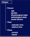
What are the patient factors and briefly discuss them
- Difference in attack rates between different ages. In the first 3 months, babies are protected by maternal antibodies that cross over the placenta particularly during the last four weeks of pregnancy, giving long term immune protection. They have a relatively short lifespan ~3 months. Then babies are very susceptible, very immune incompetent e.g. Meningitis, streptococci pneumonia etc. There is observed increased risk of infection at that age. Up until about 3 years of life
- Pathological state includes immunosuppression due to HIV, cancer treatment or transplantation – allowing the colonization of opportunistic infections.
- Physiological state includes pregnancy and puberty – at different ages women have different pHs in their vaginal flora which leads to different commensals and therefore different risks of infection.
- Calendar time: winter months have increased occurrence of norovirus (winter vomiting bug), flu
- Social factors is related to transmissible organisms: sufficiently close contact with an infected person leads to increased risk e.g. common cold, pneumonia, Ebola
- Relative time leads to incubation periods leads to incubation and infection period (if asymptomatic after a particular numbers of days, can assume to be infection free and person can be released from quarantine) as some organisms have a known incubation period.

Give examples of how patient factors can be modified
by treating pathological conditions, optimizing physiological state with e.g. adequate nutrition and hydration, modifying social factors with sanitation standards etc.
Give examples of interventions that been intended to reduce risk of infection
vaccination, prophylactic antibiotics, health examinations, provision of clean water and food, garbage collection.
What are the different mechanisms of infection and give examples of each
- Contiguous (direct) spread: peritonitis e.g. following a burst appendix, spread of E.coli from large bowel to urinary tract. In females the urethra + urethral orifice is close to rectum – higher risk of colonization of E.coli into the urethra à high occurrence of UTIs in women then men.
- Inoculation; some contaminated object inserted into part of the body e.g an eye (in the cornea) infection from a branch (fungal keratitis). Also can be as a result from a bite or stabbing.
- Haematogenous spread; travels around the bloodstream and lodges into a distal site which may be far away from site of entry e.g. endocarditis
- Ingestion: food poisoning – salmonella, faecal-oral ingestion
- Inhalation includes conditions spread by droplet (~1m) or aerosol (can move quite far distances) spread e.g. Norovirus (aerialisation from vomiting), cold, flu
- Vector: requires third party to transmit an organism e.g. Malaria mosquito, Lyme disease spread by ticks
- Vertical transmission: mother to child either during pregnancy (intrauterine transmission) or during time of delivery e.g. HIV, Hep B, syphyllis. Important to recognize mother has HIV as baby can be delivered in particular ways to protect baby from infection including C-section and starting strong anti-viral therapy immediately.

What are the general principles of the mechanism of action of an infection?

What does the management of an infection normally involve?
Management normally consists of the following steps:
- The clinician uses history, examination and investigations to make a diagnosis. Consider where is the infection and what is the infection.
- Treatment may be specific and/or supportive. Specific could involve antimicrobials and other augmentative measures such as surgery (e.g. in a perforated appendix), drainage (drainage of pus is good at relieving symptoms and sample may be useful in diagnosis), debridement (removal of damaged tissue) and dead space removal (akin to debridement e.g. in a bone infection, the damaged bone and surrounding tissue may be removed to prevent future infections etc). Supportive treatment could be symptom relief and physiological restoration e.g. restoring normal pH of blood.
- Infection preventions – in the hospital and in the community. This involves preventing infection transmission to other patients, staff and other contacts.

Do the steps involved in management have to occur in a particular sequence? Give an example of an exception
These steps commonly occur in a strict sequence but not always. These processes may be modified for example during Acute sepsis where the priority is to deal with any life-threatening complications and there are also variations if dealing with a group of people with a disease outbreak. Nevertheless the first instinct is to go through history, examination and investigation.
What are the possible outcomes of an infection?
Outcome: NB: Only smallpox has been eliminated from the world so far. Patients can die from untreatable or overwhelming infection but the aim is to cure!

Apply the model of infection to a specific example (endocarditis)
E.g. Endocarditis: heart valves may become infected during transient bacteraemia (presence of bacteria in the blood)
- Organism: bacteria may originate from the mouth, urinary tract, intravenous drug misuse or colonized intravascular lines
- Mechanism of infection: haematogenous
- Complications: local progression may lead to aortic root abscess. Valve destruction may lead to cardiac decompensation. Cerebral or limb infarction may follow septic embolus. Nephritis (inflammation of the kidneys) second to immune complex decomposition can progress rapidly if sepsis is uncontrolled or if antibiotics with renal toxicity are given without care e.g. aminoglycosides.
- Management: antibiotics (ideally given once the identity and sensitivities of the infecting organism are known), surgical management maybe required to dal with haemo-dynamic consequences of endocarditis.
- Prevention: antibiotic prophylaxis should be given to patients with damaged valves when they undergo procedures that give rise to significant bacteraemia such as dental work or urogenital surgery.
Describe antimicrobial classification based on mechanism of action
- Cell Wall Synthesis: Beta-lactams, glycopeptides (e.g. vancomycin
- Protein synthesis tetracyclines, aminoglycosides (e.g. gentamicin) and macrolides (erythromycin)
- Nucleic acid synthesis: quinolones + trimethoprim and rifampicin which affect other stages in the production of nucleic acids compared to quinolones
- Cell membrane function: polymixins (e.g. colistin)
Beta-lactams such as penicillins work by inhibiting peptidoglycan cross-linkage (responsible for cell wall rigid structure) as they bind to penicillin binding proteins which is the enzyme responsible for the formation of cross-links.
Vancomyin (glycopeptide) binds to the chains, preventing the peptidoglycan cross-links forming by preventing the penicillin binding protein enzyme from binding.
Fluoroquinolones bind to two nuclear enzymes (DNA gyrase and Topoisomerase IV), inhibiting DNA replication. Quinolones are well absorbed orally, are widely distributed and penetrate cells well.

How ca antibacterial agents be further classified?
[*] Bactericidal (kills bacteria) or bacteriostatic (inhibits bacteria, stops it growing)
[*] Spectrum: ‘broad’ (gram positive, aerobic) v. ‘narrow’ (small group e.g. gram positive cocci)
[*] Target site (mechanism of action)
[*] Chemical structure (antibacterial class)
It is best to use chemical structure and target site however as drugs are often both bactericidal and bacteriostatic depending on dosage and normally on the middle of the spectrum, not exclusively broad or narrow.
What are the ideal features of antimicrobial agents?
- Selectively toxic
- Few adverse effects (including few side effects)
- Reach site of infection (consider metabolism of drug)
- Oral / IV formulation - depending on how ill patient is, ideally able to have both oral and IV forms of the drug
- Long half-life (a dosage of once every 3 days or weekly is more convenient than 3/4 times a day)
- No interference with other drugs (minimal interactions)
Describe the Disc DIffusion method as a way of measuring antibiotic activity
- Because of convenience, efficiency and cost, the disc is probably the most widely used method for determining antimicrobial resistance.
- A growth medium is first evenly seeded throughout the plate with filter paper with the isolate of interest. Commercially prepared disks, each of which are pre-impregnated with a standard concentration of a particular antibiotic, are then evenly dispensed and lightly pressed onto the agar surface.
- The test antibiotic immediately begins to diffuse outward from the discs, creating a gradient of antibiotic concentration in the agar such that the highest concentration is found close to the disc with decreasing concentrations further away from the disc.
- After an incubation period, the bacterial growth around each disc is observed. If the test isolate is susceptible to a particular antibiotic, a clear area of “no growth” will b observed around that particular disc. The area of no growth is referred to the zone of inhibition. This is then measured in mm and compared to a standard interpretation chart.


