Session 3 Flashcards

1
Q

Describe the structure and layers of the abdominal wall

A
  • Altough continuous, the abdominal wall is subdivided into the anterior wall, right and left lateral walls and the posterior wall. The boundary between the anterior and the lateral walls is indefinite therefore the term anterolateral abdominal wall is used.
  • The anteriolateral abdominal wall is composed of skin, subcutaneous tissue (superficial fascia/fat), muscles and their aponeuroses, deep fascia, extraperitoneal fat and parietal peritoneum.
  • The anterolateral abdominal wall is bounded superiorly the cartilages of the 7th-10th ribs and the xiphoid process of the sternum, and inferiorly by the inguinal ligament and the superior margins of the anterolateral aspects of the pelvic girdle (iliac crests, pubic crests and pubic symphysis).
  • During a general physical examination, the abdominal wall is inspected and the underlying organs are palpated.
  • As access to the abdominal cavity requires surgical incision, knowledge of the composition of the abdominal wall is necessary before carrying out any physical or surgical procedure.
2
Q

What does the Superficial Fascia consist of?

A

The superficial fascia consists of fatty connective tissue. The composition of this depends on its location:

  • Above the umbilicus: a single sheet of connective tissue. This is continuous with the superficial fascia in other regions of the body.
  • Below the umbilicus: it is divided into 2 layers; the fatty superficial layer (Camper’s fascia) and the membranous deep layer (Scarpa’s fascia). Superficial vessels and nerves run between these two layers of fascia
3
Q

Describe the major landmarks of the abdominal wall part 1: umbilicus, epigastric fossa, linea alba, pubic crest and symphysis, inguinal groove, semilunar lines (linea semilunaris)

A
  • Umbilicus: at spinal level L3 / dermatome T10
  • Epigastric Fossa (pit of the stomach): slight depression in the epigastric region, just inferior to the xiphoid process. Particularly noticeable when a person is in the supine position (lying down, face up) because the abdominal organs spread out. Heartburn is commonly felt at this site.
  • Linea Alba: aponeuroses of abdominal muscles, separating the left and right rectus abdominis. Visible in lean individuals because of the vertical skin groove superficial to it. If the linea alba is lax, when the rectus abdominis contract the muscles spread apart. This is called divarication of recti
  • Pubic Crest and Symphysis: the upper margins of the pubic boens and the cartilaginous joint that unite them. Can be felt at the inferior edge of the linea alba.
  • Inguinal groove: a skin crease that is parallel and just inferior to the inguinal ligament (runs between ASIS and pubic tubercle). Marks the division between the abdominal wall and the thigh.
  • Semilunar Lines (Linea Semilunaris): slightly curved, tendinous line on either side of the rectus abdominis – running from the 9th rib to pubic tubercle. It is the lateral border of the rectus abdominus
4
Q

What are the tendinous intersections of the rectus abdominis? What is the arcuate line?

A
  • Tendinous Intersections of Rectus Abdominis: clearly visible in persons with well-developed rectus muscles. The interdigitating bellies of the serratus anterior and external oblique muscles are also visible.
  • Arcuate Line (aka Douglas’ line): where the fibrous sheath stops (inferior limit of the posterior layer of the rectus sheath), 1/3 of the way from the umbilicus to the pubic crest.
5
Q

What are the 5 muscles of the abdominal wall?

A

There are 5 bilaterally paired muscles (3 flat and 2 vertical) in the antero-lateral wall.

The flat muscles end anteriorly in a strong aponeurosis (flattened tendons) each of which interlaces in the midline at the linea alba. The flat muscles are External Oblique, Internal Oblique and Transversus Abdominis

With their opposite counterparts they form the rectus sheath which contains the two vertical muscles and neurovascular structures

6
Q

Describe the 3 pairs of flat muscles individually

A

External Oblique (origin, insertion)

  • External surfaces of the 5th to 12th ribs
  • Linea alba, pubic tubercle and anterior half of iliac crest

Internal Oblique

  • Thoracolumbar fascia, anterior 2/3ds of iliac crest and connective tissue deep to lateral third of inguinal ligament
  • Inferior borders of the10th to 12th ribs, linea alba and pectin pubis via conjoint tendon

Transversus Abdominis

  • Internal surfaces of 7th to 12th costal cartilages, thoracolumbar fascia, iliac crest and connect tissue deep to lateral third of inguinal ligament
  • Linea alba with aponeuroses of internal oblique, pubic crest and pectin pubis via conjoint tendon
7
Q

What do the flat muscles do?

A
  • The 3 flat muscles’ fibres have varying orientations with the fibres of the obliques running diagonally and perpendicular to each other, and the fibres of the transversus running transversely.
  • The three flat muscles are continued anteriorly and medially as strong, sheet-like aponeuroses. Between the mid-clavicular line and the midline, the aponeuroses form the tough, aponeutoric, tendinous rectus sheath that encloses the Rectus Abdominis
  • The aponeuroses then interweave with their fellow of the opposite side, forming the linea alba which extends from the xiphoid process to the pubic symphysis. The interweaving is not only between right and left sides but also between intermediate and deep layers.
  • The flat muscles are important in flexing, twisting and lateral flexion of the trunk. Their fibres run in differing directions and cross each other, strengthening the abdominal wall and decreasing the risk of herniaton
  • Their contraction increases intra-abdominal pressure during activities such as lifting, coughing, defecation, urination, parturition (giving birth).
8
Q

Describe the Rectus Sheath

A
  • The Rectus Sheath is formed by the aponeuroses of the three flat muscles, and encloses the rectus abdominus and pyramidalis muscles. It has an anterior and posterior wall for most of its length:
  • The Anterior wall is formed by the aponeuroses of the external oblique, and of half of the internal oblique
  • The Posterior wall is formed by the aponeuroses of half the internal oblique and of the transversus abdominus.
  • Approximately midway between the umbilicus and the pubic symphysis, all of the aponeuroses move to the anterior wall of the rectus sheath. At this point, there is no posterior wall to the sheath; the rectus abdominus is in direct contact with the transversalis fascia.
  • The area of transition between having a posterior wall, and no posterior wall is known as the arcuate line.
9
Q

Describe the vertical muscles

A

The two vertical muscles of the anterolateral abdominal wall are contained within the rectus sheath, the large Rectus Abdominis and small Pyramidalis

  • Rectus Abdominis is a long paired muscle, found either side of the midline in the abdominal wall. It is split into two by the linea alba. The lateral border of the two muscles create a surface marking called the linea semilunaris. At several places, fibrous strips, known as tendinous intersections, intersect the muscle. The tendinous intersections and the linea alba give rise to the six pack seen in individuals with low body fat.
  • As well as assisting the flat muscles in compressing the abdominal viscera, the rectus abdominis also stabilises the pelvis during walking, and depresses the ribs.
  • The pyamidalis is a small triangle-shaped muscle, found superficially to the rectus abdominis. It is located inferiorly with its base on the pubis bone and the apex of the triangle attached to the linea alba. It acts to tense the linea alba.
10
Q

What to consider when doing a surgical incision?

A
  • The location of the incision depends on the type of surgical operation, the location of abdominal viscera (organs), avoidance of nerves (particularly motor nerves), maintenance of blood supply, etc.
  • E.g. severance of a motor nerve(s) will lead to muscle paralysis, thereby causing abdominal wall weakness.
  • When design an incision, we want one that we can close and provide long-lasting strength, thus minimising the incidence of incisional herniae. Improper healing of a surgical incision or scar may become a site where herniation of the abdominal viscera can occur through the abdominal wall.
  • If we try to sew muscle together, the sutures will ‘cut out’ (think sewing butter)
  • Consider direction of muscle fibres (you want to split the muscle fibres rather than cut them)
11
Q

Describe common vertical incisions

A
  • Median/Midline: an incision that is made through the linea alba. It can be extended the whole length of the abdomen, by curving around the umbilicus. The linea alba is poroly vascularised, so blood loss is minimal and major nerves are avoided. All can be used in any procedure that requires access to the abdominal cavity.
  • Paramedian: similar to the median incision but is performed laterally to the linea alba, providing access to more lateral structures (kidney, spleen and adrenals) This method ligates (ties up) the blood and nerve supply to muscles medial to the incision, resulting in their atrophy
12
Q

Describe common transverse incisions

A
  • Transverse: this incision is made just inferior and laterally to the umbilicus. This is a commonly used procedure as it causes least damage to the nerve supply to the abdominal muscles and heals well. The incised rectus abdominus heals producing a new tendinous intersection. It is used in operations on the colon, duodenum and pancreas. Surgeons suture the external oblique aponeuroses together to provide a strong closure
  • Suprapubic (Pfannenstiel): incision is made about 5cm superior to the pubis symphysis. They are used when access to the pelvic organs is needed. When performing this incision, care must be taken not to perforate the bladder (especially if it is not catheterised) as the fascia thins around the bladder wall)
  • Subcostal: this incision starts inferior to the xiphoid process, and extends inferior parallel to the costal margin. It is mainly used on the right side to operate on the gall bladder and on the left to operate on the spleen.
13
Q

Describe the incision used in an Appendicectomy

A

Appendicectomy:

Incision at McBurney’s point

  • 2/32 of the distance between the umbilicus and ASIS
  • Through a ‘gridiron’ muscle-splitting incision

Gridiron Incision: put scissors in and open and close them to separate out the muscle fibres, followed by the next two layers. You have to separate out thee fibres of the external and internal oblique’s and the transversalis.

14
Q

Explain about Patent Urachus and Vitelline Duct (developmental abnormalities)

A
  • Patent Urachus: can present at birth or in later life in men when they develop bladder outflow obstruction due to benign prostatic hypertrophy – present clinically with urine coming out of the umbilicus
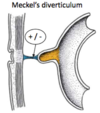
  • Vitelline Duct (joins the yolk sac to the midgut) – can persist creating a number of different abnormalities such as Meckel’s Diverticulum
15
Q

Explain about Meckel’s Diverticulum

A

Meckel’s Diverticulum: also known as Ilieal Diverticulum and is the most common GI abnormality. It is a ‘cul-de-ac’ in the ileum. Meckel’s Diverticulum follows a Rule of 2s:

  • 2% of the population affected
  • 2 feet from the ileocecal valve
  • 2 inches long
  • Usually detected in under 2’s, can be asymptomatic
  • 2:1 Male:Female

The diverticulum can contain ectopic gastric or pancreatic tissue. The ectopic tissue will secrete enzymes and acids into tissue not protected from them, causing ulceration. The reason for this is not clear.
16
Q

Explain about Vitelline Cyst and Vitelline Fistula

A
  • VItelline Cyst: the vitelline duct forms fibrous strands at either end.
  • Vitelline Fistula; there is direct communication between the umbilicus and the intestinal tract. This results in faecal matter coming out of the umbilicus.
17
Q

What is Somatic Referred pain?

A

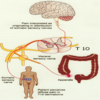
pain caused by a noxious stimulus to the proximal part of a somatic nerve that is perceived in the distal dermatome of the nerve. E.g. shingles affects nerves; pain is felt distally along the nerves from the problem
18
Q

What is Visceral referred pain?

A
  • in the thorax and abdomen, visceral afferent pain fibres follow sympathetic fibres back to the same spinal cord segments that gave rise to the preganglionic sympathetic fibres. The CNS perceives visceral pain as coming from the somatic portion of the body supplied by the relevant spinal cord segments.
  • Visceral pain is caused by ischaemia, abnormally strong muscle contraction, inflammation and stretch
  • Touch, burning, cutting and crushing does not cause visceral pain.
19
Q

Where would you feel hepatic pain?

A
20
Q

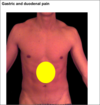
Where would you feel gastric and duodenal pain?
A
21
Q

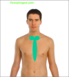
Where would you feel oesophageal pain?
A
22
Q

Where would you feel pancreatic and abdominal aorta pain?

A

Retroperitoneal structures can cause central back pain e.g. pancreas and abdominal aorta

23
Q

Where would you feel splenic pain?

A
24
Q

Where would you feel gall bladder pain?

A
25
Q

Where would you feel renal/ureteric pain?

A

patient rolls around on the floor; pain is “worse than child birth:

26
Q

Describe the acute appendicitis pain

A

Acute Appendicitis: in early appendicitis the pain begins at the umbilicus, since the innervation of the appendix enters the spine at that level (T10). Later as the appendix becomes more inflamed, it irritates the surrounding bowel wall, localising the pain to the right lower quadrant (irritation to somatic nerve).

27
Q

Describe Small and Large Bowel colic pain

A

Small/Large Bowel Colic: doubled over in pain.

Small bowel colic: periumbilical (midgut)

Large bowel colic: suprapubic (hindgut)

28
Q

Where would you feel uterine/ovarian pain?

A
29
Q

Where would you feel bladder pain?

A
30
Q

Explain Referred Diaphragmatic Irritation

A

Referred Diaphragmatic Irritation:

  • Ruptured spleen
  • Ectopic pregnancy
  • Perforated ulcer

Blood pools in pelvis, giving pain. The patient feels faint due to the loss of blood, so lies down causing the blood to rush to the diaphragm (C3,C4,C5). The presence of blood here results in referred pain to the left shoulder (Liver is in the way of blood at the right diaphragm)

Irritation of the diaphragm (e.g. as a result of inflammation of the liver, gallbladder or duodenum) may result in shoulder tip pain.

31
Q

Describe and recognize the general appearance and disposition of the major abdominal viscera: diaphragmatic representation

A
32
Q

Describe and recognize the general appearance and disposition of the major abdominal viscera: undisturbed abdominal contents

A
33
Q

Describe and recognize the general appearance and disposition of the major abdominal viscera: anterior view

A
34
Q

Explain the concept of the peritoneal cavity as a virtual space

A

The peritoneal cavity is a potential space of capillary thinness between the parietal and visceral layers of peritoneum.
It contains no organs but contains a thin film of peritoneal fluid, which consists of water, electrolytes, leukocytes and antibodies.

  • Acts as a lubricant, enabling free movement of the abdominal viscera
  • The antibodies fight infection

Lymphatic vessels particularly on the interior surface of the diaphragm absorb the peritoneal fluid.
In males the peritoneal cavity is completely closed, but in females there is a communication pathway to the exterior of the body through the uterine tubes, cavity and vagina. The communication constitutes a potential pathway of infection from the exterior.

35
Q

Describe the structure of the peritoneum

A
  • The peritoneum is a continuous, two layered membrane – the parietal peritoneum, which lines the internal surface of the abdominal wall and the visceral peritoneum, which invests viscera such as the stomach and the intestines.
  • Both layers of the peritoneum consist of mesothelium, a layer of simple squamous epithelial cells.
  • Extraperitoneal connective tissue separates the parietal peritoneum from the muscular layers of the abdominal wall. The parietal peritoneum covering the anterior abdominal wall and pelvis walls is generally attached only loosely by this tissue, allowing for the considerable change in size of the bladder and rectum.
  • Extraperitoneal tissue frequently contains a large amount of fat, especially in obese males.
  • The extraperitoneal tissue behind the linea alba and on the inferior surface of the diaphragm is dense and more firmly adherent.
36
Q

Describe the neurovascular supply to the peritoneal layers and how this relates to pain

A
  • The parietal peritoneum is served by the same blood, lymphatic and somatic nerve supply as the region of the wall it lines. Therefore, like the overlying skin, the interior of the body wall is sensitive to pressure, pain, heat and cold and laceration. Derived from somatic mesoderm in the embryo.
  • Pain is generally well localised, apart from on the inferior surface of the central part of the diaphragm, which is innervated by the phrenic nerve (C3,C4,C5, referred pain to shoulder) This explains why the pain from appendicitis shifts to over the appendix, as the parietal peritoneum becomes inflamed, localising the pain.
  • The visceral peritoneum shares the same blood, lymphatic and visceral nerve supply as the organs it covers Also, like the organs it covers the visceral peritoneum is insensitive to touch, heat and cold and laceration; it is stimulated primarily by stretching and chemical irritation. The pain produced is poorly localised, being referred to the dermatomes of the spinal ganglia providing the sensory fibres. Derived from splanchnic mesoderm.
37
Q

What is a Mesentery? And describe the Greater and Lesser Omenta

A
  • Mesentery: a mesentery is a double layer of peritoneum that occurs as a result of the invagination of the peritoneum by an organ and constitutes as a continuity of the visceral and parietal peritoneum. A mesentery connects an intraperitoneal organ to the body wall (usually the posterior abdominal wall).
  • It provides a pathway for nerves, blood vessels and lymphatics from the body wall to the viscera
  • Omentum: an omentum is a double-layered extension or fold of peritoneum that passes from the stomach and proximal part of the duodenum to adjacent organs in the abdominal cavity.
  • Greater Omentum: a prominent, four-layered peritoneal fold that hands down like an apron from the greater curve of the stomach. After descending it folds back and attaches to the anterior surface of the transverse colon and its mesentery
  • It has a role in immunity and is sometimes referred to as the ‘abdominal policeman’ – because it can migrate to infected viscera.
  • Lesser Omentum: a much smaller double-layered peritoneal fold that connects the lesser curvature of the stomach and the proximal part of the duodenum to the liver. It also connects the stomach to the portal triad. It consists of 2 parts: the hepatogastric ligament and the hepatoduodenal ligament.
38
Q

And explain about the Peritoneal Ligament

A

Peritoneal Ligament: a double layer of peritoneum that connects an organ with another organ or to the abdominal wall.

Peritoneal Ligaments Connect Liver to:

  • Anterior abdominal wall – Falciform Ligament
  • Stomach – Hepatogastric Ligament (membranous portion of lesser omentum)
  • Duodenum – Hepatoduodenal Ligament (the thickened free edge of the lesser omentum which conducts the portal triad – portal vein, hepatic artery, bile duct)

Peritoneal Ligaments Connect Stomach to:

  • Inferior surface of the diaphragm – Gastrophrenic Ligament
  • Spleen – Gastrosplenic ligament
  • Transverse colon - Gastrocolic ligament (greater omentum

Organs have bare areas, to allow the entrance and exit of neurovascular structures.

39
Q

Describe and recognize the general appearance and disposition of the major abdominal viscera: posterior view

A
40
Q

How can structures in the peritoneum be classified?

A

Intraperitoneal structures are completely covered by peritoneum, however not completely enclosed due to the mesentery (think of the concept of a fist in the balloon) e.g. stomach, spleen, liver

Retroperitoneal/extraperitoneal are outside of the peritoneal cavity and thus are only partially covered by the parietal peritoneum on their anterior surface, for example the kidneys lie between parietal peritoneum (only found on the anterior surface) and the posterior abdominal wall. SAD PUCKER

  • Suprarenal (adrenal) Glands
  • Aorta/IVC
  • Duodenum (except for the duodenal cap – first 2cm)
  • Pancreas (except the tail)
  • Ureters)
  • Colon (ascending and descending parts)
  • Kidneys
  • Oesophagus
  • Rectum
41
Q

Describe the surface regions of the abdominal wall and the planes which define them

A

The abdominal wall can be divided into 9 regions – useful clinically for describing the location of pain, location of viscera and describing surgical procedures. The 9 regiosn are formed by 2 horizontal planes and 2 vertical planes.
Horiztonal Planes:

  • Transpyloric/Subcostal plane: horizontal line halfway between the xiphoid process and the umbilicus, passing through the pylorus of the stomach.
  • Intertubercular plane: horizontal lines that joins the iliac crests.

The abdomen is divided by the midclavicular lines vertically which run from the middle of the clavicle to the mid-inguinal point (halfway between ASIS and the pubic symphysis)

42
Q

Describe the structure and relations of:

Supracolic and infracolic compartments, The greater and lesser omentum, transverse mesocolon

A
  • The Transverse Mesocolon (mesentery of the transverse colon) divides the abdominal cavity into a Supracolic Compartment, containing the stomach, liver and spleen, and an Infracolic Compartment, containing the small intestine and ascending and descending colon
  • The Infracolic Compartment lies posterior to the greater omentum and is divided into the Right and Left Infracolic Spaces by the mesentery of the small intestine.
  • Free communication occurs between the Supracolic and the Infracolic compartments through the Paracolic Gutters, the grooves between the lateral aspect of the ascending or descending colon and the posterolateral abdominal wall.
43
Q

Describe the structure and relations of Lesser and greater sac, the location of the subphrenic spaces (especially the right posterior subphrenic recess)

A
  • The Greater Sac is made up of the Supracolic and Infracolic compartments
  • The Lesser Sac, or Omental bursa, is an extensive sac-like cavity that lies posterior to the stomach, lesser omentum and adjacent structures. It has a superior recess, limited superiorly by the diaphragm and the posterior layers of the coronary ligament of the liver, and an inferior recess between the superior parts of the layers of the greater omentum.
  • Most of the inferior recess becomes sealed off from the main part (posterior to the stomach) after adhesion of the anterior and posterior layers of the greater omentum.
  • The lesser sac / omental bursa permits free movement of the stomach on the structures posterior and inferior to it because it’s anterior and posterior walls slide smoothly over one another.
  • The Greater and Lesser Sacs communicate through the Omental Foramen (epiploic foramen), an opening situated posterior to the free edge of the lesser omentum (hepatoduodenal ligament). The omental foramen can be located by running a finger along the gallbladder to free the edge of the lesser omentum, and usually admits two fingers.
44
Q

Describe the Subphrenic Spaces

A
  • The subphrenic spaces are recesses in the greater sac of the peritoneal cavity between the anteriosuperior diaphragmatic surface of the liver and the diaphragm. They are separated into right and left subphrenic spaces by the falciform ligament of the liver.
  • Subphrenic abscesses generally occur as a result of the accumulation of pus in the left or right subphrenic space as a consequence of peritonitis. They are more common on the right side due to the increased frequency of appendicitis and ruptured duodenal ulcers.
  • The Right Subphrenic Space lies between the diaphragm and the anterior, superior and right lateral surfaces of the right lobe of the liver.
  • It is bounded on the left side by the falciform ligament and behind by the upper layer of the coronary ligament. It is a relatively common site for collections of fluid after right-sided abdominal inflammation.
  • The Left Subphrenic Space lies between the diaphragm, the anterior and superior surfaces of the left lobe of the liver, the anterosuperior surface of the stomach and the diaphragmatic surface of the spleen.
  • It is bounded to the right by the falciform ligament and behind by the anterior layer of the left triangular liament. It is much enlarged in the absence of the spleen and is a common site for fluid collection, particularly after a splenectomy.
  • The left Subphrenic space is substantially larger than the right (liver is on the right)
45
Q

Describe the Recto-uterine and utero-vesicle pouch in the female

A
  • In females the peritoneum passes from the rectum to the posterior vaginal fornix and then back to the uterine cervix and body as the recto-uterine fold, which descends to form the recto-uterine pouch (of Douglas). It is the extension of the peritoneal cavity between the rectum and the posterior wall of the uterus.
  • The peritoneum spreads over the uterine fundus to its anterior surface as far as the junction of the body and cervix, from which it is reflected forwards to the upper surface of the bladder, forming a shallow vesicouterine pouch. The vesicouterine pouch is a double fold of peritoneum between the anterior surface of the uterus and the bladder.
  • The peritoneal cavity is not completely closed in females. The abdominal ostia of the uterine tubes opens into the peritoneal cavity providing a potential pathway between the female genital tract and the peritoneum.
  • Clinically, this means that infections of the vagina, uterus and uterine tubes may result in infection and inflammation of the peritoneum (peritonitis). This is, however, rare due to the presence of a mucous plug in the external os (opening) of the uterus which prevents the passage of pathogens but allows sperm to enter the uterus.
46
Q

Describe the Recto-vesical pouch in the male

A
  • In males the peritoneum leaves the junction of the middle and lower thirds of the rectum, passing forwards to the upper poles of the seminal vesicles and superior aspect of the bladder.
  • Between the rectum and bladder it forms the rectovesical pouch. The peritoneal cavity is completely closed in males.
47
Q

Describe the mesentery of the small intestine

A
  • A broad, fan shaped fold, connecting the coils of the jejunum and ileum to the posterior abdominal wall. Between the two sheets of peritoneum are blood vessels, lymph vessels and nerves. This allows this part of the intestine to move relatively freely within the abdominal cavity
  • The attached, parietal border is the root of the mesentery about 15cm from the duodenojejunal flexure at the level of left side L2, oblique (towards inferior right) to the ileocaeal junction.
  • The root of the mesentery crosses the second and third parts of the duodenum, abdominal aorta, inferior vena cava, right ureter, right psoas major muscle and right gonadal artery.
48
Q

Describe the sigmoid mesocolon

A
  • This is a peritoneal fold attaching the sigmoid colon to the pelvic wall, the attachment being an inverted V with an apex near the division of the left common iliac artery.
  • The left limb descends medial to the left psoas major and the right passes into the pelvis to end in the midline at the level of the third sacral vertebra.
  • Sigmoid and superior rectal vessels run between its layers and the left ureter descends into the pelvis behind its apex.
49
Q

Describe areas of potential weakness in the abdominal wall

A

Two potential sites of weakness exist in the inferior part of the abdominal wall. These are superficial (external) and deep (internal) inguinal rings (openings). The area between them forms the inguinal canal through which structures leave and enter the abdominopelvic cavity.

The chief sites of hernia are inguinal, femoral and umbilical. The potential areas for these hernias are the inguinal canal, femoral ring and umbilicus respectively.

50
Q

Describe the structure and borders of the inguinal canal

A
  • The inguinal canal is an oblique passage that extends in a downward and medial direction. It begins at the deep inguinal ring and continues for ~4cm ending at the superficial inguinal ring.
  • Anterior wall is formed by the aponeurosis of the external oblique, and reinforced by the internal oblique muscle laterally.
  • Posterior wall is formed by the transversalis fascia
  • The roof is formed by the transversalis fascia, internal oblique and transversus abdominis
  • The floor is formed by the inguinal ligament (a r’olled up’ portion of the external oblique aponeurosis) and thickened medially by the lacunar ligament
51
Q

What structures pass through the inguinal canal? And what happens during times of increased intra-abdominal pressure?

A
  • The canal lies in between the muscles of the anterior abdominal wall above the inguinal ligament and runs parallel and superior to the medial half of the inguinal ligament
  • The inguinal ligament is the inferior border of the aponeurosis of the external oblique muscle attached between the anterior superior iliac spine laterally and the pubic tubercle medially
  • The spermatic cord in men, and the round ligament of the uterus in women passes through the canal.
  • Additionally in both sexes, the ilioinguinal nerve passes through part of the canal and exists through the superficial inguinal ring with other contents
  • During periods of increased intra-abdominal pressure, the abdominal viscera are pushed into the inguinal canal. To prevent herniation, the muscles of the anterior and posterior wall contract and ‘clamp down’ on the canal.
52
Q

What is a hernia? And briefly describe the different types

A
  • A hernia is a protrusion of part of the abdominal viscus (organ), through a defect in the abdominal wall.
  • The hernia sac is covered by skin, subcutaneous tissues and often the layers of the abdominal wall through which the hernia passes
  • Most of the hernias occur in the inguinal, umbilical and epigastric regions.
  • Other types of hernias such as diaphragmatic hernia, hiatus hernia and rolling hiatus hernia occur within the thoraco-abdominal cavity.
53
Q

Distinguish between direct and indirect inguinal hernias

A
  • An inguinal hernia is a protrusion of the abdominal cavity contents through the inguinal canal. They are very common (Lifetime risk 27% for men, 3% for women)
  • The commonest form of hernia is the indirect inguinal hernia. Here a viscus enters the deep inguinal ring, within the diverging arms of the transversalis fascial sling, to extend either part of the way along the inguinal canal, or all the way through it, to appear in the scrotum or the labium majora. Such hernias are much more common in males than females.
  • Most indirect inguinal hernias are the result of the failure of embryonic closure of the deep inguinal ring after the testicle has passed through it.
  • The peritoneal sac originates from an area medial to the epigastric vessels and bulges into the inguinal canal via the posterior wall. As the strong conjoint tendon covers this point, a direct inguinal hernia is usually seen only as a bulge in the inguinal region.
  • A direct inguinal hernia protrudes in the inguinal canal through a weakened area in the transversalis fascia near the medial inguinal fossa within an anatomical region known as the Inguinal/Hesselbach’s triangle. The borders of the triangle are:

Inferiorly: medial half of the inguinal ligament

Medially: lower lateral border of rectus abdominis

Laterally: inferior epigastric artery

  • In inguinal hernias, the neck of the hernia sac appears above the inguinal ligament.
54
Q

Explain about femoral hernias

A
  • In femoral hernias, the hernia sac protrudes into the upper thigh medial to the femoral vein. The neck of the hernia appears, therefore, below the inguinal ligament.
  • Femoral hernias are a protrusion of abdominal viscera into the femoral canal, occurring through the femoral ring. A femoral hernia appears as a mass, often tender, in the femoral triangle.
  • Femoral hernias are bounded by the femoral vein laterally and the lacunar ligament medially. The hernia compresses the contents of the femoral canal (loose connective tissue, fat and lymphatics) and distends the wall of the canal.
  • Initially femoral hernias are small, as they are contained within the canal, but they can enlarge by passing inferiorly through the saphenous opening into the subcutaneous tissue of the thigh.
  • Femoral hernias are common in females as they have wider pelvises. Strangulation of femoral hernias may occur because of the sharp, rigid boundaries of the femoral ring.
55
Q

Explain about diaphragmatic hernias

A

In diaphragmatic hernias developmental defects of the diaphragm may allow any viscus (e.g. the large intestine) to herniate into the chest.

  • Hiatus hernias are special types of diaphragmatic hernia.
  • In a sliding hiatus hernia, the gastro-oesophageal junction may slide through the diaphragm into the chest.
  • In a rolling histus hernia, part of the fundus of the stomach may pass into the chest alongside the oesophagus.
56
Q

Explain about umbilical hernias

A
  • Umbilical hernia sometimes occurs in children due to a weakness in the umbilical scar. In adults hernia of the abdominal wall may protrude through the linea alba anywhere between xiphisternum and the umbilicus (paraumbilical and epigastric hernias).
  • They occur through the umbilical ring.
  • They are usually small and result from increased intra-abdominal pressure in the presence of weakness and incomplete closure of the anterior abdominal wall after ligation of the umbilical cord at birth.
  • Acquired umbilical hernias in adults occur most commonly in women and obese people
57
Q

Explain about incisional hernias

A

One of the most common hernias of the abdominal wall is the incisional hernia where an old surgical scar breaks down usually because of obesity or poor abdominal musculature.

58
Q

Explain about epigastric hernias? And what are the two possible broad complications of any hernia?

A

In Epigastric Hernias, they occur in the epigastric region in the midline between the xiphoid process and the umbilicus through the linea alba. The primary risk factors are obesity and pregnancy.

Hernia Complications:

  • Strangulation: the constriction of blood vessels, preventing the flow of blood to tissue
  • Incarceration: hernia cannot be reduced or pushed back into place, at least not without very much external effort?
59
Q

Explain about Hiatus Hernias?

A

Hiatal Hernias: a hiatus hernia occurs when part of the stomach protrudes into the chest through the oesophageal hiatus (T10) in the diaphragm (there are two main types). Many are asymptomatic

  • Sliding: 95% of hiatus hernias. Gastrooesophageal junction moves above the diaphragm with some stomach tissue – lower oesophageal sphincter slides superiorly. Reflux is a common complication as the diaphragm is no longer reinforcing the sphincter.
  • Rolling/Gliding/Paraoesophageal: part of the stomach (fundus) herniates through the oesophageal hiatus into the chest and lies beside the oesophagus (the lower oesophageal sphincter remains in place).