S4) The Thigh Flashcards
What is the femur?
The femur is the only bone in the thigh and acts to transmit forces from the tibia to the hip joint
![](https://s3.amazonaws.com/brainscape-prod/system/cm/280/784/004/a_image_thumb.png?1516835632)
Identify the structures visible in the proximal area of the femur
![](https://s3.amazonaws.com/brainscape-prod/system/cm/280/784/005/a_image_thumb.jpg?1516835633)
Describe the structure and function of the head in the proximal area of the femur
![](https://s3.amazonaws.com/brainscape-prod/system/cm/280/784/008/q_image_thumb.jpg?1516835633)
- Structure: smooth surface with a depression on the medial aspect (ligament of head of femur attaches)
- Function: articulates with the acetabulum of the pelvis to form the hip joint
![](https://s3.amazonaws.com/brainscape-prod/system/cm/280/784/008/a_image_thumb.jpg?1516835633)
Describe the structure and function of the neck in the proximal area of the femur
![](https://s3.amazonaws.com/brainscape-prod/system/cm/280/784/009/q_image_thumb.jpg?1516835634)
- Structure: cylindrical, projecting in a superior and medial direction
- Function: connects the head of the femur with the shaft
![](https://s3.amazonaws.com/brainscape-prod/system/cm/280/784/009/a_image_thumb.jpg?1516835635)
Describe the structure and function of the greater trochanter in the proximal area of the femur
![](https://s3.amazonaws.com/brainscape-prod/system/cm/280/784/011/q_image_thumb.jpg?1516835635)
- Structure: bony projection angled superiorly and posteriorly, lateral to the neck
- Function: attachment site for many of the muscles in the gluteal region
![](https://s3.amazonaws.com/brainscape-prod/system/cm/280/784/011/a_image_thumb.jpg?1516835635)
Describe the structure and function of the lesser trochanter in the proximal area of the femur
![](https://s3.amazonaws.com/brainscape-prod/system/cm/280/784/012/q_image_thumb.jpg?1516835636)
- Structure: smaller bony projection, on the posteromedial side of the femur, inferior to the neck-shaft junction
- Function: attachment site for the psoas major and iliacus muscles
![](https://s3.amazonaws.com/brainscape-prod/system/cm/280/784/012/a_image_thumb.jpg?1516835637)
Describe the structure and function of the intertrochanteric line in the proximal area of the femur
![](https://s3.amazonaws.com/brainscape-prod/system/cm/280/784/014/q_image_thumb.jpg?1516835637)
- Structure: a ridge of bone running inferomedially on the anterior surface of the femur, connecting the two trochanters
- Function: attachment site for the iliofemoral ligament
![](https://s3.amazonaws.com/brainscape-prod/system/cm/280/784/014/a_image_thumb.jpg?1516835637)
Describe the structure and function of the intertrochanteric crest in the proximal area of the femur
![](https://s3.amazonaws.com/brainscape-prod/system/cm/280/784/015/q_image_thumb.jpg?1516835637)
- Structure: ridge of bone that connects the two trochanters on the posterior surface of the femur
- Function: attachment site for quadratus femoris (quadrate tubercle)
![](https://s3.amazonaws.com/brainscape-prod/system/cm/280/784/015/a_image_thumb.gif?1516835638)
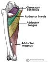
What is the pectineal line?
![](https://s3.amazonaws.com/brainscape-prod/system/cm/280/784/016/q_image_thumb.jpg?1516835639)
The pectineal line is the line formed when the intertrochanteric line passes the lesser trochanter on the posterior surface of the proximal femur
![](https://s3.amazonaws.com/brainscape-prod/system/cm/280/784/016/a_image_thumb.jpg?1516835639)
What are linea aspera?
Linea aspera are roughened ridges of bone found on the posterior surface of the femoral shaft
![](https://s3.amazonaws.com/brainscape-prod/system/cm/280/784/017/a_image_thumb.jpg?1516835640)
Describe the features of the linea aspera in the proximal region of the posterior femur
- Pectineal line → medial border of the linea aspera
- Gluteal tuberosity → lateral border of the linea aspera
![](https://s3.amazonaws.com/brainscape-prod/system/cm/280/784/020/a_image_thumb.jpg?1516835640)
Describe the features of the linea aspera in the distal region of the posterior femur
![](https://s3.amazonaws.com/brainscape-prod/system/cm/280/784/021/q_image_thumb.jpeg?1516835640)
- Linea aspera widens and forms the floor of the popliteal fossa
- The medial and lateral borders form the medial and lateral supracondylar lines
![](https://s3.amazonaws.com/brainscape-prod/system/cm/280/784/021/a_image_thumb.jpg?1516835641)
Identify the structures visible on the anterior surface of the distal femur
![](https://s3.amazonaws.com/brainscape-prod/system/cm/280/784/022/a_image_thumb.jpg?1516835641)
Identify the structures visible on the posterior surface of the distal femur
![](https://s3.amazonaws.com/brainscape-prod/system/cm/280/784/026/a_image_thumb.jpg?1516835642)
Describe the articulations of the medial and lateral condyles of the distal femur
- Posterior & inferior surfaces articulate with the tibia and menisci
- Anterior surface articulates with the patella
![](https://s3.amazonaws.com/brainscape-prod/system/cm/280/784/027/a_image_thumb.png?1516835642)
Describe the structure and function of the intercondylar fossa in the distal femur
- Structure: a depression on the posterior surface of the femur, between the two condyles
- Function: contains two facets for attachment of internal knee ligaments
![](https://s3.amazonaws.com/brainscape-prod/system/cm/280/784/028/a_image_thumb.png?1516835642)
Where do the posterior and anterior cruciate ligaments of the knee attach to on the distal femur?
- Facet for attachment of the posterior cruciate ligament – found on the medial wall of the intercondylar fossa (large, rounded, flat)
- Facet for attachment of anterior cruciate ligament – found on the lateral wall of the intercondylar fossa (smaller)
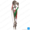
Identify the 4 muscles found in the anterior thigh
- Pectineus
- Sartorius
- Quadriceps femoris muscles
- Iliopsoas
Which two individual muscles compose the illiopsoas muscle?
- Psoas major
- Illiacus
![](https://s3.amazonaws.com/brainscape-prod/system/cm/280/784/031/a_image_thumb.png?1516835643)
Describe the structure, function and innervation of the psoas major muscle
![](https://s3.amazonaws.com/brainscape-prod/system/cm/280/784/032/q_image_thumb.jpg?1516835643)
- Structure: insert as the same tendon with the illiacus
- Function: hip flexion, lateral rotation (both muscles)
- Innervation: anterior rami of L1-3
![](https://s3.amazonaws.com/brainscape-prod/system/cm/280/784/032/a_image_thumb.jpeg?1516835644)
State the origin and attachment of the psoas major
![](https://s3.amazonaws.com/brainscape-prod/system/cm/280/784/033/q_image_thumb.jpg?1516835645)
- Origin: lumbar vertebrae
- Attachment: lesser trochanter of the femur (with illiacus)
![](https://s3.amazonaws.com/brainscape-prod/system/cm/280/784/033/a_image_thumb.jpeg?1516835645)
Describe the structure, function and innervation of the illiacus muscle
![](https://s3.amazonaws.com/brainscape-prod/system/cm/280/784/034/q_image_thumb.jpg?1516835646)
- Structure: insert as the same tendon with the psoas major
- Function: hip flexion, lateral rotation (both muscles)
- Innervation: femoral nerve
![](https://s3.amazonaws.com/brainscape-prod/system/cm/280/784/034/a_image_thumb.jpeg?1516835646)
State the origin and attachment of the illiacus muscle
![](https://s3.amazonaws.com/brainscape-prod/system/cm/280/784/036/q_image_thumb.jpg?1516835647)
- Origin: iliac fossa of the pelvis
- Attachment: lesser trochanter of the femur (with psoas major)
![](https://s3.amazonaws.com/brainscape-prod/system/cm/280/784/036/a_image_thumb.jpeg?1516835647)
Which 4 muscles compose the quadriceps femoris?
- Rectus femoris
- Vastus medialis
- Vastus intermedius
- Vastus lateralis
State the function and innervation of the vastus lateralis muscle
![](https://s3.amazonaws.com/brainscape-prod/system/cm/280/784/038/q_image_thumb.jpg?1516835648)
- Function: knee extension and stabilises the patella
- Innervation: femoral nerve
![](https://s3.amazonaws.com/brainscape-prod/system/cm/280/784/038/a_image_thumb.jpeg?1516835648)
State the origin and attachment of the vastus lateralis muscle
![](https://s3.amazonaws.com/brainscape-prod/system/cm/280/784/040/q_image_thumb.jpg?1516835649)
- Origin: greater trochanter and the lateral lip of linea aspera
- Attachment: base of patella
![](https://s3.amazonaws.com/brainscape-prod/system/cm/280/784/040/a_image_thumb.jpeg?1516835649)
State the function and innervation of the vastus intermedius muscle
![](https://s3.amazonaws.com/brainscape-prod/system/cm/280/784/041/q_image_thumb.jpg?1516835650)
- Function: knee extension and stabilises the patella
- Innervation: femoral nerve
![](https://s3.amazonaws.com/brainscape-prod/system/cm/280/784/041/a_image_thumb.jpeg?1516835650)
State the origin and attachment of the vastus intermedius muscle
![](https://s3.amazonaws.com/brainscape-prod/system/cm/280/784/042/q_image_thumb.jpg?1516835651)
- Origin: anterior and lateral surfaces of the femoral shaft
- Attachment: base of the patella
![](https://s3.amazonaws.com/brainscape-prod/system/cm/280/784/042/a_image_thumb.jpeg?1516835651)
State the function and innervation of the vastus medialis muscle
![](https://s3.amazonaws.com/brainscape-prod/system/cm/280/784/043/q_image_thumb.jpg?1516835652)
- Function: knee extension and stabilises the patella
- Innervation: femoral nerve
![](https://s3.amazonaws.com/brainscape-prod/system/cm/280/784/043/a_image_thumb.jpeg?1516835652)
State the origin and attachment of the vastus medialis muscle
![](https://s3.amazonaws.com/brainscape-prod/system/cm/280/784/044/q_image_thumb.jpg?1516835653)
- Origin: anterior and lateral surfaces of the femoral shaft
- Attachment: base of patella
![](https://s3.amazonaws.com/brainscape-prod/system/cm/280/784/044/a_image_thumb.jpeg?1516835653)