Review Q's Week 6 Flashcards
1. patho: vasculitis (1-33) 2. pharma dyslipidemia control (34-59) 3. patho lab: vascular diseases (60-68) 4. biochem: energy utilization (69-100) 5. patho lab: lipid disorders (101-114) 6. clinical medicine: choronic myocardial ischemia (115-152)
T/F: in vasculitis, the ability of the vessel to hold more blood is increased due to the thickening of blood vessels
false, the elastic property of the vessels is lost so it’s blood holding capabilities are hindered
Which of the following occur in small vessels?
a. Giant Cell Arteritis
b. Kawasaki Disease
c. Leukocytoclastic Vasculitis
d. Takayasu Arteritis
c. Leukocytoclastic Vasculitis
60 year-old women comes in complaining of headaches, joint and facial pain, fever, and difficulties with vision. Whats the diagnosis?
Giant cell arteritis
(other symptoms include jaw and tongue claudication- pain when eating or chewing)
What are different names of giant cell arteritis?
Granulomatous arteritis
arteritis cranialis
Horton disease
arteritis of the aged
Mechanism of giant cell arteritis. What findings are used to diagnose (4)?
Autoimmune reaction to elastic fibers in vessel wall

Panartheritis, aka inflammation involving all layers of an artery
Multinucleated giant cells
thick wall due to infiltrates (neutrophils, etc.)
Fragmented IEL (black coil in picture)
In Panartheritis, where does the inflammation start.
The inflammation starts in the media, and then spreads to the intima and adventitia- ending up in all the layers
media ⇨ intima & adventitia
A 30 year-old women comes in with painful, cool, and pale extremities, dizziness, headaches, chest pain, hypertension, and abdominal pain. Her renin levels are high and angiography is shown below. Diagnosis?

takayasu arteritis
(low perfusion to kidneys caused high renin, which increased BP)

What is characteristic of Takayasu arteritis?
Aneurysm (formation in the coronary vessels)
takayasu arteritis is also known as pulseless disease. Why?
When it affects the subclavian branch of the aorta, you won’t be able to feel a pulse due to the thickening of the wall, hence the name
Describe the vessels of takayasu arteritis
thick sclerosing vessels with a narrow lumen
Sclerosing arteritis of media ⇨ adventitia
Target population of
giant cell arteritis VS Takayasu arteritis
giant cell arteritis= older women (50+)
Takayasu arteritis= younger women (50-)
Polyarteritis Nodosa is
a. Granulomas
b. Necrotizing
b. Necrotizing
(giant cell arteritis and Takayasu arteritis are A)
What causes the inflammation in Polyarteritis Nodosa?
its immune (complex) mediated inflammation
polyarteritis nodosa affects which parts of the vessel wall?
Affects the whole vessel wall; transmural necrotizing arteritis
What causes the microaneurisms in polyarteritis nodosa?
Segmental inflammation & necrosis ⇨ macrophages clean the necrosis ⇨ healing occurs (fibrin mesh, platelets, collagen) ⇨ the scar tissue is not as flexible/elastic ⇨ microaneurysmsor ruptures ⇨ nodules
Whats a characteristic of polyarteritis nodosa?
Mural fibrinoid necrosis

A four year olf child comes in with conjunctivits, lymphadenopathy, and a strawberry looking toungue + cracked lips. Diagnosis?
Kawasaki Disease
A biopsy of a 2 year old girl shows bulges on the heart. diagnose and explain.
multiple coronary aneurysms formed the bulges on the heart
kawasaki disease
Necrotizing arteritis with VS without mucocutaneous lymph node (MCLN)
with MCLN= kawasaki disease
without MCLN= Polyarteritis nodosa
Symptoms of Kawasaki disease
(+ how do diagnose?)

What does Wegener granulomatosis affect?
upper/lower respireatory tract
lungs
ear, nose, throat
kidney
Patient comes in with nasal passage inflammation, numbness, weakness, scarrying on the foot, and a history of asthma. After further investigation, eosinophil count is high and the scar tissue contains granulomas. Diagnose.
Churg Strauss Syndrome
(patients often misdiagnosed with allergies)
Antineutrophil Cytoplasmic Antibodies (ANCA) are used to diagnose which types of vasculitis?
Wegener granulomatosis
Churg Strauss Syndrome
Leukocytoclastic Vasculitis
Microscopic polyangiitis
Patient comes in with palpable purpura. Further tests reveal high ANCA levels. diagnose.
Leukocytoclastic Vasculitis
vasculitis + respiratory granuloma + no eosinophils=
wegner granulomaotosis

vasculitis + respiratory granuloma + eosinophils+ asthma=
churg strauss syndrome

vasculitis + no respiratory symptoms + skin purpura
Leukocytoclastic vasculitis

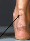
T/F: Buerger Disease (Thromboangiitis Obliterans) only affects arteries and veins
false, its called “angiitis” and not Arteritis because it affects all kinds of blood vessels, arteries, veins, nerves. the pain is due to the nerve inflammation.
Who does Buerger Disease (Thromboangiitis Obliterans) most likely affect? Where?
Heavy smokers + young males
affects hands and legs (blood flow blocked due to inflammation, may be blocked to the point of gangrene and amputation)
T/F: Thromboangiitis Obliterans is reversible at early stages of the disease if the smokers quit
true
T/F: Raynaud’s cannot cause ischemia
false, secondary Raynaud’s or Raynaud’s Phenomenon can because its accompanied by other conditions
Diagnose

Buerger Disease
Why does Raynaud’s Disease occur?
overreaction to cold
When is endogenous cholesterol synthesis highest?
at night
Mechanism of action of Statins.
Inhibitors of Cholesterol Biosynthesis
the rate-limiting enzyme in cholesterol biosynthesis is …
HMG-COA reductase
How do statins cause a decrease in LDL?
they stop cholesterol synthesis and the body compensates by making more LDL receptors so they can utilize the LDL’s of the circulation (higher LDL uptake)
When are statins contraindicated?
(adverse side effects=2)
in pregnancy

What’s the most pronounced effect of fibrates?
decrease in plasma triglyceride
first line treatment of primary hypertriglyceridemia.
Fibrates
T/F: Fibrates affect both LDL and HDL concentrations
true, decrease LDL and increase HDL
Fibrates are agonists of which receptors? What are these receptors?
α peroxisomal-proliferator activator receptors (PPARα), which are nuclear transcription factors that play a role in lipid metabolism
(+ PPARα= + lipid metabolism)
3 side-effects of fibrates
- Cholelithiasis (formation of gallstones),
- weight gain
- alopecia.
Mechanism of action of bile acid-binding resins.
Bile acid binding agents are large polymers of hydrocarbons that can bind bile salts and block intestinal absorbtion. (decrease bils salt reabsorbtion)
This causes the liver to start to convert cholesterol into bile acids to compensate, thus decreasing hepatic cholesterol. (LDL is taken from the circulation by the liver)
bile acid-binding resins mostly affect which of the following?
a. HDL
b. LDL
b. LDL
(May raise HDL levels slightly)
Side effects of Cholestyramine + Colestipol
GI disturbances such as constipation and bloating
Interfere with absorption of some drugs like warfarin and digitalis
Mechanism of action of Ezetimibe
inhibit Cholesterol Absorption in the brush border of enterocytes in the duodenum.
How does Nicotinic Acid reduce plasma levels of VLDL?
by inhibiting the synthesis of fatty acids in the liver
How does Nicotinic Acid reduce plasma TG levels?
By reducing lipolysis in adipose tissue
What’s the most effective agent for increasing HDL?
Nicotinic Acid (Niacin)
2 side effects of Nicotinic Acid (Niacin)
Weight gain due to inhibition of lipolysis
Hyperuricemia precipitates gout.
Mechanism of action of Omega-3-fatty acids
Inhibit VLDL and TG synthesis in the liver.
What is Proprotein Convertase Subtilisin/Kexin type 9 (PCSK9)?
a serine protease involved in cholesterol homeostasis- reducing serum LDL clearance.
(PCSK9 is a serine protease that reduces both hepatic and extrahepatic LDL receptors)
Mechanism of action of Evolocumab
Monoclonal antibodies that inhibit PCSK9 have emerged as a new class of drugs that very effectively lower LDL cholesterol levels (by stopping what inhibits the LDL clearance)
Which of the following most effectively lowers LDL?
a. Evolocumab
b. Simvastatin
c. Clofibrate
d. Nicotinic Acid (Niacin)
e. Lovastatin
f. Ezetimibe
g. Cholestyramine
b. Simvastatin

Which of the following most effectively lowers HDL?
a. Evolocumab
b. Simvastatin
c. Clofibrate
d. Nicotinic Acid (Niacin)
e. Lovastatin
f. Ezetimibe
g. Cholestyramine
d. Nicotinic Acid (Niacin)

Which of the following most effectively lowers triglycerides?
a. Evolocumab
b. Simvastatin
c. Clofibrate
d. Nicotinic Acid (Niacin)
e. Lovastatin
f. Ezetimibe
g. Cholestyramine
c. Clofibrate

Which of the following is used to treat combined hyperlipidemia?
a. Evolocumab
b. Simvastatin
c. Clofibrate
d. Nicotinic Acid (Niacin)
e. Lovastatin
f. Ezetimibe
g. Cholestyramine
d. Nicotinic Acid (Niacin)
When should Simvastatin be taken and why?
at night because that when the endogenous cholesterol synthesis is highest
(they inhibits cholesterol synthesis)
Describe the shown coronary lesion
a. a predominantly fibrous plaque
b. a central lipid core covered with a thin fibrous cap
c. a mural thrombus incorporated in a plaque
d. inflammatory infiltrate with giant cells
e. fibrinoid necrosis

b. a central lipid core covered with a thin fibrous cap
What is the pathological process
in this lesion?
a. Inflammation
b. Edema
c. Congestion
d. Neoplasia
e. Infarction

e. Infarction
How old is this MI?
a. 1 day
b. 5 days
c. 10 days
d. 30 days

c. 10 days
What is the lesion pointed to by the arrows?
a. Cardiac hypertrophy
b. Cardiac metastases
c. Cardiac aneurysm and thrombosis
d. Myocarditis
e. Myocardial infarction

c.Cardiac aneurysm and thrombosis
What is the lesion shown in this coronary section?
a. Intramural thrombus
b. ruptured plaque and occlusive thrombus
c. hemorrhage in a plaque
d. calcified plaque
e. fibro-fatty plaque

b.ruptured plaque and occlusive thrombus
A patient came in with squeezing chest pain and a first angiogram showed a severe occlusion. A couple of minutes later the pain was gone and a second angiography showed no occlusion. How is this possible?

Prinzmetal Angina (Variant Angina)
Describe the lesion?

largely fibrous atheromatous lesion
describe the lesion

fatty atheromatous lesion
What is Creatine kinase? When does CK increase after an MI?
an enzyme found primarily in heart muscle cells
increases in serum within 3 to 6 hours; the peak levels occur between 16 and 30 hours.
What precent of the bodies oxygen is used up by the heart?
10% of body oxygen consumption
Which of the following cardiac energy stores are short-term?
– phosphocreatine
– glycogen
– triacylglycerols
– phosphocreatine
keep the heart going for 5 to 10 sec.
Which becomes an important energy source in heart failure?
a–Glucose
b–Fatty acids
c– Lactate and Pyruvate
d– Amino acids
e– Ketone bodies
e– Ketone bodies
What are two heart functions that cause it to need high amounts of energy?
ions pumps
+
contractile apparatus
Which is insulin insensitive?
GLUT 1 or GLUT 4
GLUT 1
T/F: lactate is a waste product that’s produced when the heart uses glucose to make ATP
false, it’s not a waste product because the heart has O2 and can oxidize lactate to pyruvate, then make more ATP from it via to kerbs cycle
(its a waste product in muscle)
How does glucose enter cardiac cells?
via GLUT1/4 receptors
they go from high to low concentrations so no energy is needed
Low does lactate enter cardiac cells?
vis monocarboxylate transporters
What’s the main way of cardiac cells to make ATP?
oxidative phosphorylation
How can the heart utilize triacylglycerol?
via lipoprotein lipase activity (has high levels of this enzyme)
Which of the following needs oxygen to be able to be converted into ATP?
a. glucose
b. fatty acids
b. fatty acids
(A=produce some ATP without using O2)
On average, how does the heart produce most of the energy it uses (which method)? Where does it get the energy come from mostly?
90% of total myocardial energy produced by oxidative phosphorylation (mitochondria)
20% of energy from glucose and the remainder of energy from fatty acids
T/F: as a fetus develops, his heart relies less on fatty acids and more on glucose
false, the opposite is true. In fetal life the circulatory load is low and the heart uses glucose (limited oxygen availability), but then with development, the heart load increases and fatty acids are used for energy.
Explain what causes the heart to use more glucose as fuel when we’re fed.
higher insulin causes GLUT4 (glucose transporters) to bring more glucose into cardiac tissue
the insulin also decreases free fatty acid release from adipose tissue
the insulin also activates ↑ acetyl-CoA carboxylation and thus ↑ malonyl-CoA- this inhibits CPT-1 (which is used to transport fatty acids into the mitochondria for beta oxidization)
Explain how the heart uses more fatty acids for fuel when patient is exercising.
Exercise uses up ATP→AMP levels increase→that increases AMPK→AMPK inhibits acetyl-CoA carboxylase (which makes acetyl CoA into malonyl CoA)→malonyl CoA decreases (malonyl CoA inhibits CPT-1, which helps fatty acids get into mitochondria)→CPT-1 is more acitve→ more fatty acid beta-oxidation
T/F: CPT-1 in the heart is much more sensitive to inhibition by malonyl CoA than liver isoform
true
What two things activate VS what two things inhibit
Pyruvate dehydrogenase (PDH)
activate= Ca + Mg
inhibit= Acetyl-CoA + NADH
Pyruvate dehydrogenase (PDH) controls the hearts oxidization of two things.
oxidation of glucose and lactate by the TCA cycle in the myocardium
Which amino acid gives us pyruvate?
a. aspartate
b. glutamine
c. alanine
c. alanine
Which type of fuel does the heart work best while using?
the heart works best when using a mixture of metabolic fuels
Which amino acid gives us fumarate?
a. aspartate
b. glutamine
c. alanine
a. aspartate
Does oxidative phosphorylation need oxygen?
yes
(oxygen is the electron acceptor of complex IV of the electron transport chain…)
Which amino acid gives us alpha-ketoglutarate?
a. aspartate
b. glutamine
c. alanine
b. glutamine
(glutamine/ glutamate give us alpha ketoglutarate)
How does cardiac ischemia cause calcium overload?
when oxygen supply drops, we cannot use fatty acid oxidization for fuel, so we use glycolysis (which doesn’t use oxygen and doesn’t produce as much ATP)
Glycolysis produces NADH, which we then re-oxidize into NAD+ by making lactate. The problem starts when lactate (which is acidic) build-up and affects the ion pumps, which then lead to Ca overload.
The pumps that aid in Ca efflux (Na-Ca exchange) also don’t get enough energy to function, so that causes the Ca to stay in the cell and accumulate.
Explain reperfusion damage.
(ROS involvement)
oxygenated cardiac cells produce antioxidants to protect itself from the free radicals (which damage the cell membrane). But if a patient has an MI (example), the cardiac cells are unable to keep producing the antioxidants- which causes the free radicals (O2) to damage the cell membrane.
When reperfusion occurs and the cells get highly oxygenated blood, free radicals are formed (O2) and they end up damaging the cell membrane even more (no antioxidants to balance out the high oxygen-the antioxidants don’t have time ti be made)
T/F: during ischemia, the surrounding cardiac tissue uses fatty acid as fuel
true; Fatty acids dominate as a substrate for residual oxidative metabolism due to increased plasma levels of fatty acids
During reperfusion which acts as a fuel
a. fatty acid oxidation
b. glucose oxidation
a. fatty acid oxidation
The dominance of fatty acid oxidation during reperfusion inhibits glucose oxidation
in heart failure, which is the cardiac fuel source?
a. glycolysis
b. fatty acid oxidation
a. glycolysis
(due to decreased oxygen availability)
Describe the fetal heart metabolic profile.
high glycolytic capacity
low mitochondrial oxidation capacity
What two conditions copy the fetal heart’s metabolic profile.
cardiac hypertrophy and cardiac failure
(high glycolytic capacity+ low mitochondrial oxidation capacity)
How is shifting oxidative metabolism from fatty acids to glucose beneficial?
– Reduced oxygen requirements
– Reduced accumulation of fatty acids and toxic fatty acid metabolites (in a failing heart, the mitochondria is damaged and cannot oxidize FA, the FA intermediates build up. We switch to glucose use so we stop this build-up)
What lifestyle choices may help preserve the heart activity of patients with heart failure?
ketogenic diet
(heart then uses ketone oxidization for energy)
** not clear if this is beneficial yet- its in a tafreeg so just ignore this I guess
What is this? What does it indicate?

arcus senilis
may be a sign of hyperlipidemia
What is this?

Xanthelasma
(sign of hyperlipidemia)
Xanthoma
How do you calculate the LDL of a fasting patient with triglycerides less than 4.5mM?

How do you calculate the LDL of a fasting patient with triglycerides more than than 4.5mM?

Primary VS Secondary dyslipidemia
Primary= a genetic defect in the lipid metabolism as a cause of the problem
Secondary= due to another condition (ex/ diabetes)
What are the three ways to diagnose acute coronary syndrome?
history
ECG
clinical biochemistry
What are the three cardiac biomarkers that are important for diagnosing MI?
Creatine Kinase (CK)
Aspartate transaminase (AST)
Lactate dehydrogenase (LDH)
Which of the following cardiac markers are the first to increase after MI?
a. Creatine Kinase (CK)
b. Aspartate transaminase (AST)
c. Lactate dehydrogenase (LDH)
a. Creatine Kinase (CK)
3-8 hours after MI
Which of the following cardiac markers is the last to get back to normal after an MI?
a. Creatine Kinase (CK)
b. Aspartate transaminase (AST)
c. Lactate dehydrogenase (LDH)
c. Lactate dehydrogenase (LDH)
8-14 days
Which Lactate dehydrogenase (LDH) isoform is cardiospecific?
LDH1
When does AST increase after an MI?
6-10 hours
Which cardiac biomarkers are used for ACS diagnosis?
CK-MB
Troponins
When is a troponin test most sensitive?
A Troponin T or I taken at 12 hrs post-onset of chest pain is very sensitive
stable coronary artery disease vs. acute coronary syndrome
which one has a demand and supply mismatch?
both!
but in stable coronary artery disease, the mismatch occurs slowly while in ACS it happens suddenly
How do each of these plaque features affect vulnerability?
thickness of fibrous cap
lipid pool size
amount of inflammatory cells
fibrous cap= thinner the cap, more vulnerable to rupture
lipid pool size= bigger pool size, more vulnerable to rupture
more inflammatory cells, the more vulnerable to rupture
How does mitral regurgitation affect supply/demand of the heart?
mitral regurgitation→ increase contractility→ increases the hearts demand for blood
How does hypertension affect supply/demand of the heart?
hypertension → increasing systolic wall tension→ increases demand
How long does the angina have to last for it to be labeled as chronic?
2 months
What’s the best evaluation method of stable CAD?
history!
“It is possible to make a confident diagnosis on the basis of history alone”
Typical duration fo ischemic chest pain
1-5 minutes
What are the three characteristics of typical angina?
(if patient meets all three then definite angina)
- substernal chest pain (quality= heavy, duration=1-5min)
- provoked by exertion
- relieved by rest or nitroglycerin
What two cardiovascular conditions mimic angina? why?
Aortic Stenosis + Hypertrophic Cardiomyopathy
both cause obstruction of flow to outside of the ventricle, causing non CAD angina
How do you diagnose patients with stable CAD? What are some obstacles?
patients with stable CAD often have normal physical exams and ECG’s
That’s why we do functional tests- to provoke cardiac ischemia by stressing the patient.
We also have anatomical tests (ex/angiography) so we can see the arteries
What’s the strongest predictor of long-term survival?
LVEF is the strongest predictor of long-term survival as LVEF declines, mortality increases
(Patient with an LVEF <50% is already at high risk for CV death)
What patients are recommended for ECG exercise testing?
- Patients with an intermediate pretest probability of IHD (not a high probability- if high then we don’t need the test)
- No major ECG changes or LBBB
- Are able to exercise
blood pressure changes before and after exercise
in normal patients VS in patients with ischemic heart
in normal patients= after exercise BP increase
ischemic heart= BP decreases because heart doesn’t have strength to contract and raise BP
You suspect patient of having stable CAD. When do you use…
Coronary computed tomography angiography
VS.
Catheterization & Coronary Angiography
CT angiography= its less invasive, so its done for patients with low/intermediate likelihood of CAD- when you’re not certain
Catheter coronary angiography= more invasive and involves catheterization, do it when patient has high CAD risk or taking optimal medical therapy (OMT) fails
Which of the following are more sensitive at detecting ischemia?
a. imaging techniques
b. ECG
c. symptoms (ex/angina)
a. imaging techniques
(imaging techniques based on perfusion, metabolism or wall motion are more sensitive)

patient comes in with substernal chest pain (quality= heavy, duration=1-5min) that is provoked by exertion but NOT relieved by rest. diagnose.
atypical angina (2/3 of the three characteristics)

calssify the severity of the angina:
patient has marked limitation when climbing one flight of stairs
class III

Patient does a Stress Radionuclide Imaging and most of the anterior heart tissue appears red. What does this mean?
heart muscle tissue with a good blood flow will emit more gamma rays than areas with a poor blood flow or damaged tissue. So his heart has a good blood supply.

Stress Radionuclide Image. diagnose.

with rest all areas get O2= complete horseshoe
with stress= the apex isn’t getting enough blood
How does Stress Echocardiography work?

areas of the heart are given numbers (divided into segments), and when patient is exercising we see how each segment moves. (bad movement means ishemia)
hypokinetic means not moving well
akinetic means not moving at all
Risk stratification (of CAD) is done by evaluating 4 factors. What are they? how are they tested?
1-Risk stratification by clinical evaluation.
2-Risk stratification by ventricular function. (echocardiogram)
3-Risk stratification by response to stress testing.
4-Risk stratification by coronary anatomy. (angiography)
Patient has ECG abnormalities, which is a preferred test?
stress imaging OR exercise ECG test
stress imaging
St ress Imaging has advantages over conventional Exercise ECG Test because it can be done even if there are resting ECG abnormalities
(exercise ECG test is done if ECG is normal****)
Four ways stress imaging is better than exercise ECG
- can be done even with ECG abnormalities
- can be used if patient can’t exercise
- better sensitivity and specificity
- can quantify and localise ischemia areas
Coronary artery bypass surgery uses which two vessels to bypass the blockage?
internal mammary artery
saphenous vein
Which of the following relief of anginal symptoms by increasing blood supply? How?
• Beta-blockers
• Calcium antagonists
• Nitrates
• Nitrates
by decreasing systemic will tension
How does each of the following relief angina symptoms?
• Beta-blockers
• Calcium antagonists
they both decrease demand of blood by increasing heart rate and contractility
first step to treating chronic angina? What happens if patient is resistant?
give OMT (optimal medical therapy)
if resistant, revascularize by PCI or CABG
can revascularization therapy be used as first line treatment of CAD? if yes, when?
As first-line treatment in patients with :
Left main coronary artery disease (50% stenosis or more ).
Multivessel disease with severe or extensive ischemia.
Left ventricular dysfunction with EF of 35–50
Why do people suffer silent ischemia? (3 reasons)
higher pain threshold
silent episodes reflect less severe ischmia
defect in pain preception
How should silent ischemia be managed?
managed as angina
T/F: Silent ischemia reflects less severe ischemia.
true
