Anatomy Review Q's (same as above but via subject) Flashcards
the anatomy notes (1-207) anatomy lab 1 (208-230)
When a patient is lying supine, at which vertebral level is the heart situated in?
a. T5-T7
b. T5-T8
c. T6-T8
d. T6-T9
b. T5-T8
Where is the heart located?
a. superior mediastinum
b. middle mediastinum
c. inferior mediastinum
b. middle mediastinum
What two things separate the heart from the lungs?
pericardium and pleura
When a patient is standing, at which vertebral level is the heart situated in?
a. T5-T7
b. T5-T8
c. T6-T8
d. T6-T9
d. T6-T9
What two (main) structures of the heart bring blood to the heart?
superior and inferior vena cava
What two (main) structures take blood away from the heart?
pulmonary trunk and aorta
Which side of the body is the apex of the heart pointing towards?
to the left
What’s found anteriorly to the heart?
sternum, muscles, ribs
What’s found laterally to the heart?
lungs
What’s found posteriorly to the heart?
aorta, esophagus, and the left pulmonary vein
The pericardial cavity is a space between which two structures?
between the parietal pericardium and the visceral pericardium
Which of the following is closest to the heart?
a. fibrous pericardium
b. parietal pericardium
c. visceral pericardium
c. visceral pericardium
Which of the following protects the heart?
a. fibrous pericardium
b. parietal pericardium
c. visceral pericardium
a. fibrous pericardium
Which of the following does the transverse pericardial sinus lie anterior of?
a. aorta
b. vena cava
c. pulmonary trunk
b. vena cava
(it lies behind/posterior to the other two options)
Which of the following is used to perform ligation in surgery?
a. transverse pericardial sinus
b. oblique pericardial sinus
a. transverse pericardial sinus
Where is the oblique pericardial sinus located?
between the pulmonary veins (and the inferior vena cava)
Which of the following provides space for an enlarging heart?
a. transverse pericardial sinus
b. oblique pericardial sinus
b. oblique pericardial sinus
Describe the hearts position in comparison to the midline.
2/3 shift to the left (the rest of 1/3 to the right)
The visceral pericardium is also known as
the epicardium OR (part of the) serous pericardium
Which artery accompanies the phrenic nerve?
pericardiacophrenic artery
the pericardiacophrenic artery is a branch of the
internal thoracic artery
Which vein carries blood from the pericardium to the brachiocephalic veins?
Pericariacophrenic veins
The left phrenic nerve senses pain near the heart, where is this pain referred to?
the skin of the supraclavicular region of the left side
Which of the following borders of the heart are mainly made up of the right atrium? (one or more)
a. left border
b. right border
c. apex
d. superior border
e. inferior border
f. base of heart
b. right border
Which of the following borders of the heart are mainly made up of the left ventricle? (one or more)
a. left border
b. right border
c. apex
d. superior border
e. inferior border
f. base of heart
a. left border
+
c. apex
The outside portion of Crista terminalis is called?
Sulcus terminalis
musculi pectinati originates from which of the following?
a. limbus fossa ovalis
b. Sulcus terminalis
c. fossa ovalis
d. crista terminalis
d. crista terminalis
Which of the following borders of the heart are mainly made up of the left atrium?
a. left border
b. right border
c. apex
d. superior border
e. inferior border
f. base of heart
d. superior border
+
f. base of heart
Which of the following is a muscular groove?
a. sulcus terminalis
b. crista terminalis
a. sulcus terminalis
(crista terminalis is a muscular ridge)
Which of the following borders of the heart are mainly made up of the right ventricle? (one or more)
a. left border
b. right border
c. apex
d. superior border
e. inferior border
f. base of heart
e. inferior border
What three structures bring blood to the right atrium?
IVC, SVC, coronary sinus
Which part of the right atrium is smooth? what is it called?
the posterior part called sinus venarum
posterior= smooth
anterior= rough
Which part of the right ventricle is rough and which is smooth?
posterior= rough
anterior= smooth
What structure allows from communication between atria and ventricles?
atrioventricular orifice
(protected by the tricuspid valve on the left side and the bicuspid valve on the right)
What provides blood to the left atrium?
the four pulmonary veins
How many papillary muscles are in the right ventricle?
three (anterior, posterior and septal)
The smooth outflow part of the right ventricle is called
The smooth outflow part of the left ventricle is called
R-> the infundibulum
L-> Aortic vestibule
The blood is pumped in the right ventricle to go to ___ via ___
the lungs via the pulmonary trunk
What prevents the prolapse of the cusps into atria during systole?
chordae tendinae
Where is the left auricle found? What is its function?
on the left atrium, it provides extra space for blood
What is the oval depression with a margin that is found on the left atria called?
fossa lunata
(indicates fossa ovalis on the right atria)
Which cusp of the mitral valve has more surface area?
a. anterior
b. posterior
c. septal
b. posterior
Which part of the left ventricle is rough and which is smooth? explain.
posterior= rough
anterior= smooth
(the posterior part is rough because it has to diffuse the pressure that’s exerted by the blood pooling in from the atria)
What are three functions of the fibrous skeleton of the heart?
keeps orifices/valves intact (no dilation or contraction)
provides attachment of muscles
separates atria from ventricles
Describe the shape of the heart muscle fibers?
in spirals resembling the number 8
Which of the following has two cusps?
a. aortic valve
b. pulmonary valve
c. both
d. neither
d. neither
Which of the following has two anterior cusps
a. aortic valve
b. pulmonary valve
c. both
d. neither
a. aortic valve
When does blood enter arteries? What is the exception to the rule? explain.
blood enter arteries during systole, except the coronary arteries, which get blood during diastole. This is because the (right and left) coronary arteries are branches of the aorta, and during systole, blood rushes through it at a very high pressure and speed, so it doesn’t have time to turn perpendicularly and go to the branches of the aorta. When the aortic valve closes, the pressure decreases, allowing blood to go to the coronary arteries.
Which of the following occurs when the aortic and pulmonary valves are open?
a. systole
b. diastole
a. systole
Which of the following has two posterior cusps
a. aortic valve
b. pulmonary valve
c. both
d. neither
b. pulmonary valve
Which of the following sounds are classically heard in diastole?
a. S1
b. S2
c. S3
d. S4
b. S2
Which of the following occurs when the mitral valve is open?
a. systole
b. diastole
b. diastole
Which of the following leads to hypertrophy?
a. valve incompetence
b. valve stenosis
b. valve stenosis
Which of the following is more likely to be caused by rheumatic fever?
a. pulmonary valve stenosis
b. aortic valve stenosis
b. aortic valve stenosis
Which of the following sounds is heard when both the mitral and tricuspid valves are closed?
a. S1
b. S2
c. S3
d. S4
a. S1
(at systole)
Where does blood flow during arterial septal defects? Why?
flows from left to right (at the beginning) because the left has higher pressure
Which part of the interventricular septum is more likely to be defective?
membranous part (not muscular part)
How do you locate the superior border of the heart on a patient?
it’s from the second costal cartilage of the left side to the third costal cartilage of the right side
How do you locate the inferior border of the heart on a patient?
from the fifth intercostal space of the left side to the sixth costal cartilage
identify

right coronary artery
Where is the crux of the heart?
“crux” meaning “cross”; it is the area on the lower back side of the heart where the coronary sulcus (the groove separating the atria from the ventricles) and the posterior interventricular sulcus (the groove separating the left from the right ventricle) meet.

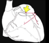
identify the black arrow

Right marginal artery
(aka acute marginal artery)
What artery goes around the pulmonary trunk?
right conus artery
aka annulus of vieussens

identify

sinoatrial nodal artery
identify the artery. Which artery does it arise from?

PDA posterior descending artery (aka posterior interventricular artery)
its a branch of the right coronary artery
Which artery is the yellow arrow pointing at?

left coronary artery
identify

left anterior descending artery (LAD)
(aka anterior interventricular artery)
identify
left circumflex artery (LCX)
identify (black arrow)

left diagonal artery (branch of LAD)
identify (blue circle)

Left conus artery
(goes around the pulmonary trunk along with right conus artery)
What’s a branch of the circumflex artery?
left marginal artery (or obtuse marginal artery)

Where does the right coronary artery (RCA) originate?
Above the right cusp of the aortic valve
What artery supplies the posterior third of the interventricular septum?
PDA posterior descending artery
The left coronary artery splits into
LAD (left anterior descending artery)
+
LCX (left circumflex artery)
Where is the coronary sinus?


Describe the location of the small cardiac vein. What is it adjacent to?
between the right atrium and ventricle

Find the Oblique vein of the left atrium


Find the Posterior vein of the left ventricle


What supplies the anterior 2/3rd of the interventricular septum?
The anterior interventricular artery (LAD artery)
What supplies blood to the left branch of the AV bundle?
Left coronary artery
What supplies blood to the majority of the hearts conducting system?
Right coronary artery
What supplies the right ventricle at the anterior interventricular groove?
Left coronary artery
Which of the following makes the patient LESS susceptible to ischemia?
a. right dominant coronary circulation
b. left dominant coronary circulation
c. balanced coronary circulation
c. balanced coronary circulation
(if blockages happen they’re better off because they have a back up)
Which coronary artery is larger?
left coronary artery
Where does Kugels anastomotic artery arise from and where does it transverse?
Arises from the proximal left circumflex artery and ends up in the distal right coronary artery
Which is located anteriorly in the sulcus between the ventricles?
a. small cardiac vein
b. middle cardiac vein
c. great cardiac vein
c. great cardiac vein
Which is located posteriorly in the groove between the ventricles?
a. small cardiac vein
b. middle cardiac vein
c. great cardiac vein
b. middle cardiac vein
What structure is found in “a”?

SA node
What is the “b” location called? What structure is also found there?

the triangle of Koch
AV bundle/bundle of His is located in it
A patient comes in with weakness, dizziness, and perspiration. He compains of pain in his chest and left arm. Which nerves are conveying the pain? What can you give the patient to relief symptoms?
Pain sensation conveyed through sympathetic nerves of the heart (T1-T5 segment of the spinal cord)
Sublingual nitroglycerin is placed under the tongue (rapid absorption) to dilate the coronary arteries.
What’s the most common artery involved in MI?
LAD/anterior interventricular artery
When do we use coronary angiography?
to localize the site of the coronary artery block
(catheter inserted though femoral to intect the dye)
What’s the location of the superficial cardiac plexus?
below the arch of aorta and in front of the right pulmonary artery
Where is the deep cardiac plexus
in front of bifurcation of trachea and behind arch of aorta
Which vegal branch supplies the superficial cardiac plexus?
inferior branch of left vagus
(the rest supply the deep cardiac plexus)
Which sympathetic chain branch supplies the superficial cardiac plexus?
left superior cervical ganglion
(the rest supply the deep cardiac plexus)
How do sympathetic and parasympathetic nerves control cardiac output?
by controlling the SA node (the pacemaker)
What is the only cause of heart pain?
ischemic injury
What are the four ways the sympathetic fibers stimulate the action of the heart?
↑ heart rate
↑ impulse conduction
↑ contraction force
↑ blood flow
What’s the only cause of pain of abdominal organs?
excessive destination
Which dermatomes are responsible for the pain in the medial side of the arm and the forearm?
T1 and T2
Which bundle branch receives blood from the left coronary artery?
both bundle branches
(right bundle branch blood from both right & left coronary arteries, while the left only from the left coronary artery)
Damage to which node is called heart block?
AV node (if defective, conductance will not reach the ventricles)
Patient has left coronary dominance, what supplies blood to the interventricular septum of the patient?
left coronary artery
(duh! it’s an important concept tho)
Describe the production of the first heart sound.
papillary muscle contracts tightens the chordae tendinae and drawing the cusps of AV valve together in order to close the mitral and tricuspid valves (lub)
What causes the semilunar valves to open?
when ventricular pressure exceeds diastole pressure in pulmonary trunk and ascending aorta
What produces the second heart sound?
In ventricular diastole, when the pressure is low in the ventricle and high in aorta and pulmonary trunk. The blood wants to backflow, but instead the close the semilunar valves are shut. This shutting is what causes the dub
Which of the following is thickest in veins?
a. tunica intima
b. tunica media
c. tunica externa
c. tunica externa
Which of the following is thickest in large arteries?
a. tunica intima
b. tunica media
c. tunica externa
b. tunica media
T/F: the tunica media of the aorta is mostly made up of elastic fibers
true
What kind of cells make up the tunica intima?
simple squamous
T/F: the tunica intermedia of capillaries is very thin
false, capillaries have only an endothelium, with no subendothelial layer or other tunics.
(Because capillaries are only one cell layer thick, they only have a tunica intima.)
Which of the following has a tunica media that has more smooth muscles?
a. aorta
b. vena cava
b. vena cava
What two layers make up the tunica intima?
subendothelial layer
+
endothelium
What two layers make up the inner and outer limit of the tunica media (surround it)?
internal elastic lamina
+
external elastic lamina
Which layer of veins makes folds to form valves?
tunica intima
Where are vasa vasorum found?
in arteries and larger veins
(found specifically in the adventitia of the aorta.)
How is the tunica intima separated from the tunica media?
by the internal elastic lamina (IEL), a prominent sheet of elastin.
T/F: elastic fibers are only found in the tunica media
false, elastic fibers are also present in the tunica adventitia (which is bigger in veins)
Why do the large arteries need a blood supply (vasa vasorum)?
Their walls are so thick that the blood they carry cannot diffuse through- they need another source of blood to supply their outer side
What kind of nerves are found in the adventitia, along vasa vasorum? What do they do?
small sympathetic nerves for vasoconstriction.
T/F: Large-sized and middle-sized arteries both have vasa vasorum
false, most middle-sized arteries don’t have a vasa vasorum (the largest of the middle-sized have it)
What separates the tunica media from tunica externa/adventitia?
external elastic laminae
The tunica media of which of the following has more elongated nuclei?
a. Large-sized artery
b. Medium-sized artery
b. Medium-sized artery
(more smooth muscle)
Which of the following allow blood to enter in a pulsatile fashion?
a. thoroughfare channel
b. precapillary sphincters
c. metarterioles
d. postcapillary venules
b. precapillary sphincters
Which of the following lacks any smooth muscle cells.?
a. thoroughfare channel
b. precapillary sphincters
c. metarterioles
d. postcapillary venules
a. thoroughfare channel
Which of the following has the highest diameter?
a. arterioles
b. venules
c. small capillaries
b. venules
T/F: tight junctions between the endothelial cells of the arterioles allow fluid to pass between them to allow diffusion
false, the tight junctions stop the leakage of fluids
Which doesn’t have a tunica intima?
a. arterioles
b. capillaries
c. both
d. neither
d. neither
(Because capillaries are only one cell layer thick, they only have a tunica intima.)
What are pericytes? What do they do?
they’re extra cells in the periphery of blood vessels that proliferate when there’s endothelial damage. They can proliferate to become endothelial cells or smooth muscles of the capillaries.
What are transcytotic vesicles used for?
to transport things through the epithelial cells of the capillaries to the surrounding tissue
Which is most likely found in BM?
a. Fenestrated capillaries
b. Sinusoids
c. Continuous capillaries
b. Sinusoids
Which forces molecules to cross via diffusion/transcytosis?
a. Fenestrated capillaries
b. Sinusoids
c. Continuous capillaries
c. Continuous capillaries
Which is found in the choroid plexus?
a. Fenestrated capillaries
b. Sinusoids
c. Continuous capillaries
a. Fenestrated capillaries
Which of the following are closed by diaphragms?
a. Fenestrated capillaries
b. Sinusoids
c. Continuous capillaries
a. Fenestrated capillaries
Which of the following doesn’t have s continuous external lamina?
a. Fenestrated capillaries
b. Sinusoids
c. Continuous capillaries
b. Sinusoids
(the rest have continuous basement membrane)
Which of the following has the largest diameter?
a. Fenestrated capillaries
b. Sinusoids
c. Continuous capillaries
b. Sinusoids
Which acts as a valve in veins?
a. tunica intima
b. tunica media
c. tunica externa
a. tunica intima
Which has more pericytes?
a. Postcapillary venules
b. Large collecting venules
b. Large collecting venules
Which is thin in small and medium-sized veins?
a. tunica intima
b. tunica media
c. tunica externa
a. tunica intima
Which is thin in large-sized veins?
a. tunica intima
b. tunica media
c. tunica externa
b. tunica media
How do you differentiate between normal versus lymphatic capillaries
lymphatic capillaries don’t have blood cells (usually)
Which are bigger, endothelial cells of blood vessels OR endothelial cells of lymphatic vessels?
endothelial cells of lymphatic vessels
How are the spaces between the endothelial cells formed?
they’re anchored by anchoring filaments (made up of elastin and endothelium)
Which of the following depolarizes?
a. sheath cells
b. glomus cells
b. glomus cells
(What induces the depolarization? low oxygen, high carbon dioxide, low pH)
What information do glomus cells rely to the brain? Through which nerve does this occur?
Changes in the CO2, O2, and H+ concentrations
via glossopharyngeal nerve
Where are baroreceptor-sensory nerve terminals located?
in the tunica adventitia
The heart tube is derived from what structure?
angioblastic cords
Initially, before the three folds occur, the heart tube is in which relation to the pericardial cavity?
a. dorsal
b. ventral
b. ventral
(becomes dorsal after the folds)
What gives rise to the pericardium?
mesoderm
(also gives rise to the heart)
Whats the origin of heart muscles?
cardiac jelly
What structure develops to become the ascending aorta and the pulmonary trunks?
truncus arteriosus

Which early structure is absorbed and becomes the right atrium (smooth part of R atrium)?
sinus venosus

What allows communication between the primordial atrium and ventricle?
atrioventricular canal
What two things does the sinoatrial valve separate?
sinus venosus and primordial atrium

During the folding of the heart tube, which structure is dorsal?
a. atria
b. ventricles
a. atria
Where does the septum perimum grow towards?
fused endocardial cushions
foramen primum allows the communication between what two structures?
the right and left atrium
After birth, increased blood return from the lungs closes foramen ovale. Which is true about this mechanism?
a. the blood pressure pushes the upper limb of the septum primum
b. the blood pressure pushes the lower limb of the septum primum
c. the blood pressure pushes the upper limb of the septum secundum
d. the blood pressure pushes the lower limb of the septum secundum
b. the blood pressure pushes the lower limb of the septum primum

Which end of the heart tube is located dorsally?
a. atrial end
b. venous end
b. out flow end
b. venous end
(its the tail end and is the inflow region)
Which structure splits the atrioventricular canal into right and left sections?
endocardial cushions that grow towards each other and fuse to form “fused endocardial cushion”
T/F: foramen secondum is formed due to perforations in septum secondum
false, it’s formed due to perforations in septum primum
Which of the following veins bring blood from the body of the embryo?
a. Umbilical vein
b. Viteline veins
c. Cardinal veins
c. Cardinal veins
What’s the origin of the smooth part of right vs smooth part of left atrium
Right= Sinus venarum
Left= pulmonary vein
What’s the origin of the rough part of right vs smooth part of left atrium
Both develop from the primordial atrium
Which of the following veins bring blood from the yolk sac?
a. Umbilical vein
b. Viteline veins
c. Cardinal veins
b. Viteline veins
(Umbilical vein brings from placenta)
What’s the most common type of congenital heart disease?
Ostium secundum types of septal defects
What occurs if both the septum primum and septum secundum fail to develop?
The interatrial septum is absent and a common atrium is formed
Which of the following forms ligamentum teres after birth?
a. ductus venosus
b. foramen ovale
c. ductus arteriosus
d. umbilical veins
d. umbilical veins
What type of defect is located in the superior part of the interatrial septum?
sinus venosus defect

The ascending aorta and pulmonary trunk develop from…
trucus arterious
After birth what do these structures develop into?
ductus venosus
ductus arteriosus
ductus venosus-> ligamentum venosum
ductus arteriosus-> ligamentum arteriosum
Which conditon occurs when truncus arteriosus is not divided? Which other condition is associated with it
Persistent truncus arteriosus (PTA)
associated with ventricular septal defects
What are the three things that late form the membranous part of the interventricular septum?
left and right bulbar ridges
downward projection of the endocardial cushion
Which of the following is used to bypass the liver in fetal circulation?
a. ductus venosus
b. foramen ovale
c. ductus arteriosus
a. ductus venosus
(the rest are used to bypass the pulmonary circulation)
What defect leads to the development of a single ventricle?
failure of interventricular septum formation
excessive cavitation of myocardial tissue during development of the ventricular walls leads to
Muscular ventricular septal defects
T/F: the heart in Ectopia Cordis is developed normally
true, the only problem is its location
What causes pulmonary atresia?
unequal division of truncus arteriosus
(pulmonary trunk has no lumen or orifice at the level of pulmonary valve.)
What’s Dextrocardia?
developing heart tube bends to the left side instead of right side (can be a part of part of situ in-versus)
What’s the most common cause of cyanosis?
a. tetralogy
b. patent ductus arteriosus
b. patent ductus arteriosus
How does AV canal/ AV septal defect occur?
Endocardial cushions fail to fuse with each other
An aortic window is an opening between which two structures? What is this defect called?
opening between aorta and pulmonary trunk near the arotic valve
Aorticopulmonary septal defect
Both the right and left ventricles have smooth outflow parts. Where are these regions derived from?
Bulbus cordis
right ventricle = conus arteriosus / Infundibulum
left ventricle = aortic vestibule
How does the patent foramen primum defect occur?
Failure of fusion of septum primum with endocardial cushions
What will the incomplete closure of interventricular foramen cause?
membrane ventricular septal defects
What causes aortic atresia?
edges of the valve are usually fuse
Which site do vasodilators mostly act on?
mostly in arteries and arterioles (bind to vascular smooth muscle cell and relax them)
identify 1-3

1 Superior vena cava
2 Right atrium
3 Inferior vena cava
identify 4-7

4 Aortic arch
5 Main pulmonary artery
6 Left atrial appendage
7 Left ventricle
compare A to B

A is the endocardium, its more pale, less dense, and has less intercalated discs
B is the myocardium, its more dense, has branched fibers, and more intercalated discs
What are the nuclei identified in the figure?

they’re the nuclei of parasympathetic ganglionic cells
Is this ventricular septal defect a membranous or muscular defect?

muscular septal defect
Identify A, B, C

A= brachiocephalic trunk
B= L common carotid artery
C= L subclavian artery
Which veins of the heart drain directly into the right atrium? (instead of the to the coronary sinus)
anterior cardiac veins

moderator band?
A thick muscular ridge only found on the right ventricle. Travels from the ventricular septum to the base of the anterior papillary muscles.
* the right branch of the AV bundle does through the moderator band

Where is the coronary sulcus? What two things does it divide?
The atria of the heart are separated from the ventricles by the coronary sulcus
(AKA coronary groove, auriculoventricular groove, atrioventricular groove, AV groove).
What is the interventricular groove and what two structures does it separate?
It’s a furrow on the anterior and posterior surfaces of the heart that marks the boundary between the right and left ventricles
Which is a direct branch of the aorta?
a. Right common carotid artery
b. Left common carotid artery
b. Left common carotid artery
(and the Left subclavian artery)
the right’s come from the brachiocephalic trunk
What are the two prominent anastomoses in the heart?
between the anterior and posterior interventricular arteries
between the circumflex artery and the terminal branch of the right coronary artery
between which layers of the heart do the arteries lie?
between the epicardium and muscle
Why do the papillary muscles get conduction signals before the rest of the ventricle?
they need to be used to keep the tricuspid valve closed before the contraction of the ventricles occurs.
