Radiography Practical Flashcards
WHAT VIEWS ARE THESE?

Patient number 240208 - Normal Canine tarsus
Mediolateral and dorsoplantar projections are provided.
Note the physes (‘growth plates’) of the tibia and fibula. They are almost closed.
The physis of the tuber calcanei is already completely closed.
Note that despite the complex anatomy of the tarsus, the borders of each joint space and each bone
are smooth.
Can you name all boney structures on the images?
Note the close interdigitation of the trochlear ridges of the talus, with the distal aspect of the tibia.


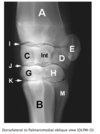
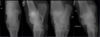
Patient number 219349 - Normal Canine Carpus
Mediolateral and dorsopalmar projections of the carpus of a dog.
Note the smooth articular surfaces and boney contours.
Can you name all boney structures on the images

Patient number 244290 - Normal Canine Shoulder
WHICH PROJECTIONS ARE THESE?
Note the presence of an endotracheal tube within the trachea, which indicates the dog was
under general anaesthesia when the radiographs were taken. This facilitates accurate patient
positioning.
Note the relatively large joint space on the mediolateral projection. This is a non-weight
bearing exam and the width of the joint space depends on the amount of traction applied to the
limb for positioning.
Note the smooth contours of the joint surfaces of both the humeral head and the glenoid
cavity.

Mediolateral (ML) and caudocranial (CdCr) projections are provided.
Normal canine elbow
WHICH PROJECTIONS ARE THESE?
Assess positioning:
•
Mediolateral projection
•
avoid rotation of the humeral condyle
•
note the humeral trochlea (medial portion) articulates more distally than the capitulum (lateral portion)
Craniocaudal projections
•
On a well-positioned craniocaudal projection (in this case, of the left elbow), the olecranon of the ulna is
positioned on midline, bisecting the humeral metaphysis and epiphysis.
•
On a cranio-10°-lateral-caudomedial oblique view (in this case, of the right elbow), the olecranon is
superimposed over the lateral aspect of the distal humerus. This view is sometimes used to free-project the
medial coronoid process.
In addition a ‘neutral’ position mediolateral view may be taken, and is useful for evaluation of the elbow joint congruity.
With this view however, the anconeal process is obscured by the medial epicondyle of the humerus.
Evaluate:
•
On the mediolateral projection:
•
The width of the joint space between the humerus and radius, as well as the humerus and ulna
•
The congruency of the ulna notch and the humeral condyle
•
The anconeal process for osteophyte formation, and union with the olecranon tuber
•
The base of the medial coronoid process for sclerosis
On the craniocaudal projections:
•
The cranial and medial border of the medial coronoid process for osteophytes or fragmentation
•
The contour of the joint surface of the humerus, for osteochondrosis lesions and sclerosis
•
The radial head and humeral epicondyles for osteophyte formation
•
The humeral epicondyles for enthesiophyte formation

Flexed medio-lateral (ML), craniocaudal (CrCd), and Cr10LCdM oblique projections are provided.


Patient number 245702 - Normal Canine Pelvis
WHAT VIEWS ARE THESE?
Assess Positioning:
•
On the lateral view, use the transverse processes of the vertebrae, and alignment of the two hemipelvises
•
On the VD view, use the dorsal spinous processes, size and shape in the iliac wings, and symmetry of the
obturator foramina. The femurs should be parallel to each other and parallel to the table (how would you
judge this?). The patellae should be superimposed over the femoral trochlea, within the sagittal plane of the
femur. The fabellae should be roughly located over the femoral cortices.
The lateral projection:
•
is useful for evaluation of the position of femoral heads compared to acetabulae
•
is useful for evaluation of the lumbosacral area, especially when looking for the presence of transitional
vertebrae
The VD hip extended projection:
•
is the official view (by the Australian Veterinary Association, also the OFA in North America, BSAVA in Great
Britain, and FCI in Europe) for screening for hip dysplasia, so correct positioning is very important
•
is useful for evaluating the position of the femoral heads relative to the acetabulae
•
is useful for assessment of pelvic fractures
•
is useful for assessment of other boney pathology of the pelvis

Lateral and ventrodorsal (VD) hip-extended projection.
Patient number 245702 - Normal Feline Pelvis
WHAT VIEWS ARE THESE?
Use the same criteria for evaluation as in dogs.
Compare these radiographs to the canine pelvis.
•
Notice the difference in the shape of the bones between the two species, with the feline long bones
being straighter than the canine bones.
•
Feline vertebral bodies are longer and narrower when compared to canine vertebral bodies.
•
Make sure you can recognise the difference between the two species, based on the radiographic
appearance.
Did you notice the smooth solid bone ventral to and bridging L5-6 vertebral bodies? This is called ventral
vertebral spondylosis. Although this is not a normal finding, the smooth and solid nature of the bone
indicates this is an inactive process, and may be an incidental finding without any clinical significance.

Lateral and ventrodorsal (VD) hip extended projection.

- medial coronoid process 8. lateral coronoid process 9. trochlear notch 10. head of radius lateral flexed view
Patient number 242727 - Normal Hip and Elbow Screening Radiographs
These radiographs were taken to screen the patient for the presence of hip and elbow dysplasia.
VIEW?
Notice:
1.
The collimated field includes the entire pelvis and femoral condyles/stifle joints
2.
The symmetry of the pelvis, with similar appearance to the iliac wings and symmetric size of the obturator foramina
3.
The femurs are parallel
4.
The patellas are superimposed over the femoral trochlea.
5.
These radiographs are normal, but when evaluating hip radiographs:
note the congruence of the coxofemoral joints
look for evidence of osteoarthritis
6.
These factors are considered when allocating a final hip ‘score’.
The elbows are taken as mediolateral projections with the elbows in full flexion.
Notice:
1.
The medial and lateral portions of the humeral condyle are superimposed, forming parallel curved surfaces.
2.
When the elbow is in full flexion, the anconeal process is readily visible.
3.
These elbows are normal, but in a patient with elbow dysplasia there will be early signs of arthritis
osteophyes on the anconeal process
sclerosis of the ulna trochlea notch
4.
In more advanced cases of arthritis, osteophytes are seen associated with all aspects of the elbow joint

The pelvis view is a ‘Ventrodorsal, hip extended’ projection.
Case 1: Patient 239711
VIEW?
Compare these radiographs to the example of a normal tarsus.
Please note:
•
mild soft tissue swelling associated with the tarsocrural joint, best identified on the plantar aspect of the joint as
seen on the mediolateral view
•
widening of the medial aspect of the tarsocrural joint space
•
flattening of the medial trochlear ridge of the talus - you can see this on both dorsoplantar and mediolateral views
•
the small mineralised fragment present in the joint space proximal to the medial trochlear ridge of the talus, as
seen on the dorsoplantar view.
This is a case of osteochondritis dissecans (OCD) of the medial trochlear ridge of the talus. What are other
predilection sites for osteochondrosis in the dog?

Mediolateral and dorsoplantar projections of the tarsus of a skeletally immature dog.
Case 2: Patient 227876
VIEW?
Compare these radiographs to the normal example of a carpus.
Please identify the following:
•
Soft tissue swelling centred on the distal aspect of the radius
•
The reduced opacity and loss of trabecular structure of the distal radial metaphysis
•
The large area of irregularly margined lucency in the distal metaphysis, consistent
with moth-eaten lysis
•
the long transition zone proximally with multiple small radiolucencies extending
toward the diaphysis, consistent with permeative lysis
•
Lysis of the distal radial cortex
•
Note that although the bone lysis extends into the distal radial epiphysis, it does not
breech the thin sub-chondral bone plate and does not cross the joint
•
Periosteal new bone (PNB) formation along the cranial and medial aspect of the
distal radial metaphysis. This PNB is continuous and solid on the cranial aspect,
and palisading on the medial aspect.
How many bones are affected?
Although the periosteal reaction identified in this lesion is continuous, the patterns of
bone lysis are aggressive. A lesion should be classified by it’s more aggressive
characteristics, thus this is an aggressive bone lesion.
What are you two main groups of differential diagnosis?
Which one is more likely and why?
How would you proceed with management of this case?

Mediolateral and dorsopalmar projections of the carpus of a skeletally mature dog.
Case 3: Patient 231270
VIEW?
What other projection(s) would you request to make this a complete radiographic study?
Please identify the following:
•
Marked soft tissue swelling around the distal aspect of the crus
•
The aggressive bone lesion in the distal tibia.
What features of this lesion allow you to determine it is an aggressive bone lesion?
•
The pathologic fracture in the distal tibial diaphysis - note the lucent lines through the cranial and
caudal cortex
What is your presumptive diagnosis?
What could you do to confirm this diagnosis?
How would you manage this patient? Is surgical stabilisation of the fracture appropriate in this case?
Why?

Mediolateral projection of the tarsus
Case 4: Patient 246004
VIEW? Mature?
What features allow you to determine this patient is skeletally immature?
Please note the presence of an endotracheal tube indicating the dog is anaesthetised.
Please identify the following:
•
Irregular contour of the articular surface of the humeral head, with a defect present in
the caudal aspect of the articular surface.
•
Underlying this defect there is increased bone opacity indicating sclerosis of the
subchondral bone.
These findings are typical for osteochondrosis of the shoulder in dogs. A cartilage flap
or nom-mineralised joint mice can only be detected radiographically, using arthrography
ie. injecting iodinated contrast medium into the shoulder joint.

Mediolateral projection of the shoulder of a skeletally immature dog.
Common sites of osteosarcoma in the dog

Case 5: Patient 239711
VIEW??
Please refer to the normal elbow for comparison.
Review the normal anatomy of the elbow joint. In this patient, note the conical shape of the distal ulna physis.
Note the growth plate of the tuber olecrani - this separate centre of ossification is called an apophysis. What
is the difference between an apophysis and an epiphysis?
Please identify the following:
•
The surgical plate on the cranial aspect of the radius, which stabilised a diaphyseal fracture, now healed - a
fracture line is no longer visible.
•
Note that the most proximal screw crosses to the ulna, fixing the radius to the ulna. Note that this screw has
partially ‘backed out’, with angulation of this screw and the screw head no longer flush with the plate surface.
•
The elbow joint is incongruent, with a large ‘step’ between the radial head and medial coronoid process of
the ulna, and subluxation of the humeral condyle from the ulna trochlea notch.
•
The distal ulna physis is located slightly more proximally than the distal radial physis - normally they are
located at a similar level. The distal ulna physis is more indistinct, as it is beginning to radiographically close.
This is an example of acquired elbow incongruity. The ulna is shortened relative to what it should be: this is
probably due to premature closure of the distal ulna physis, probably due to the same traumatic event that
caused the fracture to the radius.

Mediolateral and craniocaudal projections of the antebrachium, and flexed mediolateral projection of the
elbow of a skeletally mature dog.
Case 6: Patient 237244
VIEW?
Compare these radiographs to the example of a normal elbow.
Please identify the following:
•
The irregular radiolucent line separating the anconeal process from the olecranon. This is
called an ununited anconeal process.
•
The following findings:
sclerosis at the base of the medial coronoid process of the ulna
irregular border to the medial coronoid process, as seen on the craniocaudal view
what problem do these findings indicate?
•
The following findings:
osteophyte formation along the proximal contour of the anconeal process
sclerosis of the ulna trochlear notch
what process is happening in this elbow joint?
Ununited anconeal process is one of the causes of elbow dysplasia. What are the other
potential causes of elbow dysplasia? What is the expected sequela of elbow dysplasia?

Flexed mediolateral and craniocaudal projections of the elbow of a dog.
Case 7: Patient 237968
VIEW??
Is patient positioning adequate, or should the study be repeated?
If this radiograph was taken to investigate hindlimb lameness, is positioning adequate or would you repeat the
radiograph?
Compare this radiograph to the normal canine pelvis.
Please identify the following:
•
Asymmetry of the coxofemoral joint spaces, and the location of the centre of the femoral head compared to the
dorsal acetabular rim, indicating subluxation.
This dog has bilateral hip dysplasia with coxofemoral joint subluxation, but only mild osteoarthritis is present.
Is there a difference in severity of subluxation between the left and right side?
Also note:
•
The patchy increase in opacity of the right femoral diaphysis (compare to the left femur)
•
The smooth, solid, continuous periosteal new bone production around the medial and lateral aspect of the right
femur.
These findings are consistent with a non-aggressive bone lesion, and are indicative of panosteitis.
Are you surprised to find two diseases in one patient?

A ventrodorsal legs extended projection of the hips is provided to submit for screening for hip dysplasia.


Case 8: Patient 239099
SPECIES? VIEW?
Consider patient positioning; is positioning adequate or do you have to repeat the radiograph?
How old is this cat? How can you judge the age of an animal?
Compare this radiograph to the normal feline pelvis.
Please identify the following:
•
thin cortices of the long bones
•
generally poor mineralisation of the skeleton
•
deformed shape of the lumbosacral area on the lateral projection
•
folding fracture of the left ileum
This cat has a polyostotic osteoporotic disease, therefore the disease must be systemic. Most
systemic diseases affecting the skeleton are of metabolic origin - the ‘metabolic bone
diseases’. This cat was on a meat only diet and has nutritional secondary
hyperparathyroidism, which develops secondary to a calcium-posphorous imbalance in the
food. Dietary calcium deficiency cases a decrease in serum calcium, which triggers an
increase in PTH secretion and induces bone resorption from the skeleton, causing diffuse
osteoporosis.

Lateral and ventrodorsal legs extended projections of the pelvis of a cat.
Case 9: Patient 239268
VIEW?
Comment on patient positioning. Is positioning adequate, or do you need to repeat the radiograph?
!
Remember to use the dorsal spinous processes, size & shape of the ilial wings, and size and shape of the
obturator foramen to evaluate positioning. The femurs should be parallel to each other and parallel to the table.
The patellae should be superimposed over the femoral trochlea, and fabellae roughly divided in half by the
femoral metaphyses.
Evaluate:
•
The coxofemoral joint space - is subluxation present?
•
The cranial acetabular rim and acetabular subchondral bone
•
The depth of the acetabulum
•
The femoral head and neck for presence of osteophytes and remodelling
•
The amount of soft tissue development around the thighs
•
The remaining soft tissue structures on the images
Have you noticed the shallow acetabulae and luxated femoral heads? There is bilateral severe osteophyte
formation around teh femoral head and neck and around the acetabulum, and marked remodelling of the
femoral necks.
This is a case of severe bilateral hip dysplasia and osteoarthritis.

A ventrodorsal legs-extended projection of the pelvis is provided.
apophysis vs. epiphysis
apophysis: any outgrowth or swelling, especially a bony outgrowth that has never been entirely separated from the bone of which it forms a part, such as a process, tubercle, or tuberosity.
epiphysis: the end part of a long bone, initially growing separately from the shaft.
Case 10: 238671
VIEW?
Compare the open-mouth and the intra-oral projections - why might the nose appear different in length
on both images?
Please note the following:
•
On the lateral view
There is almost perfect superimposition of the left and right structures indicating good patient
positioning.
Revise the dental formula of the dog: Note that mandibular premolars 3 and 4 are missing.
•
On the VD/DV views, note the biateral symmetry of the turbinates of the nasal conchae and the
ethmoid.
•
On the oblique open-mouth view of the maxillary teeth
consider normal dental anatomy - what parts of the teeth and surrounding structures should you be
able to see?
note the alveolar bone lysis resulting in widened periodontal space around the roots of premolar 4.
What is your diagnosis in this dog?

Multiple projections of the skull are provided: lateral, rostroventral-caudodorsal open mouth projection,
dorsoventral intra-oral projection, and an oblique open-mouth view of the left maxilla, collimated to the
caudal cheek teeth.


Normal equine stifle
Can you name the projections of the stifle included in this study?
Is the patient well positioned for each of these views?
Can you name the bones of the stifle joint?
Evaluate the bone margins, trabecular structures, joint spaces, and soft tissues


Ununited anconeal process
Flexed lateral view

A- Radius
E- Accessory carpal bone
Int- Intermediate carpal bone
H- Fourth carpal bone
C- radial carpal bone
G- third carpal bone
B- third metacarpal bone

Lateral view

A- Radius
I- Antebrachiocarpal joint
E- Accessory carpal bone
J- Middle carpal joint
G- third carpal bone
K- carpometacarpal joint
B- third metacarpal bone
L- second metacarpal bone
M- fourth metacarpal bone


A - Radius
B - Third metacarpal bone
C - Radial carpal bone
D - Ulnar carpal bone
E - Accessory carpal bone
F - Second carpal bone
G - Third carpal bone
H - Fourth carpal bone
I - Antebrachiocarpal joint
J - Middle carpal joint
K - Carpometacarpal joint
L - Second metacarpal bone
M - Fourth metacarpal bone
Int - Intermediate carpal bone
Normal equine tarsus
Evaluate the positioning.
•
Can you name each projection?
•
Are all the radiographs correctly positioned?
•
Can you name each bone of the hock on each projection?
Evaluate the bone margins, trabecular structures, joint spaces, and soft tissues.
Normal equine carpus
Can you name each of the projections provided?
Is the patient correctly positioned in each of these radiographs?
Can you name each bone of the carpus?
If the oblique radiographs were not labelled as DMPLO/DLPMO, using your knowledge of anatomy how would
you tell which projection you are looking at? (ie how do you tell medial from lateral when looking at a carpus?)
Use one of the handouts to help you answer this question.
Evaluate the bone margins, trabecular structures, joint spaces, and soft tissues.

Normal equine fetlock
Name each projection provided within this series.
Evaluate the positioning - are all the radiographs correctly positioned?
Can you name each bone of the fetlock joint?
Is the dorsopalmar projection a true horizontal beam projection? Refer to the lateromedial view
to help answer this question)
Evaluate the bone margins, trabecular structures, joint spaces, and soft tissues.
What is the ovoid soft tissue opacity seen at the proximal aspect of the proximal phalanx?
Can you see this opacity on any other projections?
What is another name for the proximal phalanx?
Without labels, are you able to tell which projection is the DLPMO and which one is the DMPLO?
(ie how do you tell lateral from medial when looking at the fetlock joint?)

Normal equine shoulder
A single mediolateral projection of the shoulder is provided.
Consider the difficulty in obtaining the orthogonal (caudocranial) projection of the shoulder of a horse. In this
species, the mediolateral projection is usually sufficient to make most diagnoses.
Note the normal osseous anatomy - the cervical spine, the scapula, the glenoid cavity and the humerus.
Evaluate the bone margins & joint spaces

Case 1: 239244
VIEW??
This is a standard fetlock ‘series’ of radiographs, but may be supplemented by other views such as a
flexed lateromedial, or oblique views angled in a proximo-distal direction.
How can you distinguish between medial and lateral sides of the fetlock joint?
Compare to the normal fetlock example.
Please identify the following:
•
Soft tissue swelling at the level of the metacarpophalangeal joint
•
Multiple fragments proximal to the lateral sesamoid bone
•
The irregular plantarolateral and distal contours of the lateral sesamoid bone
•
The radiolucency of the lateral sesamoid bone, compared to the normal medial sesamoid bone which
has a fine trabecular pattern
This horse has sesamoiditis and apical fractures of the lateral proximal sesamoid bone. Which soft
tissue structure is most likely involved with these injuries?
Is the soft tissue swelling on the dorsal aspect of the fetlock joint consistent with direct sesamoid injury?
What else might it indicate?

Right hind fetlock, dorsoplantar, lateromedial, dorsolateral-plantaromedial oblique (DLPlMO) and
dorsomedial-plantarolateral oblique(DMPlLO) projections.
Case 2: 246029
VIEW??
horse. In this species, the mediolateral projection is usually sufficient to make most diagnoses.
Note the normal osseous anatomy - the cervical spine, the scapula, the glenoid cavity and the
humerus.
Is this a young or an old horse? What features are you using to make this decision?
Carefully examine the glenoid cavity and the shape of the scapula neck. Identify the large
osseous fragment at the cranial aspect of the scapula - this horse has an intra-articular fracture of
the supragelnoid tubercle.

A single mediolateral projection of the left shoulder is provided.
Consider the difficulty in obtaining the orthogonal (caudocranial) projection of the shoulder of a
Case 3: 214891
VIEW??
Compare these radiographs to the normal stifle.
Please identify the following:
•
Note the complex geometry of the normal distal femoral physis, and the normal appearance of the tibial
tuberosity apophysis. The tibial tuberosity growth plate is the last growth plate to close in most species.
•
Identify the remnant of the fibula - horses have an incomplete fibula; compare and contrast this with small
animal species
•
Identify the concave defect in the lateral trochlear ridge of the femur, and the fragments located within this
defect (best seen on the caudo-lateral-craniomedial oblique projection.
Can you see the lesion on other projections?
This is an example of osteochondrosis of the equine stifle. Stifle OC can also affect the patella

Standing lateromedial, flexed lateromedial, caudolateral-craniomedial oblique, and caudocranial projections
of the stifle are provided.
Case 4: 237176
VIEWS???
Compare these radiographs to the normal elbow.
Please identify the following:
•
The large, ovoid, well-marginated radiolucency on the proximo-medial aspect of the radius,
surrounded by a rim of sclerosis.
This is an example of geographic bone lysis.
•
On the craniocaudal projection, note the solid, irregular periosteal new bone on the medial
aspect of the proximal radial metaphysis.
•
Identify the lucent defect in the subchondral bone plate of the proximal radius, connecting with
the ovoid lucency, indicating the lesion communicates with the elbow joint.
Is this an aggressive or non-aggressive bone lesion?
What features help you make this decision?
This is an example of a bone cyst, with communication with the elbow joint.

Mediolateral and craniocaudal projections of the elbow of a horse are provided.
Case 5: 236329
VIEWS??
Compare these radiographs to the normal fetlock.
Please identify the following:
•
The soft tissue swelling at the level of the fetlock joint, most pronounced at the plantar aspect of the metatarsus
What soft tissue structures are located in this region?
•
The hyperextension of the fetlock joint, with distal displacement of the sesamoid bones and subluxation of the
metatarsophalangeal joint
These findings are most consistent with complete rupture of the suspensory ligament

Left hind fetlock, dorsoplantar, lateromedial, dorsolateral-plantaromedial oblique (DLPlMO) and dorsomedial-
plantarolateral oblique(DMPlLO) projections.
Case 6: 233152
VIEWS??
Compare these radiographs to the normal tarsus.
Please identify the following:
•
the soft tissue swelling, most pronounced at the level of the tarsocrural joint
•
The separate osseous fragment associated with the distal intermediate ridge of the tibia (DIRT)
•
The associated flattening or defect in the DIRT
On which views can you see this lesion?
This lesion is typical of osteochondrosis of the equine tarsus.
What are the other predilection sites for osteochondrosis of the equine tarsus?

Right tarsus, dorsoplantar, lateromedial, dorsolateral-plantaromedial oblique, and dorsomedial-plantarolateral
oblique projections are provided.
Case 7: 242372
VIEWS??
Compare this study to the normal carpus.
Please identify the following:
•
The soft tissue swelling, most pronounced at the level of the distal row of carpal bones.
•
the slab fracture of the third carpal bone
note how the slab fracture is reduced on the flexed lateromedial projection
What is the difference between a slab fracture and a chip fracture?
•
Identify the sclerotic and radiolucent areas of the third carpal bone

Right carpus, lateromedial, flexed lateromedial, dorsomedial-palmarolateral oblique (DMPLO), dorsolateral-
palmaromedial obilque (DLPMO), and dorsoproximal-dorsodistal (“skyline”) projections are provided.
