Primary immunodeficiencies 2 - adaptive immune system Flashcards
What are the different types of SCID?
Which is the most common?
- Reticular dysgenesis
- X- linked SCID
- ADA deficiency
Most common = x linked scid!!
Why is it called SCID?
Combined: affects B and T cells
Reticular dysgenesis:
a) molecular mechanism
b) blood cell abormalities
c) management
a) molecular mechanism
- autosomal recessive severe SCID (most severe form)
- mutation in mitochondrial energy metabolism enzyme AK2
- affects haemoatpoietic stem cells–> failure of production of myeloid and lymphoid cells
b) cells affected (low):
- lymphocytes - B and T
- neutrophils
- monocytes/macrophages
- platelets
Treatment:
Stem cell transplant - fatal in early life otherwise
X-linked SCID
a) molecular mechanism
b) blood cell abormalities
c) clinical phenotype
a) molecular mechanism
- mutation in Xq13.2
- mutation in cytokine receptor - failure to respond to cytokines and produce cells
b) blood cell abnormalities
- low T cells - CD4 and CD8
- low NK cells
- Normal B cell counts - but LOW IMMUNOGLOBULIN (failure of production of MATURE b cells)
c) clinical phenotype
- presents in early childhood with severe sepsis
- eczema like rash
- systemic candida infections
- failure to thrive
ADA deficiency
a) molecular mechanism
b) blood cell abnormlaities
c) tx
a) molecular mechanism
- deficiency of adenosine deaminase enzyme
- enzyme required by lymphocytes for cell metabolism
- failure of maturation along any of the cell lineages
b) blood cell abnormalities
- low/absent T cells
- low/absent B cells
- low/absent NK cells
What age does SCID present? Typical clinical features?
- presents at 3-6 months of age
- before this they are protected by maternal IgG (through placenta)
- typical clinical features
- Infections of all types
- Failure to thrive
- Persistent diarrhoea
- Unusual skin disease:
- Colonisation of infant’s empty bone marrow by maternal lymphocytes
- This is sort of like graft-versus-host disease because the maternal lymphocytes are in the baby’s bone marrow
- Family history of early death
Classes of T cell disorders
- disorders of t cell maturation
- disorders of selection of CD4 and CD8 positive T lymphocytes
- disorders of t cell effector function
What disorder causes defect in T cell maturation
Di george syndrome
Di George syndrome
a) clinical features
b) cell counts
Autosomal dominant deletion of 22q11.2 (but can also be sporadic- in most cases)
CATCH 22
- Cardiac defects: truncus arteriosus, TOF
-
Abnormal facies and atresia
- Facies
- High forehead
- Low set, abnormally folded ears
- Cleft palate
- Small mouth and jaw
- Oesophageal atresia
- Facies
- thymic hypoplasia: absence of thymic shadow on x ray
- Cleft palate
- Hypocalcaemia - due to underdevelopment of parathyroid gland
- 22: 22q11.2 deletion
b) cell counts
- low T cells
- Normal B cells
- Low IgA/IgG/IgE (AGE) - as defect in T cells leads to lack of maturation of B cells
What happens to T cell counts in Di George syndrome over time?
Homeostatic proliferation
so immune function increases with age
Which disorder leads to defect in selection of CD4 and CD8 lymphocytes?
Bare lymphocyte syndrome
Two types of Bare lymphocyte syndrome
Pathophysiology/cell counts
1. Bare lymphocyte syndrome type II
- defect in protein involved in expression of MHC II
- MHC II is not expressed at all –> so CD4 T cells are not selected
- Cell counts
- Low/absent CD4+ T cells
- CD8+ T cells usually normal
- Antibodies
- IgM: normal (as this does not require T cells)
- IgA and IgG: lOW - as these require T cells to develop
2. Bare lymphocyte syndrome type I
- MHC I is absent
- CD8+ T cells fail to develop
- Cell counts
- Low/absent CD8+ T cells
- Normal CD4+ T cells
- Normal immuoglobulins
- as CD4 T cells are intact
Clinical features of BLS
Unwell by 3 months of age
Infections of all types
Failure to thrive
Family history of early infant death
Sclerosing cholangitis
hepatomegaly
jaundice
Outline some disorders of T cell effector function
Could affect:
- Cytokine production (e.g. IFN-gamma)
- Cytokine receptors (e.g. IL2 receptor)
- Cytotoxicity
- T-B cell communication
- involving CD40 ligand and CD40
Overall clinical features of T cell deficiencies
- viral infections
- some fungal infections: eg cryptosporidium, PCP
- some bacterial ifnections: especially intracellular bacteria such as salmonella and myobacterium TB
- early malignancy
What technique is used to do a lymphocyte differential?
FACS - fluorescence activated cell sorting
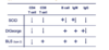
Differentiating between SCID, Di George and BLS (type II) via cell counts
what are the 4 defects that can occur in B cell maturation?
- bruton’s x linked hypogammaglobulinaemia
- hyper IgM syndrome
- common variable immunodeficiency
- selective IgA deficiency
Bruton’s X-linked Hypogammaglobulinaemia
- x linked recessive
- mutation in BTK gene
- affects B cell maturation - at the point where pre- b cells enter the circulation from the bone marrow
- so you don’t get any mature B cells
- so no antibody production
- all immunoglobulins affected: IgM, IgA and IgG
- T cells - CD4 and CD8 ae present
- B cells are absent
- affects children after 6 months - before this they have maternal antibodies
Clinical phenotype (typical presentation)
- 1 year old boy
- Recurrent bacterial infections
- CD4 and CD8 T cells present
- B cells absent
- IgG, IgA and IgM absent
What vaccines must be avoided in Bruton’s X linked hypogammaglobulinamiea?
Live attenuated vaccines must be avoided
Hyper IgM syndrome
Pathophysiology
- X linked recessive
- Mutation in CD40ligand on T cells
- Technically a T cell problem
- Results in failure of maturation of B cells
Cell counts
- Excess IgM
- low IgA and IgG
- T cells and B cell count is normal
Typical presentation
- recurrent bacterial infections as a child
- episode of PCP
- high IgM
- absent IgA
- and absent IgG
Recurrent bacterial infections in a 1yo child wih episode of PCP
Hyper IgM syndrome
1 year old boy
Recurrent bacterial infections
CD4 and CD8 T cells present
B cells absent
IgG, IgA and IgM absent
Bruton’s X linked hypogammaglobulinaemia
- as T cells are unaffected
- B cells are absent
- all antibodies are absent

