Autoimmune and autoinflammatory conditions Flashcards
What is immunopathology?
The damage to the host caused by the immune response
Can be due to innate or adaptive immune response
May or may not be an obvious pathogen involved

Difference between autoinflammatory and autoimmune response
Autoinflammatory: innate response
Autoimmune: Adaptive response
Mixed: both innate and adaptive
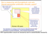
Do autoinflammatory disease tend to be localised or diffuse?
Tend to be localised eg ankylosing spondylitis
- Local factors at sites predisposed to disease lead to activation of innate immune cells such as macrophages and neutrophils, which results in tissue damage
Mechanisms of autoimmune disease
- Molecular mimicry
- Defective immunoregulation
- T- cell bypass
- Release of “hidden self antigen”
- Cytokines
Give an example of molecular mimicry causing autoimmune disease
Post streptococcal rheumatic fever
Antibodies to M protein on surface of Strep - cross reacts with cardiac myosin .
Give an example of defective immunoregulation leading to autoimmune disease
QUant or qual defect in T reg cells
Observed in: thyroid, islet cell and liver autoimmune disease
Give an example of T cell bypass causing autoimmune disease
-involves the generation of a NEW autoantigen/epitope - can be triggered by drugs or infection
Mycoplasma pneumoniae
Modifies antigen on surface of RBC - so it’s susceptible to attack by immune response - autoimmune haemolytic anaemia
Give an example of release of hidden self antigen causing autoimmune disease
Mechanism: some insult leads to release of antigen from an organ that has never been exposed to the immune system before
eg post MI - antigens within cardiac myocyte sget released– > Dressler’s syndrome (autoimmune pericarditis)
Give an example of a cytokine causing autoimmune disease
IL-2
Associated with breakdown of peripheral tolerance
Explain the genetics of autoimmune vs autoinflammatory diseases: polygenic vs monogenic
Autoimmune and autoinflammatory disorders can be monogenic or polygenic.
Mixed disorders are always polygenic.
Give some examples of monogenic autoinflammatory conditions
Important ones: FMF

What gene is affected in FMF?
Loss of function mtation in MEFV gene (Lof) >> Genes encode Pyrin-Marenostrin
Autosomal recessive
- Bacteria, urate, toxins etc. will activate cryopyrin.
- This activates ASC and in turn upregulates procaspase 1.
- This leads to increased expression of IL-1, NFkB and apoptosis
- The increased NFkB expression leads to increased TNFa production.
- Pyrin-Marenostrin is a negative regulator of this pathway.
- So:
- A GoF mutation in cryopyrin >> more inflammation
A LoF mutation in Pyrin-Marenostrin >>> more inflammation

Which cells are overactive in FMF?
Neutrophils
pyrin-manenostrin -expressed by neutrophils mainly
This is because there’s REDUCED activity of pyrin-manenostrin - a negative regulator of the INFLAMMASOME compelx.
–> increased activity of inflammasome complex
–> inflammation
Clinical presentation of FMF
Periodic fevers lasting 2-4 days associated with:
Abdominal pain due to peritonitis
Chest pain due to pleurisy and pericarditis
Arthritis
Rash
**SEROSITIS- peritonitis, pleurisy, pericarditis**
think Ps- periodic fevers, peritonitis, pleurisy, pericarditis
What are the complications of FMF?
AA amyloidosis: this is because in episodes of inflammation the liver produces acute phase proteins such as amyloid protein
This deposits in:
- liver
- kidney - nephortic syndrome and renal failure
- spleen
investigations + Treatment of FMF
Colchicine 500ug bd - binds to tubulin in neutrophils and disrupts neutrophil functions including migration and chemokine secretion
then: if is ongoing inflammation and ongoing high levels of serum amyloid:
- Anakinra (Interleukin 1 receptor antagonist) >> Will block cytokines more specifically
- Etanercept (TNF alpha inhibitor)

Which pathway is affected by monogenic auto-inflammatory conditions?
Inflammasome pathway
Increased activity of inflammasome complex
3 mechanisms responsible for monogenic autoimmune diseases - and examples of each
- failure of tolerance: APCED/APS-1
- Abnormality in regulatory T cells- IPEX
- Abnormlaity in lymphocyte apoptosis - APLS 1
Mechanism of APECED
- autosomal recessive
- mutation in AIRE - transcription factor involved in promoting expression of self antigen on thymic T cells - this enables identification and apoptosis of autoreactive T cells in the thymus
- Defect in autoimmune regulator AIRE - less expression of self-antigen in thymus, defect in central tolerance - MORE AUTOREACTIVE T CELLS
- Some autoreactive B cells (but < autoreactive T cells)
some autoreactive B cells because- B cell tolerance is T cell dependent but there is a limited repertoire of autoreactive B cells
Clinical features of APECED
Autoimmune polyendocrinopathy candidasis and ectodermal dystrophy
- hypoparathyoridism
- addison’s disease
- hypothyoridism
- diabetes
- candidiais - due to antibodies against cytokines that protect against candidasis (IL-17 and IL22)

Mechanism of IPEX
X linked recessive mutation in FOXP3 gene
Leads to lack of Treg cells
- autoreactive B cells - that produce autoantibodies
- lack of CD 25+ T cells
(FoxP3 positive cells also express CD25 so a normal person will have CD25+ve cells)
affects PERIPHERAL T cell tolerance
Which CD marker to FOXP3 positive cells express?
CD25
**by the age of 25 you are able to regulate your emotions- T reg cells….**
Symptoms of IPEX
Immune dysregulation, polyendocrinopathy, enteropathy, X-linked syndrome
3Ds:
diarrhoea
dermatitis
diabetes
- Enteropathy >> diarrhoea
- Diabetes mellitus
- Hypothyroidism
- Dermatitis

Mechanism of APLS
- mutation in FAS pathway: Fas binds to Fas Ligand which then signals to activate the death pathway: APOPTOSIS
- defect in apoptosis of T cells- lymphocytes, especially autoreactive T cells
- this leads to failure of tolerance and failure of homeostasis (more and more lymphocytes)

Clinical features of ALPS
Autoimmune lymphoproliferative syndrome
- High lymphocyte count:
- Double negative CD4-ve and CD8-ve T cells
- Large spleen
- large lymph node
- Autoimmune diseases:
- commonly autoimmune cytopaenias
- Lymphoma- high risk
Examples of polygenic autoinflammatory diseases
- Crohns
- UC
- osteoarthritis
- Takayasu arteritis
- Giant cell arteritis
Genetics of polygenic autoinflammatory disorders
Multiple genes affected:
- Mutations in the genes encoding proteins involved in pathways associated with innate immune cell function
Affect the innate immune system
- lead to activation of innate immune cells such as macrophages and neutrophils, with resulting tissue damage
HLA associations are less strong
Generally not characterised by the presence of autoantibodies
Examples of mixed pattern diseases
Ankylosing spondylitis
Psoriatic arthritis
Bechets
BAP
Genetics of mixed pattern diseases
- can affect innate and adaptive immune response
- Mutations in genes encoding proteins involved in pathways associated with innate immune cell function AND adaptive immune cell function
- HLA associations may be present
- usually NO AUTOANTIBODIES - usually autoimmune disease
**H for HLA, H for halfway. between autoimmune and autoinflammatory- there are some HLA associations in mixed pattern diseases**
Which mutations may be present in ankylosing spondylitis?
- HLA-B27 - most common mutation
* 50% of the heritability of AS comes down to HLA-B27 – associated with developing ankylosing spondylitis - IL23 Receptor:
- IL receptor type 2

Features of ankylosing spondylitis + clinicap resentaiton + treatment
- tends to occur at specific sites - at ENTHESES - where tendon/ligament inserts into bone
- localised pattern - eg sacroiliac joints
- can get bone formation eg bamboo spine
feature of localisation is characteristic of auto-inflammatory disease
Clinical presentation
- Low back pain and stiffness
- Enthesitis i.e. plantar fasciitis, Achilles tendonitis
- Large joint arthritis
- Sacroiliitis
Treatment
- first line: NSAIDS
- immunosuppression if not responding to NSAIDS - aniti TNF alpha, anti IL17

Pathogenesis of polygenic autoimmune diseases
- genetic polymorphisms
- Mutations in genes encoding proteins involved with pathways associated with adaptive immune cell function
- loss of tolerance >> development of immune reacivity towards self antigens
- generation of autoantibodies
-
HLA associations are very common
- as HLA is involved in presentation of antigen by T cells to B cells
Examples of polygenic autoimmune diseases
genetic polymorphisms (HLA polymorphisms), that lead to loss of tolerance and autoreactive B cells, T cells and autoantibodies
“DRS GG”
diabetes
rheuamtoid
sle
goodpastures
graves

Goodpasture’s disease: HLA association
HLA DR15
HLA-DR2
**these are actually the same**
Graves disease HLA association
HLA-DR3
grave–>serious–>odd number–>HLA DR 3
SLE: HLA association
HLA-DR3
**SLE has 3 letters**
T1DM: HLA association
HLA DR3 or HLA DR 4
RA: HLA association
HLA-DR4
Name two genes that may be mutated in polygenic autoimmune disease - that lead to inappropriate T cell activation
- CTLA4 >>> inappropriate T cell activation.
- PTPN 22 >> failure to control T cell activation >>> more reactive T cells >> autoimmune disease
These are negative regulators of T cell activity
Mutations in these genes lead to overactive T cells/inappropriately activated T cells –> autoimmunity

WHat is the Gel and Coombs classification system for immunopathology?
Classifies immunopathology based on whether it’s predominantly T cell or antibody mediated
-
Type I: immediate hypersensitivity which is IgE mediated (this is allergy)
- Rarely directed at self-antigens so rarely autoimmune
-
Type II: antibody reacts with cellular antigen
- Can be part of autoimmune reaction
- Antibody mediated
-
Type III: antibody reacts with soluble antigen to form an immune complex (which circulate)
- Antibody reacting with soluble antigen forming immune complex and inflammation
- Type IV: delayed-type hypersensitivity, T cell mediated response

Anaphylaxis: Gel and Coombs?
atopic dermatitis?
Type 1 hypersensiivity
*both*
Atopic asthma: gel and coombs?
Type 1 hypersensiivity
Goodpasture disease: gel and coombs?
Type 2 hypersensitivity
Graves disease: gel and coombs?
Type 2
SLE: gel and coombs?
Type 3
Serum sickness: gel and coombs?
Type 3
T1DM: gel and coombs?
Type IV
T!DM- 4 letters; type 4
allergic contact dermatitis: gel and coombs?
Type 4
Multiple sclerosis: gel and coombs?
Type 4
Which Gel and Coombs reactions are autoimmune?
Type 2 is very often autoimmune
Type 3 and 4 are sometimes autoimmune
Aetiology of multiple scleorsis
Th17 cells attack myelin basic protein and proteolipid protein in brain
Features of MS
- Nonspecific neurological symptoms
- → optic neuritis, weakness, blurred vision
- worse vision after bath = Uhthoff’s phenomenon
- tiredness
- weight loss
Diagnosis of MS
Oligoclonal bands of IgG in CSF
Management of MS
Natalizumab - prevents lymphocyte migration into CNS
What can you divide polygenic autoimmune disorders into?List some examples of each
- organ specific
- Graves
- Hashimotos
- Diabetes
- Pernicious anaemia
- Myasthaenia gravis
- Goodpasture’s disease
- organ non-specific
- Rheumatoid arthritis
- SLE
- Sjogren’s
- systemic sclerosis
- dermato/polymyositis
- ANCA associated vasculitis

Clincal picture of graves disease and explain pathology + type of hypersensitivity reaction
type 2 hypersensitivity
signs and symptoms of hyperthyroidisim
- IgG antibodies which stimulate the TSH Receptor
- This stimulates the receptor >> excess production of thyroxine
Pathophsyiology of graves disease
IgG antibodies that stimulate TSH receptor
Type II hypersensitivity reaction - Antibody-driven Autoimmune Disease
HLA association of graves disease
HLA DR 3
Clincial picture of hashimoto’s thyroiditis
Hypothyroidism: Lethargic, dry skin and hair, constipation, cold intolerance
- Result in a goitre >> enlarged thyroid infiltrated by T and B cells
What type of hypersensitivity reaction is hashimoto’s thyoriditis?
Type II and Type IV
Pathophyisology of Hashimoto’s thyoriidtis
-
Anti Thyroid Peroxidase antibodies- MORE SPECIFIC >>
- Presence correlates with thyroid damage and lymphocyte infiltration
- Anti thyroglobuin antibodies - could be present due to consequence of thyroid damage rather than pathogenic
- destruction of thyroid gland cells
- infiltration of thyroid gland by T and B cells –> GOITRE

What type of hypersensitivity is T1DM?
Type IV
- The antigen is within the pancreatic beta cells
- CD8 mediated destruction of beta cells
Pathophsyiology of T1DM as an immune reaction
- MHC I on beta cells presents self peptide to CD8+ T cells
- self-peptide found in Beta cells: GAD, Islet cell antigen 2
- CD8+ T cells attack Beta cells –> can’t produce insulin
- T cell clones have specificity for islet antigens

Which antibodies are associated with T1DM? what is the significance?
Anti-GAD + Anti- islet cell, others include:
- Anti-insulin
- Anti-IA2
NOTE: individuals with 3-4 of the above are highly likely to develop T1DM
*presence of autoantibodies predates the onset of symptoms by a lot
*currently has no role in diagnosis >> based on clinical manifestation + glucose levels
Which antibodies are present in pernicious anaemia?
Anti intrinisc factor– seems like this is more speciifc (from haem lecture)
Anti parietal cell
- antibodies against intrinsic factor >>>> failure of absorption of vitamin B12.
Features of perncious anaemia
- Vit B12 deficiency
- Macrocytic anaemia- Raised MCV
- hypersegmented neutrophils
clinical features: Tired,pale, mild numbness of feet, macrocytic anaemia, vitamin B12 deficiency, normal folate
- Neurological features
- subacute combined degeneration of the spinal cord
- peripheral neuropathy
- optic neuropathy
What is subacute combined degeneration of the spinal cord
- affects posterior and lateral columns- degeneration - sensory function
- bilateral weakness
- bilateral paraesthesia
- progressive ataxia
- babinski and rhomberg’s test positive
Reversible with B12
peripheral neuropathy + optic neuropathy
Treatment of pernicious anaemia
if antibodies are positive need B12 injections
no point giving PO supplements, need to give injections: Need parenteral vitamin B12
What type of hypersensitivty reaction is myasthenia gravis?
Type II
Pathophsyiolgy of MG + clinical features
- antibodies against nAChr–> failure of depolarisation >>> do NOT get muscle action potential which results in muscle weakness
- results in clinical features: FLUCTUATING MUSCLE WEAKNESS
- drooping eyelids - Ptsosis
- repetitive movements –> fatiguing
- weakness - worse at the end of the day
Differentiating between dermatomyositis vs MG in clinical features
MG: ptosis (drooping eyelids)
Dermatomyositis: ocular muscles are spared
Diagnosis of MG
- AChr antibody - 75% of patients
-
tensilon test- definitive diagnosis
- administering a very short-acting anticholinesterase (edrophonium bromide) >>> RAPID IMPROVEMENT IN SYMPTOMS.
What type of hypersensitivity reaction is goodpasture’s disease?
Type 2 hypersensitiivty
Antibodies in Goodpasture’s disease + presentation
Anti-GBM antibody
- basement membrane collagen type IV which is present in the lungs- haemorrhage and kidneys- glomerulonephritis
Clinical features of goodpasture’s disease
- lung disease: widespread crackles, widespread shadowing on x ray, pulmonary haemorrhage
- kidney disease: nephritic syndrome: oedema, proteinuria, haemturia, hypertension
Diagnosis of goodpasture’s disease
- anti GBM antibodies - smooth linear deposition of antibodies along the basement membrane - detected using fluorescein conjugated anti-human immunoglobulin
- crescenteric nephritis on kidney biopsy

clinical features of RA, genetic + environmental predispositions in rheumatoid arthritis
- Genetic predisposition:
- HLA DR4 (DRB1 0401, 0404, 0405)
- HLA DR1 (DRB1 0101)
- PTPN22
- Polymorphisms affecting TNF, IL-1, IL-6 and IL-1
- PAD2 and PAD4 polymorphisms
Basically there is a genetic predispositon to increased citrulinsation (removal of arginine and conversion to citruline)
-
Environmental Factors
- Smoking >> increased citrulinsation
- Gum infection with Porphyromonas gingivalis >>> only bacterium known to express PAD, thereby promoting citrullination

Antibodies in rheumatoid arthirtis
1. Anti CCP antibodies are diagnostic: - antibodies against citrullinated peptide
- **Anti-cyclic Citrullinated Peptide Antibodies**
- Bind to peptides in which arginine has been converted to citrulline by PAD
- rheumatoid factor - outdated - An antibody directed against the Fc region of human IgG
Which enzyme leads to formation of citrulline?
PAD- Peptidyl Arginine Deiminases (PAD2 and 4)
Converts arginine to citruline
This is what antibodies in rheumatoid arthritis bind to
Anything that increases cirtullination increases the risk of rheumatoid arthritis i..e smoking, gum infections
What type of hypersensitivity is rhuemtaoid arthritis?
- Type II
- antibodies bind to citrullinated peptide
- immune complexes activate classical pathway of complement
- also activates macrophages and NK cells
- Type III
* immune complexes deposit in areas >> leads to compliment activation - Type IV
-
APCs will present peptides to CD4 T cells >>> production of IFN gamma and IL-17 – > activates macrophages and and fibroblasts >>> production of:
- MMPs
- IL-1
- TNFa
DEscribe the joint in rheumatoid arthritis
the synovium becomes very inflamed forming a pannus
Type of extra growth in joint that causes pain, swelling and damage
invades the articular cartilage and adjacent bone + increase in synovial fluid volume
Result of CH50 test in rheumatoid arthritis
High CH50
As complement factors are activated
HLA association of goodpasture’s disease
HLA-DR2 and HLA DR 15
Which organ non specific diseases are associated with ANA antibodies?
SLE
Systemic sclerosis
Dermatomyositis
HLA associations of SLE
HLA- DR3
What joint manifestation do you get in SLE?
Jaccoud Arthropathy - correctible joint deformity
What immunological abnormalities are observed in SLE?
- abnormalities clearing apoptotic cells: Lots of dying cells which are not cleared and lots nuclear debris
- abmormalities in cellular activation: Increased B and T cell activations >> B cell hyperactivity and loss of tolerance occurs
Combination of nuclear debris and hyperactive immune cells = development of antibodies against nuclear proteins
What type of hypersensitivity reaction is SLE + explain mechanism
Type 3:
- Antibodies bind to antigens forming immune complexes
Immune complexes can deposit in small blood vessels leading to skin, joints, kidneys etc.
They activate complement via classical pathway + stimulate cells that express Fc (macrophages) and complement receptors >>>>
Cascade of inflammation
Comparison of Type 2 vs Type 3 hypersensiivity
Type 2: antibody mediated. Linear deposition of antibodies at specific sites e.g. Goodpasture’s syndrome - anti GBM antibodies
- GP: get antibody binding in a linear fashion at the basement membrane – evenly distributed. As antibodies a_gainst cellular antigens_
Type 3: immune complex mediated. Immune complexes deposit in a granular pattern e.g. lupus nephritis :lumpy-bumpy distribution of antibody formation

How do you interpret ANA levels?
Higher the dilution, higher the antibody level
eg 1:340 means higher antibody level than 1:10
It is represented as the greatest dilution at which the antibody can be detected. So if it can be detected at a very high dilution then the concentration must be high.
HIGH titres are associated with SEVERE disease >> Titre is important in disease monitoring
Classification of ANA antibodies
- anti dsDNA
-
components within the nucleus: ENA:
- anti ribonucleoprotein- anti-ro, anti-la, anti-sm, anti-u1rnp
- SCL 70- anti topoisomerase
- anti centromere - cytoplasmic
t-rna synthetase (Jo1)

Two types of staining patterns for ANA and which is good for disease monitoring
- homogenous: anti ds-DNA >> HIGH titres are associated with SEVERE disease >> Titre is important in disease monitoring
- speckled: anti- ENA: extractable nuclear antigens (ENA) >> Specificity is for some ribonucleoproteins (e.g. Ro, La, Sm, UR1NP >>> Titres are NOT helpful in monitoring disease activity – so only need to measure once to see if they are or not

What is anti-ds DNA specific for?
SLE
Of the two groups of ANA, which one is useful in disease monitoring?
anti DS DNA is useful for monitoring SLE activity
but anti ENA is not useful
Describe what happens to complement in SLE
- immune complex formation triggers classical pathway of complement activation by exposing the binding site for C1q
- this leads to consumption of C3 and C4 if constantly consumed
- C4 is consumed before C3
- this can be used to monitor disease activity/severity
- if both are low - VERY severe disease
- Quantification of C3 and C4 acts as a surrogate marker for DISEASE ACTIVITY
- NOTE: we measure UNACTIVATED complement proteins rather than activated ones

CRP and ESR in SLE
CRP: normal
ESR: raised
Which other antibody should you test for in SLE?
anti phospholipid Ab
Which 3 antibodies are tested for in anti phospholipid syndrome?
lupus anticoagulant: Prolongation of phospholipid-dependent coagulation tests
anti cardiolipin: Immunoglobulins directed against phospholipids and b2 glycoprotein-1
Anti-β2 glycoprotein-1 antibody
**lupus anticoagulant: can’t be done if patient is on anticoagulant medication
note: Some aspects of coagulation pathway are phospholipid dependent. If there is auto antibodies against this then there is prolongation of phospholipid dependent coagulation tests

Which cells are involved in sclroderma?
Th2 and Th17 cells
- Inflammation with Th2 and Th17 cells predominating
Polymorphisms in scleroderma
Type I collagen α2 chains
fibrillin 1
TGF-β
What is the effect ofthe polymorphisms in scleroderma?
Cytokines >>> activation of fibroblasts >>> development of fibrosis
Cytokines >>> activation of endothelial cells >>> microvascular disease
Loss of B cell tolerance to nuclear antigens
Features of limited cutaneous systemic sclerosis
- limited skin involvement: does not progress beyond forearms (and maybe perioral area)
- CREST features
Calcionisis
Raynauds
Erythroderma
Sclerodactyly- tightening of the skin
Telangictasia
- Also Esophageal dysmotility + primary pulmonary hypertension
Features of diffuse cutaneous systemic sclerosis
- skin involvement beyond forearms
- CREST features:
- calcinosis
- raynauds phenomenon
- erythoderma
- Sclerodactyly
- Telangiectasia
- EXTENSIVE gastrointestinal involvement
- interstitial pulmonary disease
- kidney disease/renal crisis >> vascular renal problem- involves endothelial cells proliferating leading to a small lumen and poor blood supply >>> severe hypertension)
Antibodies in limited cutaneous systemic sclerosis?
anti-centromere
antibodies in diffuse cutaneous systemic sclerosis
Anti-topoisomerase antibodies (anti-Scl-70) are good for diagnosing
Anti-RNA polymerase
Anti-Fibrillarin
pattern of staining in diffuse cutaneous systemic sclerosis
nucleolar

what is an important progostic indicator in systemic scleorsis?
ANA staining
clinical presentation of dermatomyositis + type of hypersensitivity reaction
- muscles
- skeletal muscle- fatigue
- lung - dyspnoea
- heart - arrythmia
- oesophagus - dysmotility
- spares ocular muscles - unlike myasthenia gravis
-
skin involvement
- heliotrope skin rash
- gottron papules
o Immune complex mediated vasculitis (type III response)
Blood results in dermatomyositis
- High serum creatine kinase/ CK
- high aldolase, myoglobin, LDH, AST, ALT
What type of hypersensitivty is dermatomyositis?
Type III- immune complex mediated
*in the skin
What type of hypersensitivity is polymyositis? + clinical features
Type IV- CD8 T cell mediated
o Not much skin involvement - unlike Dermatomyositis
o CD8 T cells kill myofibres via perforin/granzyme - type IV response
Specific antibodies for dermatomyositis?
anti Jo 1
o Dermatomyositis > Polymyositis: Anti-Mi2 (nuclear)
o Anti-aminoacyl transfer RNA synthetase antibody (e.g. anti-Jo-1) (cytoplasmic)*
antibodies for polymyositis
Anti-signal recognition peptide antibody (nuclear and cytoplasmic)
Clinical features of polymyositis
- proixmal muscle weakness - no skin involvement
histology of dermatomyositis vs polymyositis
- dermatomyositis
- speckled ANA pattern
- can see immune complex deposition
- polymyositis
* can see CD8+ T cells surrounding HLA class I myofibres
Which types of vasculitis are ANCA mediated?
Small vessel vasculitis

Key features of polyarteritis nodosa
- rare medium vessel vasculitis
- usually associated with Hep B
- Type III hypersensitivity reaction - immune complexes
- anuerysm formation
- Rosary bead appearance on histopathology
What are the small vessel. vasuclitides?
- Microscopic polyangiitis/ microscopic polyarteritis/ MPA
- Granulomatosis with polyangitis/ Wegner’s Granulomatosis
- Churg-Strauss syndrome/ eosinophilic granulomatosis with polyangiitis/ eGPs
Clinical features of wegner’s granulomatosis
- Saddle nose
- recurrent nosebleeds
- haemoptysis
- breathless
- rapidly progressive glomerulonephritis → rapid decline in kidney function
- constitutional symptoms
- peripheral neuropathy
Antibody tested for in wegner’s granulomatosis
c-ANCA: Cytoplasmic fluorescence
antibodies to the enzyme proteinase 3
- > 90% of patients with granulomatous polyangiitis with renal involvement
Staining pattern of wegner’s
Cytoplasmic fluorescence
(c-ANCA- c for cytoplasm)

Primary target for c-ANCA
enzyme proteinase 3
Histology of wegner’s
accumulation of macrophages in bowman’s capsule
Clinical presentation of churg-strauss syndrome + treatment + associated conditions
- severe allergies
- sinus problems
- → progress to severe asthma prior to systemic vasculitis
-
Eosinophilic Granulomatosis with Polyangiitis (Churg-Strauss syndrome)
- Asthma
- Vasculitis
- Eosinophilia
- Treated with corticosteroids
- Other conditions associated:
- UC
- PSC
**eosinophilic granulomatosis with polyangiitis
antibodies in churg strauss syndrome
p-anca
staining pattern of churg strauss syndrome?
perinuclear (p-anca; p for perinuclear)

primary target for p-anca
myeloperoxidase
What is ANCA? + their use
anti-neutrophil cytoplasmic antibody
Antibodies specific for antigens located in primary granules within the cytoplasm of neutrophils
- Inflammation may lead to expression of these antigens on the cell surface of neutrophils >>> inflammatory response
Anti-neutrophil cytoplasmic antibodies help in diagnosis and monitoring of disease activity
What type of hypersensitivity reaction is vasculitis?
Type 2
- Antibody engagement with cell surface antigens may lead to neutrophil activation (Type II hypersensitivity)
- Activated neutrophils interact with endothelial cells causing damage to vessels- vasculitis
**except polycarteritis nodosa which is type 3 hypersenstiivty**
Conditions associated with p-ANCA
- microscopic polyangiitis (MPA)
- eosinophilic granulomatous polyangiitis (eGPA)/ churg strauss syndrome
- UC/ Primary Sclerosing Cholangitis
p-anca vs c-anca: specificty and sensiitivity?
c-anca is more sensitive and specific
which group of disorders is ANA screening for?
connective tissue disorders:
- SLE
- Sjrojens
- diffuse and limited systemic sclerosis
- myositis
which group of disorders is anca screening for?
small vessel vasculitides
Antibodies in autoimmune thrombocytopaenic purpura?
Anti GLP-2b, 2a
Antibodies in pemphigus vulgaris
Anti-Desmoglein 1
autoantigen - epidermal cadherin
Which cytokines are usually involved in monogenic autoinflammatory conditions?
IL1 and TNF
What mutation is associated with crohn’s disease?
NOD2 (CARD-15)
card-15, caspase activating recruitment domain-15
Are autoantibodies present in autoimmne or autoinflammatory diseases?
autoimmune diseases
*not in mixed pattern diseases either*
Are HLA associations more common in autoinflammatory or autoimmune disease?
more common in autoimmune disease
*can also get them in mixed pattern diseases*
1.Young male patient with intractable diarrhoea, type 1 diabetes and severe eczema. Diagnosis?
IPEX
- 15 year old with recurrent candidiasis, nail pitting and hypoparathyroidism. Name the causative mutation.
Mutation in TF AIRE
- 10 year old with abdominal fullness and splenomegaly. FBC reveals high lymphocytes. History of autoimmune thrombocytopenia. Diagnosis?
ALPS
Auto-immune diseases
- Adaptive immune response- T and B cells
- Aberrant T and B cell responses in primary (cell development) and secondary lymphoid (where cells respond) organs which lead to the breaking of tolerance with development of immune reactivity towards self-antigens
is mixed innate and adaptive response polygnic or monogenic
- However, for the mixed innate and adaptive response, it is just polygenic.

define the two types of mutations that can lead to autoinflammatory or autoimmune conditions
- Germ line mutations- affect DNA, and are in germ line cells
- Somatic mutation- do not affect germ cells, not inherited, occurs after birth and present later in life
Genetics, epigenetics and miRNA - explain
- Genetics- mutations affecting protein sequence
-
Epigenetics- (heritable) change in gene expression (e.g. via DNA methylation). May have the same sequence but different expression
- Just because you have/do not have mutation does not mean you express it.
- MicroRNA (miRNA) is small, non-coding, single stranded RNA: These bind to target mRNA and stop it from being able to produce protein.

mongenic diseases or polygenic diseases- which is more common
polygenic diseases are common whereas monogenic diseases are rare
Spectrum of immunological disease

SBA 1: ALPS, Familial Mediterranean Fever, IPEX- match up to the following description:
- A- A single gene mutation involving MEFV, affecting the inflammasome complex and resulting in recurrent episodes of serositis
- B- A mutation in the Fas pathway associated with lymphocytosis, lymphomas and autoimmune cytopenias
- C- A single gene mutation involving Foxp3 resulting in abnormality of T reg cells
ALPS= B
Familial Mediterranean Fever= A
IPEX= C
genetic mutaitons invovled in Crohns + mehcanisms
IBD1 gene on chromosome 16, also known as NOD2 (card-15, caspase activating recruitment domain-15)
3 different mutations on this gene- associated with Crohns
Note: . they are NOT necessary for development of crohn’s but are there in those with Crohns
- NOD2 is expressed in the cytoplasm of myeloid cells- macrophages, neutrophils and dendritic cells
- NOD2 acts as a microbial sensor + stimulates NFκB
- Activation of NOD2 induces autophagy (orderly degradation of cellular components) in dendritic cells.
In Crohns: there is disordered degradation which leads to inflammation:expression of inflammation proteins + release of proteases + free radicals >>granulomata formation and tissue damage

treatment of Crohns
Overactive innate immune response so need to suppress the inflammatory response
- 1st LINE- Corticosteroids
- Azathioprine
- Anti-TNFa antibodies
- Anti-IL12/23 antibodies
SBA 2: Autoinflammatory disease, Mixed pattern disease, Autoimmune disease:
- A- Aberrant T cell and B cell responses in primary and secondary lymphoid organs lead to breaking of tolerance with development of immune reactivity towards self antigens
- B- Mutations/ polymorphisms in genes encoding proteins involved in innate and adaptive T cell function
- C- Mutations in genes encoding proteins involved in pathways associated with innate immune cell function
A- autoimmune
B- Mixed pattern diseas e
C- autoinflammatory
PTNP22 and CTLA4 summary
genetic polymorphisms in T cell activation

There are THREE forms of peripheral tolerance:
- T cells requiring co-stimulation for activation
- Regulatory T cells
- Immune privileged sites
Antibody driven Immune Reactions (Type II hypersensitivity) - mechanism of action
- antibodies bind to cell-associated antigens,
- this will expose the binding site for C1q for complement
- this will lead to to antibody-dependent destruction:
- Antibodies can activate COMPLEMENT (by binding to C1q)
- Antibodies can also bind to Fc receptors on NK cells or to receptors on macrophages which will become activated and lead to inflammation >>>> cell death
- Binding of antibodies could also interfere with receptors >> receptor activation OR blockade
Binding of antibody to receptor protein- sometimes referred as type 5 but usually just part of type 2 = can lead to activation and stimulation
explain pathopyhsiology of Type III hypersensitivity reactions in Autoimmune Disease
- Antibody binds to soluble antigens forming immune complexes
- The immune complexes deposit in the blood vessels (especially the kidneys, joints and skin
>>>>>
- skin >>> tiny bleeds in the skin, which gives rise to the purpuric rash
- joints >>> arthritis
- kidneys >>>> glomerulonephritis: haematuria, proteinuria and renal failure
classic example of type 3 hypersenstivity autoimmune disorder
SLE
SLE, antibodies against double stranded DNA, deposit particularly in the kidneys

recall the antibodies of the following conditions:
Goodpastures syndrome
myasthena gravis
Graves disease
perniceaous anaemia
diabetes mellitus
Hashimotos thyroditis
Rheumatoid arthritis
- anti basement membrane antibodies against Type 4 collage on basement membrane
- anti acyetylcholine receptor antibodies: blocks depolarisation signls = fluctuating muscle weakness
- Anti Tsh receptor antibodies- IgG antibodies against TSH receptor = hyperstimulation = overproudciotn of thyroxine
Anti gastric parietal cells antibodies + anti intrinsic factor antibodies >> causes vitamin B12 deficiency >> subacute combined degeneration
anti islet cell antibodies, anti GAD antibodies, anti IA2 >> not diagnostic for diagnosis
anti thyroid peroxidase antiboides + antithyrglobulin antibodies >> hypothyroidism
Anti cyclic citrulinated peptide antibodies- diagnostic + Rheumatoid factor- outdated
examples of type 4 autoimmune hypersensitivty reactions

example of type 2 hypersnesitivty reactions that is not type 2 hypersnesitivty autoimmune
exception with transplantation so have foreign cell-
antibody is against foreign cell not self cell
Match Type I, II, II, IV hypersensitivity:
- A- Goodpasture disease
- B- Eczema
- C- SLE
- D- Multiple sclerosis
A- type 2
B- type 1
C- type 3
D- type 4
summarise autoantibodies for liver diseases

summarise autoantibodies for gastroenterology disease

what is the diagnosis

good pastures syndrome
High TLCO- gas transferring factor- indicates bleeding
Summary of Investigations for Connective Tissue Disease

list the antibodies in rheumatology

SBA 5: Investigations show: ANA+ve, Anti-dsDNA +ve, Low C3 and C4, High ESR. Negative results for:
Ro, La, Sm, RNP, Scl70, Centromere, Jo1. What is the diagnosis?
SLE
The problem with ANCA:
- It is NOT specific
- So need to look at the specific antigen- either proteinase 3 for cANCA or myeloperoxidase for pANCA
Wegener’s vs GBM disease?
Wegeners: more upper respiratory tract symptoms eg osebleeds and haemotysis
GBM disease: PULMONARY HAEMORRHAGE - not in wegner’s
*both can cause haematuria*