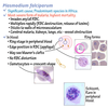
Identify the organism:
A. Penicillium
B. Blastoconidia
C. Sporothrix
D. Candida

A. Penicillium
Eosinophils in LAP (Leukocyte Alkaline Phosphatase) score:
A. Not counted
B. Double the value
C. Constant value
D. None of the above
A. Not counted
Eosinophils do not show alkaline phosphatase activity and must not be counted.
Prolonged apnea, anesthesized by succinylcholine, the enzyme responsible for the reaction is?
A. Cholinesterase
B. Aldolase
C. Pseudocholinesterase
D. AFP
C. Pseudocholinesterase
Pseudocholinesterase is a drug metabolizing enzyme responsible for hydrolysis of the muscle relaxant drugs succinylcholine and mivacurium. Deficiency from any cause can lead to prolonged apnea and paralysis following administration of succinylcholine and mivacurium.
These crystals are indicative of:
A. Liver disease
B. Nephrotic syndrome
C. Albuminuria
D. Normal

A. Liver disease
Tyrosine crystals appear as fine silky needles arranged in sheaves or bundles in acid urine. They are rarely present and may appear together with leucine and bilirubin crystals in liver disease.
The following results were obtained from a pure culture of gram negative rods recovered from the pulmonary secretions of a 10 year old cystic fibrosis patient with pneumonia:
Oxidase (+), Motility (+), Growth at 42C (+), Pigment = red (Nonfluorescent)
A. Pseudomonas aeruginosa
B. Burkholderia cepacia
C. Burkholderia pseudomallei
D. Pseudomonas stutzeri
A. Pseudomonas aeruginosa
P. aeruginosa is growing as a biofilm within the lungs of cystic fibrosis patients. In certain conditions, P. aeruginosa can secrete a variety of pigments, including pyocyanin (blue-green), pyoverdine (yellow-green and fluorescent), and pyorubin (red-brown). These can be used to identify the organism.
What to do?
Mother: Type “O”, Rh negative, no antibody to D Ag
Child: Type “O”, Rh positive
A. Transplacental transfusion
B. RhIg administration
C. Do nothing, report the results
D. DAT
B. RhIg administration
Rh immunoglobulin is a blood product that can stop your immune system from attacking Rh-positive cells. When an Rh incompatibility is identified, Rhogan will be given as a shot during week 28 of pregnancy.
(It will be given after chorionic villus sampling, amniocentesis, miscarriage, ectopic pregnancy, abortion, uterine bleeding, or any trauma during pregnancy that could leak some of the fetal cells over to you.)
Test for Paroxysmal Cold Hemoglobinuria (PCH):
A. Autohemolysis test
B. Donath-Landsteiner Test
C. Sucrose hemolysis test
D. NOTA
B. Donath-Landsteiner test
Normal = (-) hemolysis on test and control
PCH = (-) hemolysis on control but (+) hemolysis on test sample

In emphysema, the absence of alpha-1 globulin peak is due to the deficiency of:
A. A1-Antitrypsin (AAT)
B. Macroglobulin
C. Hemopexin
D. Transferrin
A. A1-Antitripsin (AAT)
Alpha-1 antitrysin deficiency is an inherited disorder that may cause lung disease and liver disease. Affected individuals often develop emphysema, which is a lung disease caused by damage to the small air sacs in the lungs (alveoli). Characteristic features of emphysema inlcude difficulty breathing, a hacking cough, and a barrel-shaped chest.

Which of the following pigments will deposit on urate and uric acid crystals to form a precipitate described as “brick dust”?
A. Urobilin
B. Bilirubin
C. Uroerythrin
D. Urochrome
C. Uroerythrin
Uroerythrin is a red pigment in the urine, where it is part of a group of yellow, brown, and red pigments generally designated as urochrome.
Mostly seen in urines that have been refrigerated, resulting in precipitation of amorphous urates.
Protein electrophoresis in pH 8.6. What proteins are closest to the cathode?
A. Albumin and Alpha 2
B. Albumin and Alpha 1
C. Beta and Gamma
D. NOTA
C. Beta and Gamma
Order of migration from Anode (+) to Cathode (-): Albumin, Alpha-1, Alpha-2, Beta, and Gamma

An ASO test can only be valid if the controls have yielded acceptable results. Which of the following indicates a valid ASO test?
A. Hemolysis in both SLO and red cell control
B. Positive control, hemolysis in all tubes
C. No hemolysis on SLO control
D. No hemolysis on red cell control

D. No hemolysis on red cell control
For an ASO tube test to be valid, the SLO control should show hemolysis and the RBC control tube should show no hemolysis.
SLO = Streptolysin O (toxin)

57% Hematocrit is normal in:
A. Male
B. 1 year old child
C. Newborn
D. Female
C. Newborn
Normal hematocrit value for a newborn is 45% to 61%.
Absorbs light and emits at longer wavelength:
A. Fluorometer
B. Nephelometer
C. AAS
D. TLC
A. Fluorometer
A fluorometer or fluorimeter is a device used to measure parameters of fluorescence: its intensity and wavelength distribution of emission spectrum after excitation by a certain spectrum of light. These parameters are used to identify the presence and amount of specific molecules in a medium. Modern fluorometers are capable of detecting fluorescent molecule concentrations as low as 1 part per trillion.

The reverse Camp test, lecithinase production, double zone hemolysis and gram stain morphology are all useful in the identification of:
A. Campylobacter jejuni
B. Staphylococcus aureus
C. Listeria monocytogenes
D. Clostridium perfringens
D. Clostridium perfringens
C. perfringens is a Gram-positive, rod-shaped, anaerobic, spore-forming pathogenic bacterium of the genus Clostridium. C. perfringens shows double-zone hemolysis on blood agar. Small area of beta hemolysis are noted (complete lysis of RBCs) surrounded by a larger zone of alpha hemolysis (partial hemolysis).

Causative agent of infectious Mononucleosis:
A. Cytomegalovirus
B. Epstein-Barr virus
C. Poxvirus
D. HPV
B. Epstein-Barr virus
EBV, also known as humna herpesvirus 4, is a member of the herpes virus family. It is one of the most common human viruses. EBV spreads most commonly through body fluids, primarily saliva. EBV can cause infectious mononucleosis (glandular fever), and other illnesses.

Anti-A = 4+
Anti-B = 4+
Weak D = 2+
Rh control = 0
A cells = 0
B cells = 0
A. ABO grouping is wrong
B. Rh grouping is wrong
C. Rh control is wrong
D. Do nothing, interpret the results

D. Do nothing, interpret the results
Patient is AB positive (Rh). Rh control should be negative.

Anti-A = 0
Anti-B = +mf
A cells = 4+
B cells = 0
A. Polyagglutination
B. Bx
C. Both
D. None of the above.
B. Bx

Smudge cells are seen in what type of leukemia?
A. Acute myelogenous leukemia (AML)
B. Chronic myelogenous leukemia (CML)
C. Acute lymphocytic leukemia (ALL)
D. Chronic lymphocytic leukemia (CLL)

D. Chronic lymphocytic leukemia (CLL)
Smudge cells are remnants of cells that lack any identifiable cytoplasmic membrane or nuclear structure. Smudge cells, also called basket cells, are most often associated with abnormally fragile lymphocytes in disorders such as CLL.

Widal and Well-Felix tests are classified under what type of serologic test?
A. Passive agglutination
B. Reverse passive agglutination
C. Direct agglutination
D. Coagglutination
C. Direct agglutination
Febrile agglutination tests, such as Widal and Well-Felix, are examples of direct agglutination.
Widal test is an agglutination test in which specific typhoid fever antibodies are detected by mixing the patient’s serum with killed bacterial suspension of Salmonella carrying specific O, H, AH and BH antigens and observed for clumping ie. Antigen-antibody reaction. The main principle of Widal test is that if homologous antibody is present in patient’s serum, it will react with respective antigen in the suspension and gives visible clumping on the test slide or card.
Weil-Felix reaction is a serological test for detecting Rickettsial antibody in the serum of a patient using heterophile antigen. This is a non specific agglutination reaction. Antibodies produced against Rickettsial antigen cross reacts with OX19 and OX2 strain of Proteus vulgaris and OXK strains of Proteus mirabilis. Patients infected with R. rickettsiae develops antibody which is reactive to Proteus vulgairis OX19 and OX2 while patients infected to R. prowazekii develops antibody which is reactive to Proteus vulgaris OX19.

In HIV:
A. increased total lymphocyte
B. increased CD8
C. decreased CD4
D. normal CD8/CD4
C. decreased CD4

Increased levels of TdT (Terminal deoxynucleotidyl transferase) activity are indicative of:
A. Acute lymphocytic leukemia
B. Acute myelocytic leukemia
C. Burkitt lymphoma
D. Eosinophilia
A. Acute lymphocytic leukemia
TdT, also known as DNA nucleotidylexotransferase (DNTT) or terminal transferase, is a specialized DNA polymerase expressed in immature, pre-B and pre-T lymphoid cells, and acute lymphoblastic leukemia/lymphoma cells.

Causative agent of pseudomembranous colitis?
A. C. perfringens
B. C. difficile
C. C. jejuni
D. All of the above

B. C. difficile
Pseudomembranous colitis refers to swelling or inflammation of the large intestine (colon) due to an overgrowth of C. difficile bacteria. This infection is a common cause of diarrhea after antibiotic use.

Excessively blue stain causes:
A. prolonged staining time
B. inadequate washing
C. too high alkalinity of stain
D. AOTA
D. AOTA
Select the best/acceptable donor for blood donation:
A. patient that received a transfusion 8 months ago
B. woman that gave birth 4 weeks ago
C. man that donated blood 10 weeks ago
D. patient with Hgb: 11 g/dL
C. man that donated blood 10 weeks ago
Only choice C is qualified to donate. The interval between whole blood donations is 8 weeks (56 days).


-
Practice Exam236
-
LabCE1
-
Review Cards - Lab Operations, Management, & Education489
-
Review Cards - Clinical Chemistry756
-
Review Cards - Microbiology1594
-
Urinalysis & Body Fluids Review310
-
Clinical Chemistry Review - Glucose, Iron, & Bilirubin138
-
Review Cards - Hematology659
-
Review Cards - Immunology548
-
Review Cards - Immunohematology523
-
Review Cards - Urinalysis & Body Fluids443
-
Review Cards - Molecular Diagnostics114
-
Urinalysis: Renal Function163
