Pictures Flashcards
What does this radiograph show?

Megaoesophagus (can be seen as large distension dorsal of trachea)
What does this radiograph show?

Oesophageal blockage just caudal to the heart base (common position)
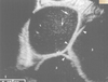
What pathology does this show?

Insulinoma
What pathology can be seen?

Pancreatic adenocarcinoma
What does this radiograph show?

Normal oesophagram
What does this scintigraphy show?

Metastatic prostatic carcinoma
What does this scintigraphy show?

Normal uptake due to growth plates etc.
What does this MRI show?

Spinal mass
What can be seen on this ultrasound?

Normal kidney
WHat can be seen in this ultrasound?

Liver (left) and spleen (right; hyperechoic)
What is this?

Splenic mass
Normal xray

Why is the right of the image so radiopaque?

The animal is thicker here -
What is the major presenting clinical sign in this cow?

Rumen bloating - left side severely distended
What procedure is carried out via this incision? What landmarks are used to locate the correct place?

Rumen puncture. Landmarks: Transverse processes of the spine, last rib, tuber coxae on the wing of the ileum
Which organs can be found superficially on the right of the cow?

Which organs can be found deep on the right side of the cow?

Which tooth is this? How can you decide?

Maxillary cheek tooth.
- 2 infundibuli
- 5 pulps
Which tooth is this? How can you tell?

What type of gag is this?

hausmans
Is there anything wrong with this horse’s mouth?

Malerrupted tooth upper left
What pathology can be seen here?

Wave mouth
What does this image show?

Points on buccal edge of maxillary arcade
What does this image show?

Buccal ulceration due to maxillary teeth
Outline the uses of these rasps

- Straight head, long length - lower cheek teeth, 3rd-6th upper cheek teeth
- Obtuse angled head, long length - caudal upper cheek teeth, curve of Spee
- Angled offset head, medium length - upper 1st-4th cheek teeth
- S float - smooth off first cheek teeth and 6th maxillary cheek teeth, bit seat, angle of curve of Spee
What do these images show?

Retained caps (deciduous teeth)
Where are the needles probing?

The pulp cavities
What does this image show?

Acquired dental displacement
What does this image show?

2-3 yo mandibular bumps (normal)
What does this image show?

Mandibular apical tooth root infection
What does this image show?

Maxillary apical tooth root infection of 06 or 07 (does NOT discharge into sinus)
What does this image show?

Apical tooth root infection
What does this image show?

Apical tooth root infection (L side of picture) - gas in bulging root, and soft tissue/fluid in sinus.
What tool is shown?

Molar grabbers
What tool is shown?

Spacers
What procedure is being carried out?

Minimally invasive tooth removal by lateral buccomotomy
What tooth can be seen?

Wolf tooth (05, 1st premolar)
What problem has been fixed here? What material has been used?

Jaw Fx, cerclage wire
What does this image show?

Megaoesophagus (air filled)
What does this image show?

FB blockage just caudal to heart base (common site of obstruction as oesophagus narrows here)
What does this image show?

Gastric ulcer (deep, almost to serosa)
What is this instrument?

Balfour retractors
What is this instrument?

Gossett retractors
What does this image show?

Omentalisation following enterotomy repair
What does this image show?

Enteroplication
What does this image show?

Bacteria contained in neutrophils (indicates septic peritonitis if found in abdomenocentsis)
What is the likely cause of this diarrhoea?

Large Intestinal Pathology
What is the likely cause of this diarrhoea?

Small Intestinal Pathology
What is the likely cause of this diarrhoea?

Small intestinal (meleana)
What is the likely cause of this diarrhoea?

Small intestinal pathology (^ volume)
What does this image show?

Normal rabbit abdomen - food everywhere!
What does this image show?

Distended bladder, distended stomach, small amount of gas in SI
What does this image show?

Stomach massively distended, stomach contents dehydrated away from stomach wall (black line inbetween stomach and contents) - surgery indicated
What does this image show?

General distended and bloated guts.
- Surgery not indicated, gut stimulants and pain relief
What does this image show?

Strangulation of gut
What does this image show?

Ulcers
What does this image show?

Self trauma caused by excessive rolling due to colic
What does this image show?

Positions for auscultation of gut sounds
What does this image show?

Septic mucous membranes - think GIT rupture if associated with colic
What does this image show?

Large intestinal pelvic flexure (?)
What does this image show?

Left: left ventral colon
Right: Left dorsal colon
Pelvic flexure
What does this image show?

Distended SI loops (equine)
What does this image show?
Cross sectional ultrasound of distended SI loops (equine)
What does this image show?

Longitudinal ultraound of distended SI loops (equine)
What does this image show?

Ventral mindline laparotomy
What does this image show?

Pelvic split
What does this image show?

Transanal surgical approach to rectal surgery
What does this image show?

Method of stapling for a colorectal resection and anastamosis
What does this image show?

Megacolon
What does this image show?

Rectal mass demarcated by gas on either side
What does this image show?

Mass in colon on ultrasound
What does this image show?

Colon and rectum following barium enema - mass -> filling defect, irregular mucosa on opposite side suggests metastasis or inflammation
What does this image show?

Liver tumours (bullseye)
What does this image show?

Enlarged sublumbar LNs
What does this image show?

Rectal poylps being treated transanally
What does this image show? Does the tissue look viable?

Rectal prolapse. Tissue looks viable
What does this image show? Does the tissue look viable?

Rectal prolapse - tissue looks necrotic and should be resected
What does this image show?

Colon sutured to transversus abdomenalis to treat repeat rectal prolapse
What does this image show?

Open anal sacculectomy
What does this image show?

Closed anal sacculectomy
What does this image show?

Anal sac apocrine gland adenocarcinoma (50% + metastasis at time of diagnosis)
What does this image show?

Anal furunculosis
