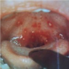
Pediatric Respiratory Infections Flashcards

1
Q

Differences between pediatric patients and adults presenting with possible respiratory illness

Basics:

A

•Basics:

–Anatomically smaller airways

–Proportionately more soft tissue in nose and mouth

–“Obligate nose breathers” in early infancy (relatively large tongue and epiglottis): significant proportion are not able to breathe orally

–Breathe with diaphragms and can’t use intercostals and other accessory muscles very well

–Less reserve than adults: can deteriorate quickly

The number one cause of cardiac arrest in children is respiratory arrest

2
Q

Differences between pediatric patients and adults presenting with possible respiratory illness

Historical features

A

•Historical features

–A toddler may present with “decreased appetite” per mother’s report instead of c/o sore throat

–A neonate may present with apnea instead of respiratory distress

–Parents often describe their child as “lethargic”

3
Q

Pediatric patients with possible respiratory illness

PE

A

• PE

–Physical exam

  • Often examined in parent’s lap
  • Must keep in mind vital sign normal values
  • Tonsils and adenoids generally diminish in size after age 5 years

–Signs of respiratory distress

  • Nasal flaring
  • Grunting
  • Head bobbing
  • Retractions
  • retractions, tachypnea, some stridor, head bobbing
4
Q

Stridor vs Wheezing

A
5
Q

Upper Respiratory Infections (Colds)

A
  • Acute, self-limiting viral syndrome of the upper respiratory tract
  • Children younger than six years have an average of six to eight colds per year (up to one per month, September through April), with a typical symptom duration of 14 days
  • Young children in daycare appear to have more colds than children cared for at home. However, when they enter primary school, children who attended daycare are less vulnerable to colds than those who did not.
  • Older children and adults have an average of two to four colds per year, with a typical symptom duration of five to seven days
6
Q

URI cont’d: symptoms

Most Common Sxs

A

•Most common sxs:

Fever may be the predominant manifestation of the common cold during the early phase of infection in young children. It is uncommon in older children and adults.

–Nasal congestion, nasal discharge, and sneezing are common in children

–Erythema and swelling of the nasal mucosa and nasal discharge. Nasal discharge may be clear initially, but often becomes colored (yellow or green) within a few days

Cough occurs in more than two-thirds of children with the common cold

7
Q

URI cont’d: symptoms

Other Sxs

A

•Other sxs:

Sore throat (typically an early manifestation), hoarseness, headache, irritability, difficulty sleeping, decreased appetite, cervical adenopathy, and conjunctival injection

8
Q

URI cont’d

-Other URI findings

A
  • Other URI findings
  • You wouldn’t really order imaging, but if you did: self-limited radiographic abnormalities of the paranasal sinuses
  • Abnormal middle ear pressures
  • viral nasopharyngitis may result in Eustachian tube dysfunction and abnormal middle ear pressure, or
  • abnormal middle ear pressure may result from the viral infection of the mucosa of the middle ear Eustachian tube

→ predisposes to otitis media

9
Q

URI cont’d

Typical viral pathogens:

A
  • Typical viral pathogens:
    • Rhinovirus (about 30-50%)
    • RSV
    • Influenza
    • Parainfluenza
    • Nonpolio enteroviruses
      • Echoviruses
      • Coxsackieviruses
    • Coronaviruses
    • Human metapneumovirus (hMTP)
10
Q

URI cont’d: transmission

A
  • Hand contact: Self-inoculation of one’s own conjunctivae or nasal mucosa after touching a person or object contaminated with cold virus
  • Inhalation of small particle droplets that become airborne from coughing (droplet transmission)
  • Deposition of large particle droplets that are expelled during sneezing and land on nasal or conjunctival mucosa (typically requires close contact with an infected person)
11
Q

Differential Diagnosis of URI

A
  • Allergic, seasonal, or vasomotor rhinitis; rhinitis medicamentosa
  • Acute bacterial sinusitis
  • Nasal foreign body
  • Inhaled foreign body
  • Pertussis - classically begins with mild cough and coryza (catarrhal phase)
  • Structural abnormalities of the nose or sinuses
  • Influenza
    • Although influenza virus may cause the common cold, it usually causes more severe illness; abrupt onset of fever (often >39°C [102.2°F]), headache, myalgia, and malaise in addition to cough, sore throat, and rhinitis
  • Bacterial pharyngitis or tonsillitis
12
Q

Complications of URI

A
  • AOM
  • Sinusitis
  • Asthma exacerbation
  • Pneumonia
  • Epistaxis
  • Conjunctivitis
  • Pharyngitis
13
Q

Epidemiologic and Clinical Features of Viruses that Cause the Common Cold in Children

A
14
Q

Acute Otitis Media

RIsk Factors

A

Most common affliction necessitating medical therapy for children younger than 5 years

Risk factors

  • Prematurity and low birth weight
  • Young age - anatomical differences of ear canal
  • Early onset
  • Family history
  • Race - Native American, Inuit, Australian aborigine
  • Altered immunity
  • Craniofacial abnormalities
  • Neuromuscular disease
  • Allergy
  • Day care
  • Crowded living conditions
  • Low socioeconomic status
  • Tobacco and pollutant exposure
  • Use of pacifier
  • Prone sleeping position
  • Fall or winter season
  • Absence of breastfeeding, prolonged bottle use
15
Q

Otitis Media cont’d

A

•Most common bacterial pathogens: S pneumoniae, H influenzae, Moraxella catarrhalis

  • Peak incidence 3-18 months
  • Presentation
  • Neonates: fussiness, poor feeding
  • Older child: fever, otalgia, ear tugging
16
Q
A
17
Q

Otitis media cont’d

A

Treatment:

amoxicillin

80-90 mg/kg/day

Prevention:

  • Avoid cigarette smoke exposure
  • Avoid bottle propping (and no bottle after age 1 year)
  • Tympanostomy tube placement for recurrent episodes
18
Q

Otitis media cont’d

Complications:

A
  • Intratemporal - Perforation of the tympanic membrane, acute coalescent mastoiditis, facial nerve palsy, acute labyrinthitis, petrositis, acute necrotic otitis, or development of chronic otitis media
  • Intracranial - Meningitis, encephalitis, brain abscess, otitis hydrocephalus, subarachnoid abscess, subdural abscess, or sigmoid sinus thrombosis
  • Systemic - Bacteremia, septic arthritis, or bacterial endocarditis
19
Q

Mastoiditis

A
20
Q

Sinusitis

A
  • Inflammation of the mucosal lining of one or more of the paranasal sinuses.
  • Acute bacterial rhinosinusitis = secondary bacterial infection of the sinuses.
21
Q

Sinusitis

Predisposing factors

A

Predisposing factors

  • URI
  • Allergic rhinitis
  • Anatomic obstruction
  • Mucosal irritants
  • Sudden changes in atmospheric pressure.
22
Q

Sinusitis

Symptoms include:

A

Symptoms include: cough, nasal symptoms, fever, headache, facial pain and swelling, sore throat, and halitosis

23
Q

Sinusitis cont’d

•Diagnosis is based on:

A

•Diagnosis is based on:

  1. Persistence of nasal discharge: if the child has a very congested and/or runny nose for 10 days without improvement, especially when it is associated with a daytime cough (may also have a nighttime cough)
  2. Severe symptoms: if the child has a high fever (over 39 C, which is 102.2 F) for 72 hours or has a high fever and is not eating or drinking and is difficult to calm
  3. Worsening symptoms: A child’s cold got better and then in a day or two the child is suddenly much more ill with a fever and/or pus-filled nasal discharge
24
Q

Characteristic Features of Viral vs Bacterial Rhinosinusitis in Children

A
25
Q

Differential Diagnosis

A
  • uncomplicated viral URI
  • allergic or non-allergic rhinitis
  • nasal foreign body
  • enlarged or infected adenoids
  • mucosal cyst of the maxillary antrum
  • pertussis
  • Did you know that kids don’t have all their sinuses developed until about 12 years old?
    • Frontal sinuses are formed by around 8 years old.
    • Maxillary, ethmoid are present at birth but develop to full size by adolescence.
26
Q

Work up?

A
  • Diagnosed clinically for routine cases
  • No routine imaging needed for routine cases, but…
    • Plain radiographic or computed tomography (CT) findings that are compatible with sinus inflammation include
      • Complete opacification
      • Mucosal thickening of at least 4 mm
      • Air-fluid level
    • However, abnormal imaging studies cannot distinguish between bacterial, viral, or other causes of sinus inflammation
  • Sinus aspiration for culture and sensitivity in complicated cases
27
Q

Complications of Sinusitis

A
  • By direct extension or by retrograde thrombophlebitis
  • Preseptal (periorbital) and orbital cellulitis
  • Septic cavernous sinus thrombosis
  • Meningitis
  • Osteomyelitis of the frontal bone
    • Pott’s Puffy tumor: subperiosteal abscess associated with osteomyelitis. It is usually seen as a complication of frontal sinusitis or trauma predominantly in the adolescent age group
  • Epidural, subdural, or brain abscess
28
Q

Herpangina

A
  • Coxsackieviruses A 1-10, 12, 16, and 22
  • Discrete erythematous-based macules à evolve into papules that vesiculate and then ulcerate centrally, creating an erythematous halo.
  • Lesions are typically smaller than 5 mm in diameter. Most cases of herpangina involve 2-12 lesions
  • Hand foot mouth is also coxsackievirus (usually A16 or enterovirus 71)
  • Not to be confused with Herpetic gingivostomatitis (herpangina: posterior, HSV is anterior)
29
Q

Herpetic gingivostomatitis

A
  • Prodrome that lasts about four days and may include fever (>38ºC (100.4ºF)), anorexia, irritability, malaise, sleeplessness, and headache
  • Enanthem begins with red, edematous marginal gingivae that bleed easily and clusters of small vesicles
  • Coalesce to form large, painful ulcers of the oral and perioral tissues
  • Cervical adenopathy often present
30
Q

Acute Pharyngitis

Viral

A
  • Viral
    • Supportive care
    • CMV, adenoviruses, HSV, influenza viruses, and enterovirus
    • EBV,
      • Monospot testing is not accurate under 4-5 years of age or before 2nd week of illness
      • remember it has a really long incubation period
      • Exudative pharyngitis is accompanied by fever, generalized adenopathy, hepatosplenomegaly, heterophile antibodies
      • Sometimes treated with steroids
31
Q

Acute Pharyngitis

Group A beta hemolytic streptococcus

A
  • Group A beta hemolytic streptococcus
    • 15 to 30 percent of all cases of pharyngitis in children between the ages of 5 and 15 years
    • Peaks during the winter and early spring
    • Rapid strep in office, back up culture
32
Q

Acute Pharyngitis
Other bacterial pathogens

A
  • Other bacterial pathogens
    • Group C and group G strep: acute rheumatic fever is not a complication of infection due to these organisms
    • Arcanobacterium hemolyticum
    • Corynebacterium diphtheriae
    • Tularemia
33
Q

Strep pharyngitis

History

A

History:

–Typically has an abrupt onset of symptoms

–Typically the school aged child

Sore throat

Fever

Headache

GI symptoms: abdominal pain, nausea, and vomiting

–Poor oral intake

NO cough or rhinorrhea!! Aka, no viral symptoms ( coryza, conjunctivitis, hoarseness, anterior stomatitis, discrete ulcerative lesions or vesicles).

34
Q

Strep pharyngitis

Physical

A

Physical:

  • Exudative pharyngitis or erythema of posterior orophayngeal mucosa
  • Enlarged tender anterior cervical lymph nodes
  • Palatal petechiae
  • Inflamed uvula
  • Scarlatiniform rash
  • Pastia’s lines
35
Q

Strep pharyngitis

Work Up

A

Work Up:

Rapid strep with back up culture if negative

  • How can you possibly get a sample??
    • Use two swabs at once; double tongue depressor for stronger kids, have smaller child pant like a puppy dog
    • Wear a mask because you’ll probably get coughed on
    • Some facilities do a RNA probe for back up instead of culture.

Can you tell by looking? → Nope!!

36
Q

Strep pharyngitis PE Findings

A
37
Q

Strep pharyngitis cont’d

A
  • Goals of antimicrobial therapy for eradication of group A streptococcus (GAS) from the pharynx in the setting of acute streptococcal pharyngitis include:
    • Reducing duration and severity of clinical signs and symptoms, including suppurative complications
    • Reducing incidence of nonsuppurative complications (eg, acute rheumatic fever)
    • Reducing transmission to close contacts by reducing infectivity
  • Initiation of treatment within 9 days of onset of illness will prevent complications
38
Q

Treatment of strep pharyngitis

A

Antibiotic options for treatment of GAS pharyngitis include penicillin (and other related agents including ampicillin and amoxicillin), cephalosporins, macrolides, and clindamycin.

  • Penicillin V

250mg PO BID or TID for 10 days for children <27kg

500 mg PO BID or TID for 10 days for children > 27 kg or adults

(liquid formulation tastes icky, by the way)

  • Amoxicillin 50mg/kg/day divided BID-TID (“low dose amox”) for 10 days
    • (bubblegum flavor!)
  • Pen G (Benzathine PCN):
    • <27 kg: 600,000U/dose IM X 1
    • >27 kg-adult: 1.2 million U/dose IM X 1
39
Q

Recurrence of strep

A
  • Persistence of streptococcus carriage in the setting of viral infection
  • Nonadherence with the prescribed antimicrobial regimen
  • New infection with GAS acquired from household or community contacts
  • Treatment failure (eg, repeat episode of pharyngitis caused by the original infecting strain); treatment failure is uncommon.
  • For patients with as many as six GAS infections in a single year or three to four episodes in two consecutive years, tonsillectomy may be an appropriate therapeutic consideration
40
Q

Deep neck infections

A
  • peritonsillar abscess
  • retropharyngeal abscess
  • lateral pharyngeal space infection
41
Q

Peritonsillar Abscess

A
  • Most common deep neck infection in children and adolescents, accounting for at least 50 percent of cases
  • A collection of pus located between the capsule of the palatine tonsil and the pharyngeal muscles.
  • Symptoms include:
    • severe sore throat (usually unilateral)
    • Fever
    • “hot potato” or muffled voice
    • Pooling of saliva or drooling may be present
    • Trismus
    • neck swelling and pain
    • ipsilateral ear pain
    • Fatigue
    • Irritability
    • decreased oral intake
42
Q

Peritonsillar abscess cont’d

A
  • requires needle aspiration or incision and drainage to determine if pus is present
  • often polymicrobial
    • Streptococcus pyogenes (group A streptococcus [GAS])
    • Staphylococcus aureus (including methicillin-resistant S. aureus [MRSA])
    • respiratory anaerobes (including Fusobacteria, Prevotella, and Veillonella species)
    • Haemophilus species
  • usually occurs in the superior pole of the tonsil, manifest by a defined collection of pus between the tonsillar capsule, the superior constrictor, and the palatopharyngeus muscle. PTA also may occur in the midpoint or inferior pole of the tonsil, or may be dispersed with multiple loculations in the peritonsillar space
  • preceded by tonsillitis or pharyngitis and progresses from cellulitis to phlegmon to abscess
  • Imaging: CT tonsils with contrast:
    • hypodense mass with ring enhancement
43
Q

Peritonsillar abscess

Treatment:

A
  • Treatment:
    • gold standard for diagnosis of peritonsillar abscess remains the collection of pus from the abscess through needle aspiration
    • antimicrobial therapy
      • amoxicillin/clavulanic, cephalosporins, and clindamycin
    • supportive care (hydration, analgesia)
44
Q

Peritonsillar abscess

Complications:

A
  • Complications:
    • Airway obstruction
    • Aspiration pneumonia if the abscess ruptures into the airway
    • Septicemia
    • Internal jugular vein thrombosis
    • Jugular vein suppurative thrombophlebitis (Lemierre syndrome)
    • Carotid artery rupture
    • Pseudoaneurysm of the carotid artery
    • Mediastinitis
    • Necrotizing fasciitis
    • Sequelae of Group A streptococcus infection
45
Q

Retropharyngeal abscess

A
  • most commonly in children between the ages of two and four years
  • The retropharyngeal space contains two chains of lymph nodes that are prominent in the young child, but atrophy before puberty
  • Usually polymicrobial, same as peritonsillar abscess:
    • Streptococcus pyogenes (group A streptococcus [GAS])
    • Staphylococcus aureus (including methicillin-resistant S. aureus [MRSA])
    • respiratory anaerobes (including Fusobacteria, Prevotella, and Veillonella species)
    • Haemophilus
46
Q

Retropharyngeal abscess cont’d

Symptoms

A

Symptoms:

  • Difficulty swallowing (dysphagia)
  • pain with swallowing (odynophagia)
  • drooling with decreased oral intake
  • Unwillingness to move the neck secondary to pain (torticollis), particularly unwillingness to extend the neck
  • Change in vocal quality (muffled, or with a “hot potato” quality [dysphonia])
  • gurgling sound, or stertor (snoring sound)
  • Respiratory distress (stridor, tachypnea, or both); stridor develops as disease progresses
  • Neck swelling, mass, or lymphadenopathy
  • Trismus (in approximately 20 percent)
  • Chest pain (if there is mediastinal extension)
47
Q

Retropharyngeal abscess cont’d

Dx & Tx

A
  • Diagnosis
    • Imaging: lateral neck film
    • CT with contrast
      • Generally, the anteroposterior diameter of the prevertebral soft tissue space in children should not exceed that of the contiguous vertebral bodies.
      • retropharyngeal soft tissue swelling as more than 7 mm at C2 and more than 22 mm at C6
  • Treatment:
    • empiric antibiotics (ampicillin-sulbactam, clindamycin, vancomycin)
    • Surgical drainage criteria
48
Q

Epiglottitis

A
  • 4 D’s:
    • Drooling
    • Dysphagia
    • Dysphonia
    • Dyspnea
  • Toxic appearing
  • “Tripod” position, “sniffing” position
  • Management if suspected:

_*Direct examination of the airway under anesthesia (with the availability of personnel who can perform a tracheostomy if needed_

49
Q

Laryngotracheitis (croup)

A
  • Inflammation of the larynx and trachea (laryngotracheobronchitis includes lower airway symptoms)
  • Most commonly occurs in children 6 to 36 months of age. It is seen in younger infants (as young as three months) and in preschool children, but it is rare beyond age six years
  • Most commonly caused by
    • parainfluenza virus
    • influenza virus
    • respiratory syncytial virus
    • adenovirus.
50
Q

Laryngotracheitis (croup)

Typical Features of croup

A
  • Typical features of croup include
    • nasal congestion
    • low-grade fever
    • barking-type cough
    • inspiratory stridor that may worsen with crying.
  • The diagnosis is made based on clinical assessment
  • Neck radiographs reveal the characteristic “steeple” sign, which reflect subglottic tracheal narrowing.
  • croup: https://www.youtube.com/watch?v=Qbn1Zw5CTbA
51
Q

Croup cont’d

Tx

A
  • Treatment:
    • Warm mist in closed bathroom with hot water running or cool night air
    • Corticosteroids, usually administered orally or parenterally, have been shown to improve symptom severity and reduce hospital length of stay (dexamethasone = Decadron).
    • Inhaled racemic epinephrine has been shown to reduce stridor within 30 minutes; therefore, it will provide the most immediate benefit and should be offered to the girl described in the vignette. ED observation period of at least 3 hours to see if symptoms return after nebulized epi treatment.
    • Children who have significant respiratory distress, hypoxemia, or inability to tolerate oral intake should be hospitalized for close observation.
    • Mildly affected children may do well with steam (sit in bathroom with hot water running until room is steamy) or cool air (step outside into cool night).
52
Q
A
53
Q

Bacterial Tracheitis

A
  • Invasive exudative bacterial infection of the soft tissues of the trachea
  • “Acute bacterial laryngotracheobronchitis”
  • Pathogens:
    • Bacterial: Staphylococcus aureus, Streptococcus pneumoniae, gram-negative enteric bacteria, Pseudomonas aeruginosa
    • Predisposing viral infections with: influenza A, influenza B, respiratory syncytial virus (RSV), parainfluenza virus, measles, and enterovirus
54
Q

Bacterial tracheitis cont’d

Symptoms

A

Symptoms:

  • Stridor, cough, and respiratory distress
  • Fever (common but not universal)
  • Stridor (inspiratory or expiratory)
  • Cough (not painful; membranous exudates may be expectorated)
  • Drooling is uncommon, but may be present
  • Preference to lie flat
  • Other less common: neck pain, orthopnea, choking, dysphagia, dysphonia, and syncope
55
Q

Bacterial tracheitis cont’d

A

Suspect in child with:

  • acute onset of airway obstruction in the setting of viral upper respiratory infection and in children with laryngotracheitis who are febrile, toxic-appearing, and have a poor response to treatment with nebulized epinephrine or glucocorticoids
  • Definitive diagnosis of bacterial tracheitis requires direct visualization of an inflamed, exudate-covered trachea
56
Q

Bronchiolitis

A
  • Bronchiolitis is a clinical syndrome that most commonly occurs in children younger than 2 years, with a peak incidence between 2 and 6 months of age and is characterized by upper respiratory symptoms (eg, rhinorrhea) followed by lower respiratory (eg, small airway/bronchiole) infection with inflammation, which results in wheezing and or crackles (rales).
  • More than half of RSV hospitalizations occur in infants younger than 6 months.
  • https://www.youtube.com/watch?v=lIE_UElOk3c
57
Q
A
58
Q

Bronchiolitis (cont’d)

A
  • one- to three-day history of upper respiratory tract symptoms, such as nasal congestion discharge and mild cough, followed by lower respiratory infection with inflammation, which results increased respiratory effort (eg, tachypnea, nasal flaring, chest retractions) and wheezing and/or crackles (rales).
  • Pathogens:
    • RSV, rhinovirus
    • less common causes include parainfluenza virus, human metapneumovirus, influenza virus, adenovirus, coronaviruses, and human bocavirus
  • Clinical diagnosis
59
Q

Bronchiolitis cont’d

DDx

A
  • Differential diagnosis:
    • recurrent viral-triggered wheezing or asthma
    • pneumonia
    • foreign body aspiration
    • chronic pulmonary disease
    • aspiration pneumonia
    • congenital heart disease
    • heart failure
    • vascular ring
  • Rapid detection (using immunofluorescent or enzyme immunoassays) of viral antigen or viral culture performed on nasopharyngeal secretions are the best tests for diagnosing RSV.
60
Q

Bronchiolitis cont’d

Management

A
  • Kids with RSV who wheeze have an increased risk of subsequent wheezing
  • Management:
    • Admit if hypoxic (oxygen saturations below 90% on room air) or dehydrated
    • IVFs if dehydrated
    • Trial of albuterol nebs→ no longer routinely recommended (new AAP guidelines 2015)
61
Q

Bronchiolitis Clinical Practice Guideline in Limerick Format

A

When treating bronchiolitis,
Refraining from films would delight us!

Please also avoid
The neb and the ‘roid…
After all it is only a virus!

  • Marion Sills, Associate Professor of Pediatrics and Emergency Medicine
    University of Colorado School of Medicine
62
Q

Pneumonia

A
  • Bacterial, viral, atypical, other
  • Presenting signs/sxs
    • Fever
    • Cough
    • tachypnea
    • increased work of breathing (retractions, nasal flaring, grunting, use of accessory muscles)
    • hypoxemia
    • adventitious lung sounds
63
Q

Pneumonia: Bacterial

A
  • Strep pneumoniae, Strep pyogenes, Haemophilus influenzae, Staph arueus, Klebsiella, anaerobes
  • Most pediatric patients who have pneumonia will recover uneventfully
  • 10% of pediatric patients with bacterial pneumonia may require hospital admission.
  • Children with pneumonia who do not respond to initial therapy after 48-72 hours should have a clinical, laboratory, and radiographic reassessment.
  • CXR may reveal a focal infiltrate (segmental or lobar consolidation).
64
Q

Pneumonia: Bacterial cont’d

A
  • Pleural and parenchymal complications can occur
  • Complications occur more commonly in patients who have bacterial pneumonia
    • bronchopleural fistulas
    • effusion/empyema
    • necrotizing pneumonia
    • pneumatocele
    • pulmonary abscess
  • Parapneumonic effusions are the most common complication occurring in up to 40% of bacterial pneumonias; progression to empyema occurs in more than 50% of these cases.
  • Moderate to large pleural effusions, especially those that impair respiratory function, should be drained.
    • pleural fluid layering greater than 10 mm on lateral decubitus
    • tube thoracostomy drainage, VATS = video assisted thoracoscopic surgery
65
Q

Differential Diagnosis of Pneumonia

A
66
Q

Pneumonia: Viral

A
  • Most likely cause of pneumonia in children
  • RSV, parainfluenza, adenoviruses, rhinoviruses, influenza viruses, varicella virus, rubeola virus
  • URI sxs followed by onset of tachypnea, cough, fever
  • PE dull or decreased breath sounds, wheezing or crackles, or normal
  • CXR with perihilar and diffuse parenchymal infiltrates (though sensitivity and specificity may be low)
67
Q

Pneumonia: Atypical and Other Pathogens

A
  • Mycoplasma pneumoniae
    • Adolescents or children over 5
    • Most commonly URI symptoms without pneumonia
    • Gradual onset and usually is heralded by headache, malaise, and low-grade fever
    • Occasionally can be more acute and mimic pneumococcal pneumonia
    • Nonproductive to mildly productive cough
    • Wheezing and dyspnea also may occur
    • Scattered rales and wheezes on lung exam may be present
  • Chlamydia pneumoniae
  • Also gradual onset of cough with low-grade fever
  • Chlamydia psittaci
  • Bordetella pertussis
  • Legionella
  • Histoplasma
  • Coccidiodes
  • Tularemia
  • Q fever
68
Q

Pertussis

A
  • Before 1940’s, one of the most common childhood diseases, with over 200,000 cases a year
  • Decreased incidence by 75% since widespread use of the pertussis vaccine
  • In 2010, over 48,000 case reported
  • Stages:
    • Cattarhal (usually 7-10 days, range 4-21 days)
    • Paroxysmal (1-6 weeks, but up to 10 weeks)
    • Convalescence (usually 7-10 days, range 4-21 days)
  • Toxin mediated disease
    • Attaches to respiratory cilia
    • Toxins paralyze cilia and causes inflammation of the respiratory tract → interferes with clearance of pulmonary secretions
69
Q

Pertussis cont’d

A
  • Infants under 12 months who are not fully vaccinated are at highest risk for serious complications and death
  • Infants don’t whoop
  • Young infants may present with apnea and minimal or no cough
  • https://www.youtube.com/watch?v=KZV4IAHbC48
70
Q

Influenza

A
  • Preventive measures: immunization!!
  • Rapid flu testing: accuracy depends upon prevalence
  • No testing or treatment unless risk factors present:
    • Age less than 2
    • Immunocompromised family member in home (including pregnant women, infants)
    • Asthma
    • Heart disease
  • If these risk factors are present, treatment or prophylaxis is indicated
71
Q

Influenza

Treatment:

A

Treatment:

  • Clinical benefit is greatest when antiviral treatment is administered early, especially within 48 hours of influenza illness onset.
  • oseltamivir (Tamiflu®) and inhaled zanamivir (Relenza®)
  • Amantadine and rimantadine → current strains of influenza A are resistant
72
Q

EV-D68

A
  • From mid-August to October 10, 2014, CDC or state public health laboratories have confirmed a total of 691 people in 46 states and the District of Columbia with respiratory illness caused by EV D68
  • Enteroviruses commonly circulate in summer and fall.
  • Almost all the confirmed cases this year of EV-D68 infection have been among children.
  • Many of the children had asthma or a history of wheezing
  • Symptoms include fever, runny nose, sneezing, cough, and body and muscle aches.
  • Severe symptoms may include wheezing and difficulty breathing
  • Cases with weakness and myelitis
73
Q

Tuberculosis in children

A
  • Pediatric cases rarely develop into contagious forms of disease; therefore, child-to-child transmission is rare.
  • This is unlike the timeline and contagiousness of TB in adults, who could have been infected a generation earlier, and who are more likely to become contagious.
  • Children who are younger than age 5 years are more likely than adults to have disease instead of just latent infection.
  • When preschool-age children have TB, the cases most often point to prolonged exposures in the household, the extended family, or the close community, for example, at a child day-care center.
74
Q

Case 1

Ima Wheezer

6 month old Caucasion female infant presents to your clinic with trouble breathing.

It is January in Parker, CO, and this is the 4th child you’ve seen today with this CC

HPI:

MOC reports that Ima has had cold symptoms for 4 days including clear rhinorrhea, cough, and congestion. Ima has been coughing more today, and MOC notes that she has been breathing fast. Cough is most concerning – she coughs until she gags herself. Doesn’t turn blue. Not sleeping well.

MOC has heard wheezing and states infant has had a fever to 102 this afternoon. Siblings have had colds recently.

Decreased oral intake. 3 wet diapers in past 24 hours.

PMH:

FT, VD, no complications; IUTD. No hosp/operations.

Developmentally doing well. Sitting, reaching for toys, putting things in mouth.

SH: Attends daycare, no smokers in the home. Two older sibs, ages 3 and 6

FH: asthma in older sibling that resolved when he was 4, but Baby A has never wheezed. There is no h/o atopic dermatitis or other family members with asthma.

ROS negative

VS: T 101, P 134, R 60, BP 88/50, sats 88% RA

General: coughing intermittently, appears in mild resp distress with tachypnea and subcostal retractions, no nasal flaring, no cyanosis, alert and interactive

HEENT: NCAT, AFSF. TM’s clear bilaterally, with good LM and LR, mobile upon insufflation. Oropharynx with mild post erythema, no exudate. MM moist.

Neck: supple, full active ROM, no adenopathy

Lungs: tachypneic, with mild subcostal retractions, moderate expiratory wheezing and crackles bilaterally in all lung fields

CV: S1 S2 with no M/R/G, pulses 2+/4.

Abdomen: soft, NT/ND, NABS, no HSM or mass

Ext: no c/c/e

Neuro: good strength and tone, moves all extremities. Grossly nonfocal

Skin: no rash, good turgor

A

•DDx and work up?

–recurrent viral-triggered wheezing or asthma

–pneumonia

–foreign body aspiration (especially if she were older: at 9 months they have a pincer grasp to pick up tiny things; older siblings fed her something?)

–chronic pulmonary disease

–aspiration pneumonia

–congenital heart disease

–heart failure

–vascular ring

•Viral panel (PCR) or antigen detection, or culture

Case 1 Diagnosis

  • Bronchiolitis
  • Treatment

–Oxygen via nasal cannula to keep sats over 90%

–Encourage adequate hydration

–Nasal suctioning q 2 hours PRN

–Tylenol/ibuprofen PRN fever