Ophthalmology Flashcards
1
Q
- Painful, light-sensitive, pupil mid-dilated, eye is red
- Often, the cornea will be hazy

A
Acute Angle-Closure Glaucoma
2
Q

A
- Corneal infection with Herpes Simplex
- Other infections of the cornea are usually bacterial, which cause round ulcers
3
Q

A
Hyphema
4
Q

A
Vitreous Hemorrhage
5
Q

A
Central retinal artery occlusion
- There’s a little cherry red spot
- The white part of the reina is from lack of ophthalmic artery blood supply
6
Q

A
Calcification causing Amaurosis Fugax
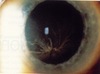
7
Q

A
Amaurosis Fugax from a temporary arterial obstruction
8
Q

A
Talc emboli in IV drug user causing Amaurosis Fugax
9
Q

A
- “Blood and Thunder” retina
- Caused by central retinal vein occlusion
10
Q

A
- Retinal Detachment
- The hazy material below the “V” shape is the detached area of the retina
11
Q

A
- Ischemic optic neuropathy: giant cell arteritis
- Note the swollen disc with indistinct borders
12
Q

A
Papillitis
-Swollen, inflamed disc that’s elevated with indistinct margins
13
Q

A
- Blephritis
- Lid crusting, redness, and loss of lashes
14
Q

A
Ectropion
15
Q

A
Entropion
16
Q

A
Hordeolum (AKA “Stye”)
17
Q

A
Orbital Cellulitis
18
Q

A
Dacryoadenitis
19
Q

A
Dacryocystitis
20
Q

A
Gonococcal Conjunctivitis
21
Q

A
Viral Conjunctivitis
22
Q

A
Allergic Conjunctivitis
23
Q

A
Subconjunctival Hemorrhage
24
Q

A
Episcleritis
25

Pinguecula
26

Pterygium
27

Corneal Abrasion
28

Acid Burn
29

Corneal Abscess
30

Iritis
31

- Primary Open-Angle Glaucoma (POAG)
- Note the large, deep cupping of the optic nerve head
32

Congenital Glaucoma
33

Cataract
34

Congenital Cataract
35

Optic (disc) Atrophy
36

Non-Proliferative Diabetic Retinopathy (NPDR)
37

- Proliferative Diabetic Retinopathy
- Note the new vessels on the retina that are very thin and fine
38

Diabetic Vitreous Hemorrhage
39

AV Nicking from HTN
40

- Hypertensive changes
- Swollen disk, hemorrhages, cotton wool spots
41

Leukemia Retinopathy
42

Sickle Cell Retinopathy
43

Sarcoidosis
44

Sarcoidosis "Candlewax drippings" in sarcoid vasculitis
45

Basal Cell Carcinoma
46

Choroidal melanoma
47

Melanoma of the iris
48

Conjunctival Lymphoma
49

Metastatic Carcinoma of the Choroid
50

Kaposi's Sarcoma
51

-Subcapsular cataract from steroid use
52

Bullseye lesion from Chloroquine
53
Whorl lesion in the corneal epithelium from Amiodarone usage
