Ophthal Flashcards
What screening should take place for patients with a family history of glaucoma?
Annual screening from the age of 40
What are the risk factors for POAG?
Genetics A-C Myopia HTN DM Steroids
What are the features of POAG?
Peripheral visual loss Reduced acuity Disc cupping +-pallor
How would you investigate for POAG?
Automated perimetry Slit lamp with dilatation Tonometry Central corneal thickness measurement Gonioscopy (measures peripheral anterior chamber configuration and depth)
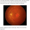
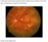
What are the fundoscopic features of ARMD?
Drusen - yellow areas of pigment deposition in and around the macula In wet ARMD, well demarcated red patches may be seen representing haemorrhages
What is the typical presentation of ARMD?
Subacute vision loss with: Reduced acuity Difficulties in dark adaptation Fluctuations day to day Photopsia
What are the key risk factors for ARMD?
Age>75 Smoking FHx HTN DM
What is the first and second line management of allergic conjunctivitis?
1st - topical or systemic antihistamines 2nd - topical mast cell stabilisers e.g. sodium cromoglicate
What is the management of ARMD?
Vit ACE for dry reduce progression VEGF useful in wet Laser photogoaculation is beneficial but risks acute visual loss
Whatis the goal of treatment in pOAG and what drugs are used?
To reduce IOP Beta blockers e.g. timolol PG analogues e.g. latanoprost Sympathomimetics e.g. brimonidine Carb anhydrase inhibitors - Dorzolamide Miotics e.g. pilocarpine
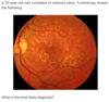
What are the different grades of hypertensive retinopathy, and name the grading system used?
Keith-Wagener classification 1 - Tortous vessels with silver wiring 2 - 1+ AV nipping 3 - 2+ Cotton wool exudates and flame/blot haemorrhages 4- 3+ papilloedema
What is seen on fundoscopy of papilloedema?
Venous engorgement Loss of venous pulsation Blurring of optic disc margin Elevation of optic disc
What are the causes of papilloedema?
SOL Malignant HTN IIH Hydrocephalus Hypercapnia
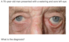
What are the features of an Argyll Robertson pupil?
Small irregular pupils which accomodate but do not respond to light
What is a Hutchinson’s pupil?
Unilaterally dilated pupil which is unresponsive to light. A result of compression of the occulomotor nerve of the same side, by an intracranial mass (e.g. tumour, haematoma)
What are the commonest causes of sudden painless vision loss?
Ischaemic/vascular (amaurosis fugax) Vitreous haemorrhage Retinal detachment Retinal migraine
What are the causes of vitreous haemorrhage?
DM bleeding diatheses Anticoagulation
What might patients with retinal detachment describe?
Dense shadowing that starts peripherally and progresses towards the centre Like a veil or curtein over the field of vision Straight lines may appear curved
What is the management of herpes zoster ophthalmicus?
Oral aciclovir
What are the two aims of treatment of acute glaucoma and how is this achieved?
Reduce aqueous secretion - acetazolomide Induce papillary constriction - topical pilocarpine
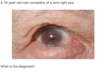
What are the features of acute closed angle glaucoma?
Severe pain Reduced acuity symptoms worse with mydriasis Hard red eye Haloing Non-reactive pupil Corneal oedema N/V +- abdo pain
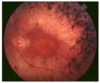
What is seen on fundoscopy of central retinal artery occlusion?
Cherry red macula on a pale retina
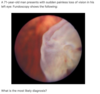
What ist he management of a stye?
Analgesia and hot compress
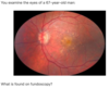
What distinguishes between non-proliferative and proliferative DMR?
Non-proliferative DMR shows microaneurysms, blot haemorrhages, hard exudates, cotton wool spots, venous bleeding et/ Proliferative DMR also shows neovascularisation +-viteous haemorrhage
What conditions are associated with anterior uveitis?
Ank spon Reactive arthritis UC/Crohns Behcets Sarcoid
What is the management of anterior uveitis?
Urgent review Cycloplegics - dilate the pupil for pain relief (e.g. Atropine) Steroid drops
What are the classical features of ret pig?
Night blindness initially Tunnel vision
What is the strongest risk factor for subcapsular cataracts?
Steroids
What are the risk factors for cataracts?
Smoking EtOH Trauma DM Steroids Radiation Myotonic dystrophy Hypocalcaemia
Flashers and floaters indicate?
Vitreous/retinal detachment
Amaurosis fugax is transient or permanent?
Transient
What is the most common cause of vitreous haemorrhage?
Proliferative DMR
What is the commonest cause of persistent watery eye in an infant, andhow is it managed?
Nasolacrimal duct obstruction manabed by teaching lacrimal duct massage - 95% of cases will resolve by 1 year
What are the side effects of prostaglandin inhibitors such as Latanoprost?
Increased eyelash length Iris pigmentation Periocular pigmentation
What investigations are used in ARMD?
Slit lamp microscopy initially Fluorsecein angiography if neovascular disease is suspected Ocular coherence tomography if needed
True or false - optic neuritis is associated with Marcus Gunn pupil?
True
What should be done for babies with a purulent eye dischagre?
Urgent swabs looking for chlamydia and gonogoccus
What are some complications of zoster ophthalmicus
Ant uveitis (commonest) Conjunctivitis Keratitis Episcleritis Ptosis Post herpetic neuralgia
Myopia and hypermetropia relevance to glaucoma?
Acute closed angle glaucoma is assocaited with hypermetropia Primary open angle glaucoma is associated with myopia
Does hyper or hypocalcaemia predispose to cataracts?
Hypocalcaemia
What is the strongest predictor of ocular involvement in zoster ophthlalmicus?
Hytchinson’s sign
When would you see a dendritic ulcer on fluorescein eye staining?
Herpes simplex keratitis
A 79-year-old gentleman presents with a 3 months history of a red swollen left upper eyelid. He remembers initially developing a bump on the eyelid which was uncomfortable but then got bigger forming a hard lump. He reports no pain currently and has not noted any problems with his vision and the eye itself appears healthy.
Chalazion/Meibomian cyst, which occurs after the progression of an internal stye (hordeolum intermum)
What is the classical presentation of optic neuritis?
Vision loss Eye pain ‘behind the eye’ Red desaturation
What type of scan would you do to assess the posterior spread of infection in orbital cellulitis?
Contrast CT of the orbits, sinuses and brain
What is a contraindication to vitamin/antioxidant therapy for ARMD?
Smoking - because beta carotene increases risk of lung cancer
Bilateral grittiness worse first thing in the morning indicates?
Blepharitis
What are the classical features of central retinal vein occlusion?
Sudden painless loss of vision with severe retinal haemorrhaging on fundoscopy
What should be done for young children with a squint?
Ophthal referral
Which medication causes brown pigmentation of the iris?
Latanoprost
What is the first line glaucoma treatment for patients with heart block?
Latanoprost
What drug causes pupillary copnstriction, blurred vision and headahces?
Pilocarpine
What conditions cause tunnel vision?
Papilloedema Glaucoma Ret Pig Choroidretinitis Optic atrophy
What type of eyedrop might cause a corneal ulceration?>
Steroids - through fungal infection
What are the features of episcleritis?
Red eye classically not painful Watering +- mild photophobia Phenylephrine induced blanching of the vessels (not the case in scleritis)
How does latanoprost work in the management of POAG?
Increases uveoscleral outflow
Stormy sunset appearance?
Central retinal vein occlusion
What clinical feature differentiates scleritis from episcleritis?
Pain only seen in scleritis
What clinical test may be used to detect a strabismus?
orneal light reflection test, where a light source is held 30cm from the child’s face to assess reflection symmetry on the pupils
What is the MOA of dorzolamide?
Carbonic anhydrase inhibitor
What type of visual impairment does POAG cause?
Peripheral visual field impairment
Long hx of diabetes in an elderly patient presenting with unilateral blurring and halos is strongly indicative of?
Cataracts

Treated diabetic retinopathy

Age related macular disease

Papilloedema

Age related macular degeneration

Retinal detachment

Central retinal artery occlusion

Proliferative retinopathy

Retinitis pigmentosa

Treated diabetic retinopathy

Entropion

Ectropion

Retinal detachment

Age related macular degeneration

Central retinal vein occlusion