Observing microorganisms through a microscope Flashcards
Acid fast staining of a patient’s sputum is a rapid reliable, and inexpensive method to diagnose tuberculosis. What color would bacterial cells appear if the patient has TB?

The bacteria will appear fuschia
what genera of bacteria require acid-fast staining?
- Mycobacterium
- Nocardia
How large are protozoa?
100 microns
how big are yeasts?
8 microns
What is the size range of bacteria?
0.2-1 microns wide and 2-8 microns long (some are much longer than wide)
Clymidia and mycoplasmas are 0.25 microns they are at the __________ of the resolution of a _____________ microscope.
- lower limit
- light
how big are viruses?
20-250 nm
What kind of flagellar arrangement does this bacteria have?

- monotrichous
what are microscopes used for?
to magnify small objects
why does size of a specimen matter when choosing which type of microscopy to use?
because different microscopes have different resolution ranges, the size of a microscope determines what type of microscopy is most effective in veiwing the microbe.
As light wave length decreases what happens?
resolution increases

A. Ocular lenses
B. Objective lenses
C. Condenser
D. Diaphragm
E. Illuminator
F. Coarse focusing knob
G. Fine focusing knob
Magnification
How much all the lenses magnify the specimen
(TM = Ocular x Objective)
Resolution
- The ability of lense to distinguish objects; resolving power
- 0.2 microns is the resolving power of a light microscope
Contrast
Stains change refractive index; an increase in contrast between bacteria and surrounding media allows for better visability
Refractive index
- how light appears as it passes through an object
- measures the light bending ability of a medium
why are filters used on light microscopes?
to increase resolution by decreasing the wave length of the light passing through the the side.
What does light do as it passes through substances?
it bends
what does air do to light?
it bends it
when is immersion oil used?
with a 100x objective lense or higher
why is oil used with an oil lens?
- to lessen the amount of light lost by bending in air; keep light from bending
- No oil; resolving power will be deminished greatly
- Light may bend so much that it misses the small high magnification lens
Brightfield microscopy
- simplest of all the optical microscopy illumination techniques
- Dark objects are viasble against a bright background
- light is reflected off specimen does not enter objective lenses

Darkfield microscopy
- Light objects visable against dark background
- used to enhance the contrast in unstained samples
- instrument of choice for spirochetes
- only light reflected off specimen enters the objective lense

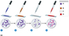
Fluorescence Microscopy
- uses UV light; energizes fluorochromes
- Flourescent substancesabsorb UV light and emit visable light
- Cells may be stained with fluorescent chemicals called fluorochromes
- immunofluorescence; fluorochromes only stain object you want to be stained, antibodies with fluorochromes attached attach to antigens on bacteria.

How immunofluorescence works:
- Fluorochromes are attached to antibodies
- the antibodies attach to the antigens on bacteria
Electron microscopy allows
detailed images of cell parts
What is used in electron microscopy that differs from light microscopy?
- heavy metal salts
- electrons; beams of electrons, not light
- electromagnetic lenses
- fluorescent screens
why does an electron microscope have a better resolution power than a light microscope?
- electron wave length is 100,000 times smaller than visable light wave length
what are the 2 types of electron microscopes?
- scanning electron microscope SEM
- transmission electron microscope TEM
What are SEMs for
to observe surface structures
What are TEMs for?
to observe internal structures
what kind of microscope was used to capture this image?

- TEM; transmission electron microscope
- allows internal structures to be seen
What kind of microscope was used to obtain this image?

- SEM; scanning electron microscope
- allows surface structures to be observed
- Scanning microscopes also give a sense of depth in images
Why are staining techniques used in light microscopy?
- staining provides contrast between a specimen and it’s background
how do you prepare a slide to be stained?
- smear the sample
- Let it dry
- Heat fix it (to cause bacteria to stick to the slide and also kill them)
What is the charge inside a bacterial cell?
negative
what do positively charged dyes do?
the enter the negatively charged bacteria
Basic dye
- Cationic chromophore
- enters the cell; because opposite charges attract.
- Basic dye is a stain that is cationic (+ve charged) and so will react with material that is (-ve) negatively charged. This dye is usually synthetic, that act as bases, and which are actually aniline dyes. Their color base is not water soluble but can be made so by converting the base into a salt.
Acidic dye
- Anionic chromophore
- stays out of the cell, staining the background because like charges repel.
- An acid dye is a dye which is a salt of a sulfuric, carboxylic or phenolic organic acid. The salts are often sodium or ammonium salts. Acid dyesare typically soluble in water and possesses affinity for amphoteric fibers while lacking direct dyes’ affinity for cellulose fibers.
What are the 3 types of staining techniques?
- Simple
- Differential
- Special
Simple stains
- Use a single basic dye
- A mordant may be used to hold the stain or coat the specimen to enlarge it
mordant
a substance, typically an inorganic oxide, that combines with a dye or stain and thereby fixes it in a material.
Differential stains
- react differently with different bacteria
- ex. Gram stain and acid-fast stain
What type of staining technique is this?

Simple stain technique
What type of staining technique is this?

negative stain technique; simple stain using acidic dye
What type of staining technique is this?

Differential staining technique; gram stain uses more than one dye.
what allows Crystal violet iodine percipitate to stay in gram positive cell walls?
The gram positive bacterium’s thick peptidoglycan cell wall.
Acid-Fast bacteria and acid fast stain
- bacterial cells that retain a basic dye in the prresence of acid-alcohol are called acid-fast.
- Non-acid fast cells lose primary stain when rinsed with acid-alcohol, and are counterstained with a different color basic stain.
- used for bacteria containing mycolic acid in their cell walls; mycobacterium and nocardia
When an acid fast stain is preformed what will happen to the cells that do not contain mycolic acid in their cell walls?
cells which don’t have mycolic acid will hold on to methylene blue stain

A. Crystal violet
B. Iodine
C. Alcohol
D. Safranin
- CV is applied
- Iodine (mordant is applied) percipitate CV-I crystals are formed
- Alcohol wash washes CV-I out of Gram negative bacteria (decolorization)
- Safranin (counterstain) is then applied to stain gram negative bacteria.
In the medical field why is it important to know if a bacterium is gram +,- or acidfast?
- its important to know in order to use the correct antibiotics to treat the infection caused by the microbes.
What are some types of special stains?
- Endospore stain
- Flagella staining
- Capsule stain
Endospore staining
- Heat is required to drive stain into the endospore
- Differential stain
- uses basic dyes

Flagella staining
- enlarges the flagella
- requires a mordant to make the flagella wide

Capsule staining
- Stain uses both basic stain and negative (acidic stain)
- shows bacterial capsules
