Neuroradiology Flashcards
Computed Tomography
–Ionizing radiation (x-rays)
–Measuresattenuation of radiation by tissues
–Revolutionized the practice of modern medicine
What are the risk of a CT scan?
- Cancer induction
- Contrast nephropathy
What are the color densities of CT scan?
–CSF
•Dark
–Grey matter
•Light grey
–White matter
•Dark grey
–Bone (Skull)
Bright (white)
Density of blood depends on age. What is the difference between acute bleed and chronic bleed?
Acute Bleed - Bright
Chronic - Dark
Epidural hematoma: acute bleed (Left picture)
Subdural hematoma: Chronic bleed (right picture)
Intravenous Contrast is used for CT scans. It is Iodinated, causes high attenuation of x-rays. How does it appear?
Appears bright
What Intravenous Contrast not used for?
Not used for head trauma
- Acute hemorrhage and contrast both appear bright
- Want to avoid confusing one for the other
What is Intravenous Contrast helpful for?
Intracranial infection or tumor
- Inflammation from infection and tumor disrupts the blood-brain barrier
- Contrastenters into diseased areas of brain and causes them to become brighter and easier to detect
What is the difference between these two images?
Images of Abscess
Left image = Without contrast
Right image - With contrast
CT scan
This is an image of?
Glioblastoma Multiforme
CT scan w/ contrast
Magnetic Resonance Imaging
–Uses large magnetic field
–No known increased cancer risk
–Complex physics; measures radiofrequency signal from protons
What are the imaging sequences of an MRI?
–T1weighted
•Fatis bright, water is dark (fat has lots of H1)
–T2weighted
•Wateris bright (think H2O)
–Diffusion weighted imaging (DWI)
•Acute stroke is bright
–Susceptibility weighted imaging (SWI)
•Blood is very dark
What are the strengths of T1 compared T2?
–T1weighted
- Anatomy
- Bone marrowpathology (such as tumor)
–T2 weighted
- Pathology
- Edema (increased water content) - bright
Infection, tumor, inflammation (good for detecting this)
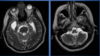
Which image was done with T1 weighed and which one is done with T2 weighed?
Left image = T1 (Cerebrospinal fluid is dark)
Right image = T2 (Cerbrospinal fluid is bright)
What type of MRI was this image obtained from?
T1
Bright fatty bone marrow of the skull
How does blood appear on MRI?
– Appearance depends on ageand composition of blood products
–SWI sequence
•Very dark in most stages of blood
IV contrast w/ MRI
•Intravenous contrast
–Gadolinium; large radiofrequency signals
–Appears very bright
Using IV contrast w/ MRI is helpful for?
–intracranial infection or tumor
- Inflammation from infection and tumor disrupts the blood-brain barrier
- Contrastenters into diseased areas of brain and causes them to become brighter and easier to detect
This radiographic test was used to obtain this image?
MRI w/ contrast
Image of Abscess
This is an image of ?
Glioblastoma Multiforme
Done with MRI w/ contrast
CT vs MRI
Which is which?
LEft
CT: Bone bright, scalp fat dark
Right
MRI: Bone bright + dark, scalp fat bright
What are the two different type of stroke a person could have?
•Hemorrhagic stroke
–Bleeding in the brain
–Usually due to hypertension
•Ischemic stroke
–Lack of blood flow to the brain
–Usually due to clot
Acute Ischemic Stroke
Timing?
Physiology?
•Timing
–Less than 2 weeks old (including acute and subacutestages)
•Physiology
–Cytotoxic edema
•Increased intracellular fluid
Decreased extracellular fluid
On CT scan, How does Acute Ischemic Stroke appear?
–Increased water in brain tissue
–Affected tissue becomes darker with time
–Very early stroke is difficult to detect
If 6 past after an Acute Ischemic Stroke, how does that affect the CT scan?
– At 6 hours, 60% of patients will have CT abnormality
–At 24 hours, all patient will have CT abnormality
Image of CT scan
Acute Left middle Cerebral Artery territory Ischemic Stroke
Image of CT scan
Acute Brainstem Ischemic Stroke
How does an Acute Ischemic Stroke appear on MRI? and what technique is used?
•Diffusion weighted imaging (DWI)
–Acute ischemic stroke is bright
What radiological testing is excellent for detecting very early ischemic strokes?
Diffusion weighted imaging (DWI)
Catches stroke between 10-60 min
Image of DWI Sequence
Left Image: Acute Right Cerebellar Ischemic Stroke
Right Image: Acute left middle cerebral artery ischemic stroke
What causes the bright signal in a diffucion weighed image?
- Brain cells swell, taking in extracellular water
- Restricted motion of free water outside cells causes the bright signal
This is an image of a Acute Ischemic Stroke done by T2 sequence MRI. WHat is the radiological test detecting?
It detects increased water in the brain, which appears as a bright signal.
How long does it take for increased water in brain tissue to appear after an ischemic stroke?
3 hours
(image of Acute Stroke MRI T2)
What is a Hemorrhagic transformation?
–Usually occurs 2-5 days after ischemic stroke onset
–Related to reperfusion of infarcted brain
–Occasionally spontaneous
Treatment with thrombolytic drugs is the most common cause of what?
Hemorrhagic transformation
Image of Hemorrhagic Transformation
Left image: Acute left middle cerebral artery territory stroke
Right image: Acute hemorrhage in infarcted tissue
Chronic Ischemic Stroke
Timing?
Physiology?
•Timing
–Greater than 2 weeks old
•Physiology
–Edema resolved
-Dead tissue is removed and is replaced by fluid
How does a Chronic Ischemic Stroke appear on CT scan compaired to MRI?
CT: it is very dark (near water) density
MRI: Very bright (near water) signal of T2 sequence
These are images of Chronic Ischemic Strokes (Left image = Old right middle cerebral artery stroke and Right= Old right basal ganglia stroke). What imaging test was used?
Note that the fluid is dark
CT scan
This is an image of a Chronic Stroke (Left = Old left occipital stroke and right= Old left middle cerebral artery stroke). What imaging study was used?
MRI (T2)
Image of Stroke Territories
These are images of what type of test?
CT angiogram on the left and CT Venogram on the right
What is occuring in these images and what test is being used?
Left image: Noncontrast CT sca
Bright clot in left middle cerebral artery
CT angiogram
Left middle cerebral artery occlusion
What is occuring in these images and what test are being used?
Left: CT angiogram
Left middle cerebral artery occlusion
Right: DWI (bright signal)
left middle cerebral artery territory
What is occuring in these images and what test are being used?
Left: CT scan
Acute Left middle cerebral artery territory stroke
Right Cerebral Blood Flow map (CT perfusion imaging)
Decreased flow to left middle cerebral artery territory
What is occuring in these images and what test are being used?
Left: CT angiogram
Occlusion of basilar artery
Right: CT scan
Acute brainstem stroke
This is an image of what type of test?
MR Angiogram
What type of damge occured in this image?
Left middle cerebral artery occlusion
What are the two types of Hydrocephalus
Communicating and Non-communicating
What is Communicating Hydrocephalus?
–All ventricles enlarged
–Usually caused by hemorrhage or infection
Whats is non-communicating hydrocephalus?
–Not all ventricles enlarged
–Structural CSF flow obstruction
–Causes include brain mass or developmental anomaly
The right image is a picture of a normal head. What is occuring in the left image? What test was used to capture this image?
Communicating Hydrocephalus
MRI: Sagittal T1
Decreased CSF absorption by arachnoid granulations – blood from subarachnoid hemorrhage, or meningitis scarring)
Normal pressure hydrocephalus – wet, wobbly, and wacky.
This is an image of what?
Communicating Hyrdocephalus
MRI: Axial T1
Looks like a Rosarch Test
This is an image of what?
Communicating Hydrocephalus
MRI: Axial T2
This is an image of?
Non-communicating Hydrocephalus
This is an image of? Also, what is the arrow pointing to?
Non-communicating Hydrocephalus
It is pointing to a large mass in posterior fossa
Fat appears bright on T1 weighed. What is it good for?
Good for detecting bone marrow pathology
DWI (MRI)
–Acute stroke is bright
–Great for detecting very early acute infarct (within 10 to 60 minutes)
SWI (MRI)
•Susceptabilityweighted
–Blood is very dark
–Very good for detecting hemorrhage
Chronic Stroke taken by MRI T2
CT angiogram
