Cranial Nerves Flashcards
What are the 3 major cranial nerves serving special sense organ?
I- olfactory
II- Optic
VIII - Vestibulocochlear
They develop in part from epidermal neurogenic placodes
are not parts of other cranial sensory-motor nerve
What are the 4 somatic motor nerves that move the eye and tongue?
III - Occulomotor (exits from vental midbrain)
IV - Trochlear (exits dorsally and decussates i.e crosses)
VI - Abducens (emerges caudal to VII)
XII - Hypoglossal (work close with C1)
- have no affiliated sensory ganglia, thus they have only efferent components (innervated striated skeletal m.)
- sensory fibers in the peripheral parts of these nerves are “hitch-hikers” from nearby mixed nerves
- are more (XII, VI, III) or less (IV) comparable to ventral roots of spinal nerves
What are the 4 mixed branchial nerves that move and sense jaws, face, pharynx, and larynx?
V - trigeminal (no taste fibers)
VII - Facial
IX - Glossopharyngeal
X - Vagus
Pharyngeal Arch Nerves
One or more sensory ganglia
This nerve is the only special motor nerve that is cranial only in a roundabout way and innervates the sternocleidomastoid and trapezius muscle
XI - Spinal Accessory
Motor nuclei in CNS
What is the Otic Placode?
(located around medulla)
produces auditory and vestibular sensory hair cells and ganglion cells (receive input and send axons centrally as nerve)
CT for inner ear
Involves Cranial nerve VIII
Placodes are regions of the surface ectoderm
What is the Optic Placode?
Produces Lens
Involves CN II (brain tract)
(not neurogenic)
What is the olfactory placode?
Central Projecting sensory cells
(produces olfactory sensory neurons (also known as the ganglion cells))
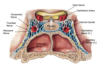
This structure is located at the diencephalon wall. What is it and what does it contain?
This is the Optic Vesicle
contain sensory cells and ganglion cells
What does CN I (Olfactory Nerve) consist of?
consists of the central-projecting axons of bipolar olfactory sensory neurons (= olfactory receptor cells) situated in the olfactory epithelium.
The axons collect as fine bundles that traverse the olfactory foramina in the cribriform plate of the ethmoid bone to reach the olfactory bulbs where they synapse on second order olfactory neurons.
Chemosensory
Receptors for Odarent molecules
Easily disrupted
This “Cranial Nerve” is actually a CNS tract, not peripheral nerve and covered by meningies. Located in the nasal cavity?
CN I (Olfactory)
It is covered with dura, arachnoids, and pia mater
The nerve is just the axons coming through cribiform plate. Bulb and tract are part of the brain
Patient comes in reporting that he has difficulty smelling after doing some drugs. What is one possible diagnosis that can be considered?
Anosmia (loss of sense of smell)
This occurs when there is Damage to the olfactory sensory epithelium or disruption of the fibers at the cribiformplate
To exam the function of CN I you will expose the patient to test odorants
Head injury or frontal lobe surgery can lead to damage
What is the path of the Optic nerve (CN II)
begin at the retina and extend to the optic chiasm (outside orbit), where approximately half of the optic fibers cross to the other side. The fibers continue as the optic tracts and most of them terminate by synapsing in the lateral geniculate nuclei (LGN is a big nucleus of the thalamus) of the thalamus.
What is different about the Optic nerve compared to some other cranial nerves?
The retina and optic nerves arise from the walls of the embryonic diencephalon. Accordingly, the optic nerves are brain tracts, not peripheral nerves.
Most of the axons that make up the optic nerve come from where?
axons of retinal ganglion cells.
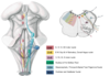
This nerve is actually made of two nerves. It enters the brain at the pontomedullary junction, just dorsal to the root of the VIIth nerve.
CN: VIII
(Vestibulocochlear Nerve)
The VIIIth nerve fibers pass peripherally to into the _____, accompanied by the VIIth nerve, via the __________.
Temporal bone (Petrous Part)
Internal Acoustic meatus
What does the cochlear nerve (portion of CN VIII) comprise of?
afferent axonal processes of cells in the spiral ganglion which innervate the auditory hair cells (mechanosensory) of the spiral organ (Organ of Corti - sensory hair cells + galnglion)
as well as,
efferent axons of central neurons located in the superior olivary/periolivary nuclei that form the olivocochlear bundle (modulate sensitivity - cochlear amplifier)
The Vestibular nerve (part of CN VIII) comprises of?
afferent axonal processes of cells in the vestibular ganglion (Scarpa’s) which innervate:
vestibular hair cells of the ampullae of the semicircular canals (3), sensing angular acceleration
vestibular hair cells of the maculae of the saccular and utricular otoliths, sensing linear acceleration
as well as,
efferent axons of central neurons located in two vestibular efferent nuclei
Where do the afferent fibers of the cochlear nerve project?
project to the lowest order brainstem auditory centers, the dorsal and ventral cochlear nuclei
The vestibular nerve afferent fibers project to where?
to the vestibular nuclei (superior, medial, lateral and descending) and to the vestibulocerebellum
The vestibular nuclei connect to what other structures besides the Vestibular nerve afferent fibers?
connect with brainstem, spinal and cerebellar centers involved in postural and gaze control (among others)
MLF and Vestibulospinal tracts are what?
Major central vestibular pathways
They lead to extrocular and cervical motor nuclie controlling eye and head movements
How do you test Vestibular function?
Routine auditory test (done with a tuning fork)
or
asking the patient to walk a straight line (balance)
or
observing eye movements during introduction of cool or warm water into the external ear canal.
What is the Caloric Test?
Observing eye movement during introduction of cool/warm water.
induces alternating eye movements (Nystagmus) by generating convection in the endolymph of the horizontal semicircular canal. The caloric vestibulo-ocular reflex is especially useful in testing brainstem function in comatose patients.
What is a complication of the CN III?
This is the oculomotor nerve and it also has parasympathetic components - to smooth muscle of the eye
What are the Embryonic Origins of Striated Cranial Muscles Innervated by Somatomotor Cranial Nerves III, IV, VI and XII?
- Dorsal head mesoderm (pre-otic mesoderm) in front of the ear (otic vesicle) produces the extraocular muscles, innervated by cranial nerves III, IV and VI
- Occipital somites 1-4 give rise to a migratory stream of myocytes that travel ventrally into the floor of the oral-pharyngeal region to produce the tongue muscles innervated by CN XII.
Which ganglion does CN III innervate?
parasympathetic ciliary ganglion.
Side note: Innervates most of the striated and smooth muscles of the eye, thus has both somatic and visceral (parasympathetic) motor fibers
The parasympathetic ciliary ganglion and the occular smooth muscles innervated by CN III are derived from what?
Neural crest
The CN III roots emerge from where?
emerge from the ventral midbrain, on either side of the posterior perforated substance, and between the mamillary bodies and the pontine fibers
Edinger-Westphal Nucleus* (GVE)
Preganglionic parasympathetic neurons in the Edinger-Westphal Nucleus innervate post-ganglionic neurons in the ciliary ganglion (located in orbit) where they synapse. Ciliary ganglia send axons into the front of the eye
Oculomotor Nucleus (GSE)
Motor neurons innervate:
4 striated muscles that move the eye
- superior rectus
- inferior rectus
- medial rectus
- inferior oblique
1 striated muscle that elevates the upper eyelid
-levator palpebrae superioris
Ciliary ganglion neurons innervate 2 smooth muscles in the eye?
- the pupillary sphincter muscle which constricts the pupil (red)
- the ciliary muscle (active when you look at things close) which regulates the curvature of the lens (blue) - recieve 95% of neurons in ciliary ganglio.
What are the characteristics signs of a unilateral occulomotor nerve palsy?
“down out syndrome”
Ptosis (drooping) of the upper eyelid, due to lack of action of the levator palpebrae superioris
Dilation of the pupil due to loss of sphincter pupillae function
Depressed and abducted resting eye position due to the IVth and VIth nerve eye muscles acting unopposed
CN IV (Trochlear) innervates only what?
ONLY the superior oblique muscle
(contains somatic motor fibers)
The trochlear nerve received that name because the superior oblique muscle passes through a pulley (L., trochlea) before inserting on the globe.
What is the direction of the Trochlear nerve roots?
roots exit dorsally at the midbrain/hindbrain junction and cross to the opposite side (right to left, left to right)
Where is the trochlear nuclei located?
trochlear nuclei are located near the midline beneath the most superior part of the fourth ventricle
(green arrow)
*look at picture*
What are the two main actions of the superior oblique muscle?
Depresses and medially rotate (in-tort) the eye
Trochlear Nerve palsy
Most notable effect of loss of SO muscle function due to trochlear nerve palsy is a persistent laterally rotated eye posiition (ex-torsion)
The laterally rotated eye position may be compensated for by tilting the head away from the direction of the non-functional SO mucle.
This nerve only innervates the lateri rectus muscle, which Abducts the eye
CN VI (Abducent nerve)
Contains only somatic sensory fibers
Where does the root of CN VI emerge from?
emerge near ventral midline at ponto-medullary junction
Loss of function of the abducens nerve leads to what?
It causes the eye to remain adducted during forward gaze (i.e. inability to abduct)
If this occurs bilaterally = crossed eye
What exactly has many oppourtunities to disrupt Cranial Nerve II, III, IV, and VI?
The Cavernous Sinus
Because of the close proximity of the pituitary and internal carotid to the optic chiasma and the nerves to extraocular muscles (III, IV, VI), visual and eye movement deficits are often involved in cases of pituitary tumors, internal carotid anurysmsor thrombosis in the cavernous sinus.
What is Cranial nerve XII
This is the hypoglossal nerver
Somatomotor innervation of the tongue muscles
This nuclei is located in the medulla oblangata, just below the ventricle
Hypoglossal motor nucleus
What is the direction of the Hypoglossal nerve?
The nerve exits the skull via the hypoglossal foramen, between the jugular foramen and the occipital condyle
The rootlets exit the brain over a long distance along the groove separating the pyramids and inferior olive. They are followed by the rootlets of C1
The motor fibers of C1 that travel with CN XII enter where together?
the ansa cervicalis (blue)
Where they innervate the thyrohyoid (red) and geniohyoid (green) muscles
What are the muscle that are innervated by the hypoglossal nerve?
Genioglossus m.
Hyoglossus m.
Styloglossus m.
Chondroglossus m.
intrinsic tongue muscles
(but not the palatoglossus m., by CN X)
This test is used for?
Patient protrudes the tongue, press left and right on the cheek against the tester’s finger pressure and make fast movements by saying “la la la”
To test the hypoglossal nerve
When is unilateral hypoglossal weakness more evident?
During tongue protrusion
the tongue deviates to the weak side
Branchiomotor cranial nerves include
V, VII, IX, X, XI
Mixed nerves each with one or more sensory ganglia
What is the embryonic origin of CN V, VII, IX, and X?
- 1st arch mesoderm: muscles of mastication, anterior digastric and 2 tensors, innervated by mandibular division of N. V
- 2nd arch mesoderm: the muscles of facial expression and the more ancient posterior digastric and stylohyoid muscles, innervated by N. VII
- 3rd and 4th arch mesoderm: stylopharyngeus, Lev. Palati, pharyngeal constrictors and other muscles innervated by N. IX and X
Which nerve is known as the grand mover of the jaw and sensor of the face?
FACIAL NERVE
What are the 3 divisions of the facial nerve?
Opthalmic V1 (senesory)
Maxillary -V2 (sensory)
Mandibular V3 (sensory and motor)
Where do the roots of the trigeminal nerve emerge from?
Exits lateral surface of pons by passing through the middle cerebellar peduncle
Motor root is largely separate and joins mandibular division
Trigeminal Senory Ganglion
(semilunar of Gasserian)
Is plastered onto the floor of the middle cranial fossa by dura
(MASSIVE)
- The ophthalmic division (V1 ) passes through the superior orbital fissure into the orbit, accompanied by cranial nerves III, IV and VI
- The maxillary division (V2) passes through the foramen rotundum into the pterygopalatine fossa
- The mandibular division (V3) passes through the foramen ovale into the infratemporal fossa
What is the funciton of the Sensory fibers for CN V?
Convey touch, pain, and temperature from skin on face and the cranial meningies
Lining and content of the major cranial cavities
V1: orbit & upper 1/2 of nasal cavity
V2: lower 1/2 of nasal cavity upper 1/2 of oral cavity
V3: lower 1/2 of oral cavity
Trigeminal motor nucleus
a large branchiomotor nucleus in the rostral pons innervating the following 1st arch muscles:
temporalis, masseter, medial & lateral pterygoids, anterior digastric, mylohyoid, tensor tympani and tensor veli palatini.
Trigeminal Sensory nuclei
Principle Trigeminal Sensory Nucleus
-in rostral pons, mainly serving touch
Nucleus of the Trigeminal Spinal Tract
- extends through caudal pons & medulla
- mainly serving temperature and pain
Mesencephalic Trigeminal Nucleus
- centrally located, peripherally projecting sensory neurons! Very Unique!
- in caudal midbrain & rostral pons
- mainly serving proprioception from muscles of mastication and teeth
Trigeminal nerve is involved in which reflex?
The jaw jerk reflex (involve Vmes and Vmot)
Corneal Relfex
How is the Jaw jerk reflex tested?
by a gentle hammer tap which activates proprioceptors in the jaw muscles
What is the corneal reflex?
involving sensory fibers of V1, relay neurons in the main V sensory nucleus and facial nerve motoneurons innervating the orbicularis oculi is tested by a gentle touch to the cornea
Hitchhiking PS Pathways
(CN V)
Although the Trigeminal Nerve has no visceral motor components, that is, no preganglionic nucleus or fibers…..
…..most of the post ganglionic parasympathetic fibers in the head travel along branches of the Trigeminal Nerve
This nerve is related to 1st pharyngeal pouch and Hyoid arch (2nd)
Facial Nerve (CN VII)
Intamately involved in the middle ear
What is the direction of the facial nerve?
- roots emerge at pontomedullary junction
- comprises a larger motor root and a smaller nervus intermedius which carries sensory and visceromotor fibers
- enters temporal bone at internal acoustic meatus
What are the functional efferent components of the Facial nerve?
Efferent
branchiomotor to hyoid arch muscles (SVE)
- muscles of facial expression, stylohyoid, posterior digastic, stapedius (Central Nucleus - VII motor nucleus )
Secretomotor (parasympathetic) to ptergopalatine and submandibular ganglia (SVE) (Central Nucleus - Suprior Salivatory nucleus)
What are the functions Afferent components of the facial nerve?
Geniculate Ganglion in temporal bone
- major gustatory component; innervates taste buds on palate and anterior 2/3 of tongue (SVA) (Project to Nuc Sal Tract)
- smaller touch and pain component (GSA) (Projects to Nuc Vsp Tract)
What are the 3 major nerves that arise from the temporal bone?
Facial nerve
Greater petrosal nerve
Chorda Tympani
What is the function of the facial nerve?
Mainly motor to muscles of facial expression, stylohyoid & posterior digastric
(exites stylomastoid foramen and give off branches to the side of the face and a small branch to the stapedius muscle)
What is the function of the greater petrosal?
PS: pterygopalatine ganglion to lacrimal gland and small mucous glands of palate and nasopharynx
Distributed on branches of V2 & V1
What is the function of the chorda tympani?
Joins the lingual branch of V3
Taste from anterior 2/3 of tongue
PS: submandibular ganglion to submand. & sublingual glands
(goes to lingual and submandibular region - gustatory and PS)
How do you test CN VII?
Sweet , sour, salty, and funny faces
What is the relationship of structures that transverse the jugular foramen?
CN IX, CN X, and CN XI are in numerical order, anterior to the internal jugular vein as they transverse the foramen. They are immediately posterior to the internal carotid artery as they emerge from it. The superior and inferior sensory ganglia of CN IX and CN X are see as thickning of those nerves immediately inferior to thei exit from the cranium
Which nerve is a Branchial nerve related to 2nd pharyngeal pouch (tonsilarfossa between stylohyoid ligament and middle constrictor)and 3rd Arch (think about the greater horn of the hyoid bone)?
CN IX - Glossopharyngeal
What is the direction of CN IX?
- roots emerge at rostral medulla, dorsal to anterior end of inferior olive and in line with adjacent vagusroots
- then they enters jugular foramen with Xth and XIth nerves
- gives off tympanic nerve into the temporal bone, which then emerges from the bone as the lesser petrosal nerve
- The nerve exits jugular foramen at base of skull
What are the funtional efferent components of the Glossophayngeal nerve?
–branchiomotor to 3rd arch muscle (stylopharyngeus)(SVE)
(Central Nucleus-rostral pole of nucleus ambiguus(SVE))
-secretomotor control of parotid gland by parasympathetic (GVE) fibers to oticganglion(GVE) via lesser petrosal nerve
(Central Nucleus -Inferior Salivatory nucleus (GVE))
What are the afferent components of the Glossopharyngeal nerve?
- superior and inferior ganglia
- taste from posterior 1/3 of tongue (SVA) and sensory from carotid body and sinus (GVA)
(Project to - Nuc Sol Tract (SVA and GVA))
- touch and pain from middle ear, tonsils, back of soft palate, posterior 1/3 of tingue and pharynx
(Project to - Nuc Vsp Tract (GSA))
Which nerve is know as the Wanderer?
CN X
VAGUS NERVE
What are the afferent components of CNX?
- superior(jugular) and inferior (nodose) ganglia
- Major sensory inputs from cervical, thoracic and abdominal viscera (GVA) and minor taste input from epiglottic region (SVA)
(Nuc Sol Tract (GVA & SVA))
-touch and pain from ear canal, auricle & meninges (GSA)
(Nuc Vsp Tract (GSA))
What are the efferent components of CN X?
-motor to 4th-6tharch muscles (SVE= branchiomotor) and more properly, motor to striated muscles of pharynx, larynx and soft palate
(nucleus ambiguus (SVE, mostly))
-Preganglionic parasympathetic (GVE) to all visceral structures from neck to upper hindgut
(dorsal vagal motor nucleus (GVE, mostly))
Describe the Vagus Nerve Pharyngeal distribution. What are the different branches motor and sensory?
- Superior auricular branch - sensory to back of ear
- Meningeal branch - sensory to dura in posterior cranial fossa
- Pharyngeal branch - motot to palatal muscles and pharyngeal constrictors and sensory from epiglottis, upper pharynx and possible aspects of soft palate
- Superior laryngeal branch - sensory from upper part of larynx and motor to cricothyroid m.
- Recurrent laryngeal branch - motor to all other laryngeal mm and to constricto and esophagous + sensory from lower larynx, upp esophagous
Describe the vagus distribution to thoracic and abdominal viscera
- Visceromotor fibers (GVE) from the Dorsal Motor Nucleus of the Vagus target parasympathetic ganglia in the heart, lungs, kidneys and abdominal viscera, as far down as the beginning of the descending colon.
- Afferent fibers (GVA) from the Vagal Sensory Ganglia (especially the inferior) bring information back to the brain from all of the organs that receive vagal efferents.
These Vagal visceral sensory fibers (GVA) terminate in the Nucleus of the Solitary Tract in the medulla.
Parasympathetic regulation the heart, lungs and all derivatives of the embryonic fore- and hind-gut, as well as the kidneys are recieved from which nerve?
Vagus Nerve
How is Cardiovascular function controlled? Which nerves are involved?
This control is Autonomic
Sensory signals pertaining to arterial pressure (CN IX) and blood oxygenation (CNX) are relayed to the nucleus of the solitary tract, which in turn organizes autonomic outflow via interneurons in the medullary reticular formation. This circuit provides negative feedback to visceral motor neurons that govern cardiovascular function.
LOOK AT PICTURE FOR MORE UNDERSTANDING
How do you test the function of CN IX?
Say AHHHHH
The gag reflex (IX), palatal elevation and vocal cord position during phonation can be readily tested (Gental touch to the pharyngeal wall)
Problems with Nerve IX and X are most evident how?
Difficulty swallowing (palal and pharyngeal muscles) and Phonation (laryngeal mm.) Although many of the IX-X muscles cannot be directly examined, careful observation of swallowing and phonation can revealing.
Also, Painful neuralgia can arise from the IX-X innervated regions of the posterior tongue, pharynx, ear canal and external ear.
What is the function of the Spinal accesory nerve (CN XI)?
Provides motor innervation to SCM and Trapezius, particularly the superior part of the latter.
XI has no permanent sensory ganglia.
Where is the motor nucleus for CN XI located?
In the cervical spinal cord from C1-C6
What is the direction of CN XI?
Roots exit laterally, they are neither ventral nor dorsal. The roots continue directly in line with the caudal vagal motor roots.
XI axons form an ascending bundle within the duralsac, enter the skull through the foramen magnum and join with caudal vagal fibers to leave the skull via the jugular foramen.
Deficits in contralateral head turn and weakness in should elevation and rotation is caused by what?
Lesion of XI
Contralateral head turn = SCM
Shoulder = Trapezius
There are two nerves that join together so that they run together, which are they and describe them?
Vagus (X) and Spinal accessory (XI) nerve
LOOK AT IMAGE
Summary of Cranial Nerve Eferent and Afferent Nucleei
Summary of Cranial Parasympathetic pathways
Special Sensory
Olfaction - CN I (SVA)
Vision - CN II (SSA)
Hearing and Balance - CN VIII (SSA)
Taste - CN II, IX, and X (SVA)