Neuroanatomy Exam 3 Flashcards

1
Q

What do somatic sensory fibers convey?

A

Information from receptive endings for pain, temperature, and mechanical stimuli in somatic structures (skin, muscles)

2
Q

What do visceral sensory fibers convey?

A

Information from receptive endings in visceral structures such as the walls of blood vessels

3
Q

What are visceral motor fibers?

A

Preganglionic autonomic axons

4
Q

What do somatic motor fibers innervate?

A

Skeletal muscle

5
Q

Somatic motor fibers are the axons of…..

A

alpha and gamma motor neurons

6
Q

How are cells concerned with visceral vs. somatic function arranged in the spinal gray matter?

A

Cells concerned with visceral function tend to be closer to the sulcus limitans (more medial). Cells concerned with somatic function tend to be more lateral.

7
Q

What muscles develop from the pharyngeal arches?

A

Striated muscles in and near the head and neck

8
Q

Which cranial nerves contain somatic motor fibers?

A

3, 4, 6, 11 and 12

9
Q

What extraocular muscles does Cranial Nerve 3 (Occulomotor Nerve) innervate?

A

1) levator Palpebrae Superioris
2) Medial, superior and inferior recti
3) Inferior oblique

10
Q

Where do fibers of cranial nerve 3 originate?

A

Occulomotor nucleus

11
Q

Where is the occulomotor nucleus?

A

The anterior edge of the periaqueductal gray in the rostral midbrain

12
Q

Describe the structure of the occulomotor nucleus. What does it consist of?

A

Consists of a series of longitudinal cell columns (referred to as subnuclei) that supply individual muscles

13
Q

The coloumn of the occulomotor nucleus that supplies the superior rectus projects to….

A

The contralateral eye

14
Q

The column of the occulomotor nucleus supplying the levator palpebrae superioris innervates this muscle….

A

Bilaterally

15
Q

The columns of the occulomotor nucleus suppling the medial rectus, inferior oblique, and inferior rectus project to…..

A

The ipsilateral eye

16
Q

What is the accessory occulomotor nucleus?

A

Also known as the Edinger-Westphal Nucleus, it is a column of the occulomotor nucleus containing preganglionic parasympathetic neurons

17
Q

What does the accessory motor nucleus project to?

A

The ipsilateral ciliary ganglion

18
Q

The ciliary ganglion innervate the…..

A

Pupillary sphincter and the ciliary muscle

19
Q

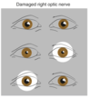
What is lateral strabismus?

A

Damage to one occulomotor nerve. The eye ipsilateral to the lesion deviates laterally.

20
Q

In lateral strabismus, what muscles are affected?

A

The medial rectus is paralyzed and the lateral rectus operates unopposed. The superior and inferior recti and inferior oblique are also paralyzed (prevents vertical movement)

21
Q

Diplopia and being unable to move your eye laterally are clinical signs of…..

A

Lateral Strabismus

22
Q

T/F The ipsilateral levator palpebrae superioris is paralyzed in lateral strabismus

A

True (results in ptosis)

23
Q

T/F The pupillary sphincter and ciliary muscle remain functional in lateral strabismus

A

False, they become non-functional (ipsilateral effect)

24
Q

In lateral strabismus, the pupil on the affected side is (dialted/undialated) as a result of the now-unopposed pupillary dialator, and it (does/does not) constrict in response to light

A

1) Dialated
2) Does not

25
What is usually the first clinically detectable sign of something pressing on the third nerve?
A dialated pupil unresponsive to light
26
What extraoccular muscle does the trochlear nerve innervate?
The superior oblique
27
Where at the cell bodies of origin for the trochlear nerve located?
In the contralateral trochlear nucleus
28
The trochlear nucleus is located at the level of the (superior/inferior) colliculus
Inferior
29
Where do fibers leaving the trochlear nucleus decussate?
Pontomedullary Junction
30
What does the superior oblique muscle do?
Helps move the adducted eye downward (like when reading or walking down stairs). Also aid in eye intorsion
31
Lesions to the trochlear nerve result in...
An extorted eye, resulting in tilting of head away from the lesioned side to compensate
32
The abduscens nerve innervates the.....
Lateral Rectus
33
Where do the fibers of the abduscens nerve originate?
The ipsilateral abduscens nucleus
34
Where is the abduscens nucleus located?
In the caudal pons beneath the floor of the fourth ventricle
35
What is the internal genu of the facial nerve?
The place where the facial nerve vibers wrap around the abduscens nucleus
36
What is responsible for the facial colliculus?
The abduscens nucleus and the internal genu
37
Damage to the abduscens NERVE causes...
Medial strabismus
38
What is medial strabismus?
Affected eye deviates medially. Individual may move the affected eye from adducted position to midposition (but not past it)
39
Damage to the abduscens NUCLEUS results in....
Lateral Gaze Paralysis
40
What is lateral gaze paralysis?
Damage to abduscens nucleus cause an inability of the ipsilateral eye to **abduct** past midposition and the contralateral eye to **adduct** past its midposition
41
Why does damage to the abduscens nucleus cause lateral gaze paralysis?
The abduscens nucleus contains not only recuts motor neurons but also internuclear neurons with axons that ascend through the MLF to the CONTRALATERAL occulomotor nucleus.
42
The abduscens nucleus also contains interneurons that project to the....
contralateral occulomotor nucleus
43
Why is it important that the abduscens nucleus projects interneurons to the contralateral occulomotor nucleus?
This makes it possible to have both eyes look in the same direction. Looking in the same direction requires one lateral rectus and the contralateral medial rectus to contract simultaneously. Simultaneous firing of abduscens motor neurons and internuclear neurons allow for coordinated lateral gaze.
44
Damage to one MLF results in.....
the ipsilateral eye (to the lesion) failing to move past midposition during attempted horizontal gaze
45
The accessory nerve innervates....
Neck and shoulder muscles
46
The hypoglossal nerve innervates....
Tongue Muscles
47
Where do the fibers of the hypoglossal nerve originate?
In the ipsilateral hypoglossal nucleus
48
If there is damage to the hypoglossal nerve, sticking your tongue out deviates.....
toward the side of the lesion
49
Branchiomeric nerves innervate....
striated muscle of branchail (pharyngeal) arch origin (they all contain pharyngeal motor fibers.
50
T/F The trigeminal nerve is the general sensory nerve for the head
True
51
What type of information (type of sensation) is the trigeminal system resposnsible for?
Transmission of tactile, proprioceptive, and pain and temperature infromation from the head to the cortex, cerebellum and reticular formation
52
What are the three divisions of the trigeminal nerve?
1) Ophthalamic 2) Maxillary 3) Mandibular
53
The main sensory nucleus of the trigeminal nerve receives information about....
Touch and jaw position
54
Tactile afferent are.....
Large diameter, heavily myelinated
55
The main sensory nucleus of the trigmenial nerve gives rise to these two tracts.....
1) Anterior Trigeminothalamic Tract (cross midline to join medial lemniscus) 2) Posterior Trigeminothalamic Tract (uncrossed, not part of medial lemniscus)
56
The spinal trigeminal nucleus receives information about....
Pain and temperature
57
What are the three subdivisions of the spinal trigmenial nucleus and which is responsible for receiving pain and temperature information from the head?
1) Most caudal: Caudal Nucleus 2) Interpolar Nucleus 3) Most rostral: Oral Nucleus \*\*\*Caudal Nucleus responsible for processing pain and temp info\*\*\*
58
Afferents conveying pain and temperature information in the spinal trigeminal tract are (small/large) diameter
Small
59
What is the systematic organization of the divisions of the trigmenial nerve with respect to the caudal spinal trigeminal nucleus?
Mandibular Division fibers are most posterior Ophthalamic Fibers are most anterior Maxillary lie somewhere in between
60
Neurons in the anterior parts of the caudal nucleus (trigeminal system) respond to areas of the face in the.....
Ophthalamic Distribution
61
The trigeminal motor nucleus innvervates muscles of....
Mastication
62
Where is the trigeminal motor nucleus located?
In the midpons at the level of attachment of the trigeminal nerve to the brainstem
63
The facial nerve innervates muscles of....
Facial Expression
64
What is the nucleus of origin for the facial nerve?
The facial motor nucleus
65
Where is the facial motor nucleus located?
In the anterolateral tegmentum of the caudal pons
66
Which nucleus is involved in the corneal blink reflex?
The facial motor nucleus
67
What is the corneal blink reflex?
If EITHER cornea is touched by a foreign object, BOTH eyes automatically blink
68
Sensory innervation of the cornea is by way of which division of the trigeminal nerve?
Opthalamic
69
The corneal blink reflex tests....
Both trigeminal nerves and both facial nerves
70
What are corticobulbar neurons?
Upper motor neurons originating in the cortex of the frontal lobe that supply the MOTOR nuclei of the cranial nerves
71
Damage to the facial nerve or the facial motor nucleus affects the...
Ipsilateral half of the face
72
A lesion of motor cortex or corticobulbar fibers results in damage to which side of the face?
The contralateral half
73
T/F The glossopharyngeal nerve conveys information from intraoral receptors
True
74
Much of the visceral sensory fibers of the inner mouth enter the ______ and synapse on in the \_\_\_\_\_\_\_
1) Solitary Tract 2) Solitary Nucleus
75
While most sensation of the tongue and throat (intraoral) enters the solitary tract and synapses on the solitary nucleus, information about pain from the pharynx and posterior part of the tongue enter the _________ tract and terminate in the __________ nucleus
1) Spinal Trigeminal Tract 2) Spinal Trigeminal Nucleus
76
T/F The vagus nerve is the principal parasympathetic nerve
True
77
A major collection of _______ \_\_\_\_\_\_\_\_ fibers travels in the vagus nerve to thoracic and abdominal viscera generally
1) Preganglionic Parasympathetic
78
Where do most of the preganglionic parasympathetic fivers that travel through the vagus nerve to thoracic and abdominal vsicera arise from?
The dorsal motor nucleus of the vagus
79
What is the principal parasympathetic nucleus of the brain?
The dorsal motor nucleus of the vagus
80
Where is the dorsal motor nucleus located?
In the floor of the fourth ventricle, just lateral to the hypoglossal nucleus
81
What underlies a structure in the floor of the fourth ventricle known as the vagal trigone?
The dorsal motor nucleus of the vagus
82
T/F Most vagal visceral sensory fibers enter the solitary tract and terminate in caudal portions of the solitary nucleus
True
83
Which vagal afferents are thought to enter the spinal trigeminal tract and terminate in the spinal trigeminal nucleus?
Afferents innvervating the larynx, esophagus, and lower pharynx
84
Vagal pharyngeal motor fibers arise in what nucleus?
Nucleus Ambiguus
85
What do vagal pharyngeal motor fibers innervate?
Most of the striated muscles of the larynx and pharynx
86
What can the gag reflex test?
The glossopharyngeal nerve (afferent limb) The Vagus Nerve (Efferent Limb)
87
Brainstem damage commonly causes deficits on one side of the head and the (same/opposite) side of the body
Opposite
88
T/F All the exiting cranial nerves are ipsilateral to the side they innervate
True
89
T/F Almost all the cranial nerve nuclei deal with contralateral structures
FALSE Almost all deal with ipsilateral structures
90
Practice Question (Ch 12 Q1): A patient reports to his physician with difficulty swallowing and slurred speech upon waking one morning. The pysician asks the patient to stick out his tongue, upon which the tongue deviates to the right. The most likely side of injury is the:
Right Hypoglossal Nucleus -Tongue points to the side of lesion
91
Practice Question (Ch 12 Q2) A patient with a hisotry of uncal herniation, in which the midbrain is compressed against the tentorial notch, is most likely to present with:
Problems moving the eyes
92
Practice Question (Ch 12 Q3) An individual with a tumor that compresses only the occulomotor nerve, will present with:
The eyes pointed down and out
93
Practice Question (Ch 12 Q4) The cell bodies responsible for control of the muscles of mastication are found in the:
Trigeminal Motor Nucleus
94
What inputs does inegrated sensations of flavor require?
1) Gustatory Stimulation (stimulating taste buds) 2) Stimulation of olfactory receptors 3) Stimulation of chmical-sensitive and somatosensory free nerve endings of the trigeminal (and other nerves)
95
What area of the frontal lobe gives the overall sensation about food and drink by integrating multiple factors?
The orbital cortex
96
Which cranial nerves innervate the taste buds?
7, 9, and 10
97
What type of papillae contain taste buds?
Fungiform, Foliate, and Circumvallate
98
T/F Taste receptor cells are modified epithelial cells with neuron-like properties
True
99
How are taste receptor cells similar to neurons?
1) Contain transduction machinery 2) Produce receptor potentials in resposne to the appropriate taste stimuli
100
Second order gustatory neurons are located in.....
The Solitary Tract
101
What are the two things second order taste fibers do?
1) Participate in reflex activities, such as salivation, swallowing, and coughing by way of cranial nerve motor nuclei 2) Project to the cerebral cortex by way of the thalamus
102
The projection of 2nd order gustatory neurons to the thalamus is (crossed/uncrossed)
Uncrossed
103
Descrie the mode of travel of 2nd order gustatory neurons
1) Ganglia of 7, 9 and 10 project to the VMP via the Central Tegmental Tract. Gustatory cortex neurons then project to the orbital cortex of the frontal lobe
104
How does taste reachthe hypothalamus and the limbic system?
Gustatory cortex projects to the amygdala and that sends taste information to the hypothalamus and the limbic ststem. However, in most mammals, taste information reaches the hypothalamus and amygdala more directly though a projection from the parabrachial nuclei.
105
Olfaction is mediated by receptors that project directly to the.....
Telencephalon
106
The axons of olfactory recpetor neurons form with cranial nerve?
Cranial Nerve 1
107
T/F Sensory nerve ending of trigeminal nerve fibers are also found in the olfactory epithelium
T
108
What is the olfactory epithelium?
The beginning of the olfactory system, it is a patch of cells occupying about 1 to 2 cm of the roof and adjacent walls of the nasal cavity on each side.
109
How many receptor cells does each olfactory epithelium have?
Around 3 million each
110
What are the trigeminal endings found in the olfactory epithelium responsible for?
Noxious senstaion (not really one of smell)
111
T/F The olfactory receptor cells, just like taste receptor cells, are semi-neurons
False, olfactory receptor cells are true neurons
112
T/F The unmyelinated axons of the olfactory receptors are among the thinnest and most slowly conductin in the entire nervous system
True
113
The axons of olfactory receptors collect into small bundles known as the....
olfactory fila
114
The olfactory fila pass through the hooles in the cribiform plate of the ehmoid bone and end in the.....
Olfactory Bulb
115
The primary neurotransmitter released by the axons of olfactory receptors in the olfactory bulb is.....
Glutamate
116
What type of receptors are used by olfactory receptor neurons to be able to detect a wide range of odors?
G Protein-Coupled
117
T/F Olfactory information bypasses the thalamus on its way to the cerebral cortex
True
118
The olfactory bulb develops as an outgrowth from the.....
Telencephalon
119
The olfactory nerve reaches the (ipsilateral/contralateral) cerebral hemisphere and does so (directly/indirectly)
1) Ipsilateral 2) Directly (skips the thalamus)
120
The olfactory nerve terminates in the.....
Olfactory bulb
121
What is the most prominent cell type of the olfactory bulb?
The mitral cell
122
Describe the configuration of the mitral cell
Similar to a cortical pyramidal cell in reverse. It has an axon that emerges from the pointed side of the pyramid and moves toward the interior of the bulb to enter the olfactory tract. A dendrite emerges from the broad side, descends to the surgace of the bulb, and receives coontacts from the incoming axons of olfactory receptors (CN 1)
123
What forms glomeruli?
The dendrites of the mitral cell, which spread out in large spherical arborizations
124
The axons of all the hundreds of olfactory receptor neurons that express a given receptor protein converge on just.......
One or two glomeruli
125
T/F Different odorants activate different sets of glomeruli in patterns that systematically map out the chemical properties of odorants across the surface of the olfactory bulb
True
126
Give a basic description of a mitral cell
Send dendrites into the glomeruli and send their axons into the olfactory tract
127
The olfactory bulb projects to....
the olfactory cortex
128
Olfactory receptor neurons wind up represented in the (ipsilateral/contralateral) cerebral hemisphere
Ipsilateral
129
T/F Neurons of the olfactory bulb and primary olfactory cortex respond to selected simulants while neurons of the olfactory association cortex are more likely to respond to multiple odorants
False, the relationship is switched. Olfactory bulb detects multiple odorants while the olfactory association cortex respond to selected stimuli
130
What are conductive olfactory deficits?
Processes that prevent odorants from reaching to olfactory epithelium
131
What are sensorineural olfactory deficits?
Processes that damage olfactory receptor neurons or parts of the olfactory CNS
132
What can cause sensorineural olfactory deficits?
Head injuries (may tear the olfactory fila loose from the olfactory bulb) or neurdegenerative diseases.
133
Practice Question (Ch 13 Q1): A lack of taste from the posterior aspect of the tongue, with a significant decrease in the ability to perceive bitter tastants may suggest damage to:
CN 9
134
Practice Question (Ch 13 Q2): The chemical neurotransmitter that is released from the depolarized taste bud to activate the cranial nerve is always...
ATP
135
Practice Question (Ch 13 Q3): The arrow is pointing to the __________ in this cross section of the human rostral medulla:
Primary affernt fibers of the tongue and viscera
136
Practice Question (Ch 13 Q4): The first cranial nerve (CN1) that detects olfaction in humans has fibers that pass through the:
Cribiform Plate
137
Practice Question (Ch 13 Q5): A patient comes to the emergency room after being in a car accident in which his vehicle was struck from behind. He claims he is having trouble smelling. The clinician detects that he is unable to smell coffee or cinnamon, but he did respond to the smell of ammonia. What cranial nerve may allow him to "smell" ammonia?
CN 5 (detects noxious smells such as methanol and ammonia)
138
Hearing and balance are dealt with by the ___ cranial nerve
8th
139
The _____ divisiion of the 8th cranial nerve conveys information about sound, while the _______ division signals position and movement of the head.
1) Cochlear 2) Vestibular
140
Auditory and Vestibular Receptor cells are located in the walls of the ___________ labyrinth
Membranous
141
Where is the membranous labyrinth?
It is suspended in the bony labyrinth, which is a cavity in the temporal bone
142
The bony labyrinth is filled with (perilymph/endolymph)
Perilymph
143
What is Perilymph?
Fluid similar in composition to CSF; low Potassium concentration high sodium concentration
144
T/F The subarachnoid space around the brain is continuous with the perilymphatic space of the bony labyrinth through a tiny canal in the temporal bone
True
145
What is the tiny canal called that connects the perilymphatic space with the rest of the subarachnoid space of the brain?
The Cochlear Aqueduct
146
The membranous labyrinth is filled with (perilymph/endolymph)
Endolymph
147
What is endolymph?
A fluid similar to intracellular fluids in ionic composition (high K+ concentration, Low Na+ concentration
148
What continuously produces endolymph?
Specialized epithelialcells called stria vascularis
149
The microvilli that project as a bundle from one end of the hair cell projects into the (endolymphatic interior/perilymphatic exterior)
Endolymphatic Interior
150
What are the three locations (labyrinth locations) where hair cells are?
Semicircular Ducts, Utricle, and the Saccule
151
What stimulus is transduced by the organ of corti?
Sound
152
What makes hair cells so rigid?
Cross-linked actin filaments
153
Briefly describe how hair cells operate based on mechanical stimuli (hint: how do tip links factor in and what causes the hair cells to open their channels)
1) Tip links connect each stereocilium to its next tallest neighbour 2) Deflection of the hair bundle **toward tallet stereocilia** stretches tip links 3) Stretching tip liinks opens mechanically gated ion channels more 4) K+ ions from endolymph flow through into the hair cells to depolarize cell 5) This cases the opening of voltage gated Ca2+ channel and increased neurotransmitter release onto 8th cranial nerve 6) Excitatory transmitter (glutamate) then causes increased firing frequency in eighth nerve fibers
154
T/F Deflection of hair cells **away from** their next tallest neighbour decreases the tension of the tip links, and causes mechanically gate ion channels to close
True
155
What are the two muscles attached to the mididle ear bones that modulate the transmission of vibrations to the inner ear?
1) Tensor Tympani 2) Stapedius
156
What ossicle does the tensor tympani attache to?
The handle of the malleus
157
When the tensory tympani contracts it (increases/decreases) the tension on the tympanic membrane and (increases/decreases) the transmission of vibrations through the ossicular chain
Increases, Decreases
158
What ossicle does the stapedius attach to?
The neck of the Stapes
159
When the stapedius contracts, it (increases/decreases) the transmission of vibrations just like what happens when the tensor tympani contracts
Decreases
160
The tensor tympani receives motor innervation from the _______ nerve
Trigeminal
161
The stapedius receives motor innervation from the ________ nerve
Facial
162
What is the auditory portion of the membranous labyrinth called?
The Cochlear Duct
163
What is the perilymphatic space above the cochlear duct called?
The Scala Vestibuli
164
What is the space below the cochlear duct called?
The scala tympani
165
What does the Reissner's Membrane function as?
A diffusion barrier between the endolymph and perilymph
166
Inner hair cells are (sensory/amplifier) cells, and outer hair cells are (sensory/amplifier) cells
Sensory, Amplifier
167
T/F Motion oof the outer hair cells can enhance the vibration of the basilar membrane
True
168
Auditoy Information is distributed (bilaterally/unilaterally) in the CNS
Bilaterally
169
Where are the cell bodies of auditory primary afferents located?
The Spiral Ganglion (of the Modiolus)
170
Auditory primary afferents are myelinated by....
Schwann cells until they enter at the pontomedullary junction
171
Where does bifurcation of auditory primary afferents occur?
At the pontomedullary junction (where they enter the brainstem)
172
After bifurcation of the auditory primary afferents, one branch is sent to the _________ \_\_\_\_\_\_\_\_\_\_ nucleus and one branch to the __________ \_\_\_\_\_\_\_\_\_\_\_ nucleus
1) Dorsal Cochlear Nucleus 2) Ventral Cochlear Nucleus
173
What is the major ascending auditory pathway of the brainstem?
The lateral Lemniscus
174
T/F Each lateral lemniscus carries information only from one ear
False, each one carries information from both ears
175
Rather than heading straight to the thalamus, where do (almost) all fivers of the lateral lemniscus terminate?
The Inferior Colliculus
176
Starting at the level of the lateral lemniscus, descirbe the projections involving auditory information
1) Lateral Lemniscus terminates in the Inferior Colliculus 2) IC projects bilaterally through the Brachium of the IC 3) Projected to the Medial Geniculate Nucleus 4) Fibers from MGN project tonotopically to the primary auditory cortex
177
Lower frequency sounds are mapped on the auditory cortex more (laterally/medially) while higher frequency sounds are mapped more (laterally/medially)
1) Laterally 2) Medially
178
Where is the primary auditory cortex located?
In the transverse temporal gyri, mostly buried in the lateral sulcus
179
Many of the fibers of the _________ cochlear nucleus are involved in sound localization and end in the superior olivary nucleus
1) Ventral
180
Fibers from what cochlear nucleus are largely involved in sound localization?
Ventral
181
Fibers of the ventral cochlear nucleus involved in sound localization end in the ________ \_\_\_\_\_\_\_\_ nucleus, at the _______ end of the facial motor nucleus in the _______ (what level of the brainstem)
1) Facial Motor 2) Rostral 3) Pons
182
Sound localization can be accomplished by comparing the time of ________ and the ______ of a sound at the two ears
1) Arrival 2) Intensity
183
The time of arrival comparison of sound is begun in the (medial/lateral) superior olive
Medial
184
What provides the anatomical substrate for binaural comparison?
Fibers from the ventral cochlear nuclei of both sides converging on the medial superior olive of each side
185
In the time-of-arrival comparison, each superior olivary nucleus (after receiving input from the ventral cochlear nuclei) projects through the lateral lemniscus to the (ipsilateral/contralateral) inferior colliculus
Ipsilateral
186
An individual with a damaged facial nerve may complain that sounds are too (loud/quiet) in their (ipsilateral/contralateral) ear
1) Loud 2) Ipsilateral
187
The vestibular division of the eight cranial nerve conveys information about _______ and ________ acceleration of the head
1) Linear 2) Angular
188
What are the two otolithic ograns and where are they located?
The utricle and the saccule; they are located in the vestibule (which is the central area of the bony labyrinth)
189
Receptors in the semicircular ducts detect (linear/angular) acceleration of the head
Angular
190
Angular acceleration with respect to the head would be like (rotating the head from left to right/nodding the head up and down)
Rotating
191
What do the receptors in the utricle and saccule detect?
Linear Acceleration and Position of the Head
192
What are examples of linear accelerating forces? What about with respect to the head?
Examples include those forces experienced elevators (up and down) and in cars (forward and backward)
193
The utricle is most sensitve to (linear/angular) acceleration in the forms of which direction?
Linear, in the forms of forward-backward and side-to-side
194
The saccule is more sensitive to linear acceleration in what directions?
Up-down and forward-backward
195
The vestibular primary afferents have their cell bodies in the....
Vestibular Ganglion
196
The peripheral processes of vestibular primary afferents receive their input from the hair cells of the......
Semicircular Ducts, Utricle, and Saccule
197
Where do the central processes of the vestibular primary afferents enter the brainstem?
At the pontomedullary junction
198
What are the four vestibular nuclei?
Inferior, medial, lateral, and superior
199
T/F Each vestibular nucleus has its own pattern of secondary connections
True
200
Other inputs to the vestibular nuclei include projections from the cerebellum by way of the.........
Juxstarestiform Body
201
The vestibular nuclei poject to the nuclei of which cranial nerves?
3,4, and 6
202
What is the main function of the vestibular system?
Regulate posture and coordinate eye and head movements
203
The lateral vestibulospinal tract arises in the (lateral/medial) vestibular nucleus
Lateral
204
Give a brief overview of the mode of travel of the lateral vestibulospinal tract
1) Start: Lateral vestibular nucleus 2) Travels through the Lateral Funiculus 3) Sends furtherexcitatory projections to the motor neurons for antigravity muscles at all ipsilateral spinal levels
205
As the lateral vestibulospinal tract travels through the lateral funiculus, it sends excitatory projections to the motor neurons for (balance/antigravity) muscles at all (ipsilateral/contralateral) spinal levels
1) Antigravity 2) Ipsilateral
206
Vestibulospinal fibers influence ________ muscles and _______ muscles
Antigravity, neck
207
The (medial/lateral) vestibulospinal tract is responsible for stabalizing head position as we walk around or when our heads move in space in other ways, as well as for coordinating head and eye movements
Medial
208
The medial vestibulospinal tract arises mainly in the (medial/lateral) vestibular nucleus, and reachs the (ipsilateral/contralateral/both) sides of the cervical spinal cord
1) Medial 2) Both
209
The medial vestibulospinal tract reaches both sides of the cervical spinal cord by projecting caudally though the (MLF/LF)
MLF
210
Gaze can stay fixed on an object even though the head is moving by means of the ____________ reflex
Vestibuloocular
211
What is the vestibuloocular reflex?
Vestibular Nuclei coordinate with the motor neurons of the oculomotor, trochlear, and abducens nuclei to allow for object tracking even while the head is moving
212
Give an example of the vestibuloocular refelx
If the head turns, the eyes will automatically counter the turn in the opposite direction thus the direction of gaze would not change
213
What is the purpose of a healthy vestibular nystagums?
To keep images from moving on the retina
214
What is a romberg sign?
Greatly increased swaying and loss of balance caused by closing the eyes
215
What does a positive romberg sign indicate?
Defective vestibular or somatosensory function
216
Where does the combination of vestibular, visual and somatosensory inputs occur to make sense of orientation and movement?
The Vestibular Nuclei
217
T/F The visual system works in tandem with the vestibular system to provide information about the position and motion of the head in space
True, the visual system is anothet major source of this information (it is dominant in most cases)
218
Practice Question (Ch 14 Q1) Hearing loss can be attributed to multiple causes, including a genetic mutation in potassium channels involved in the production of endolymph. Where in the cochlear apparatus would this genetic defect in the potassium channel be located?
Scala Media
219
Practice Question (Ch 14 Q2) An individual with significantly reduced hearing in one ear and reduced bone conduction is most likely to have injured....
Inner Hair Cells
220
Practice Question (Ch 14 Q3): Damage to the outer hair cells most often results in:
Decreased otoacoustic emissions
221
Practice Question (Ch 14 Q4): Damage to the facial nerve can result not only in the loss of facial expressions and taste perception but also in:
Increased Hearing of Low Frequencies
222
Practice Question (Ch 14 Q5): Auditory information enters the pontomedullary junction of the CNS, sending ascending information to the inferior colliculus and then off to the thalamus before arriving at the superior temporal gyrus (auditory cortex). What part of the thalamus is used for auditory stimuli?
Medial Geniculate
223
Practice Question (Ch 14 Q6) A standard test used in emergency medicine involves instilling cold water into a patient's ear and watching for eye movements. What would be the expected pattern of nystagmus in a neurologically intact individual lying on his back, if cool water were instilled into the right ear?
Nystagmus that starts out with its fast phase to the left, then slowly fades away. Direction of nystagmus is opposite to the ear in which the water is instilled
224
Practice Question (Ch 14 Q7): The ability to feel the motion of going up in an elevator is due to the hair cells in the:
Saccule
225
What is the dorsal motor nucleus of the vagus?
Nucleus of origin of most preganglionic parasympathetic neurons for thoracic and abdomnial viscera
226
The nucleus gracilis is the site of termination of the....
fasciculus gracilis
227
The nucleus gracilis is the origion of the (upper/lower) limb portion of the medial lemniscus
lower
228
What is the site of termination of the fasciculus cuneatus?
Nucleus cuneatus
229
T/F Proprioceptive primary afferents travel through the fasciculus cuneatus to reach the lateral cuneate nucleus
True
230
Uncrossed cuneocerebellar fibers from the cuneate nucleus enter the cerebellum through the (inferior/superior) cerebellar peduncle
Inferior
231
The lateral cuneate nucleus deals with proprioceptive information from the (upper/lower) limbs
Upper
232
The lateral cuneate nucleus gives rise to (crossed/uncrossed) _________ fibers that enter the cerebellum through the inferior cerebellar peduncle
1) Uncrossed 2) Cuneocerebellar
233
What nucleus is the site of termination of part of the spinal trigeminal tract?
The spinal Trigeminal Nucleus
234
At what nucleus does the anterolateral pathway originate?
Spinal Trigeminal Nucleus
235
The spinal trigeminal nucleus deals with what type of sensory information?
Pain and Temperature
236
What type of information does the spinal trigeminal tract carry, and where does it have its primary afferents?
Originates in the ipsilateral face, and carries pain and temperature information
237
The Posterior Spinocerebellar Tract originates from _______ nucleus
Clarke's
238
Describe what kind of information and from where Clarke's Nucleus processes
Proprioceptive Information from the ipsilateral lower limb
239
The posterior spinocerebellar tract, after originating from clarke's nucleus, is (crossed/uncrossed) and will carry proprioceptive information from the lower limb that will reach the (contralateral/ipsilateral) half of the cerebellum
1) Uncrossed 2) Ipsilateral
240
The posterior spinocerebellar tract enters the cerebellum through the (superior/inferior) cerebellar peduncle
Inferior
241
The anterolateral pathway originates from.....
The spinal posterior horns and intermediate gray
242
The anterolateral pathway conveys what type of sensory information?
Pain and Temperature
243
This tract carries unconscious proprioceptive information from the lower limb to the cerebellum
Anterior Spinocerebellar
244
What is the principal ascending pathway for fine touch, conscious proprioception, and vibratory formation?
Medial Lemniscus
245
This nucleus is known as containing the lower motor neurons for the ipsilateral half of the tongue
Hypoglossal
246
Primary afferents conveying visceral information from cranial nerves \_\_\_, \_\_\_\_, and _____ travel through the solitary tract to reach the solitary nucleus
7, 9, and 10
247
What is the site of origin of the medial vestibulospinal tract?
Medial Vestibular Nucleus
248
The abduscens nucleus is located in which section of the brainstem?
Caudal Pons
249
This nucleus contains the lower motor neurons for the ipsilateral lateral rectus
Abduscens
250
Some of the primary afferents of the spinal trigeminal tract carrying tactile information end in what region of the brainstem?
Caudal Pons
251
Describe the lateral lemniscus
It is the ascending auditory fibers
252
The ascending auditory fibers that form the lateral lemniscus originate from which two nuclei?
Superior Olivary and Cochlear
253
What fibers form the middle cerebellar peduncle?
The pontocerebellar fibers
254
The pontocerebellar fibers have their sources in what nuclei?
Pontine
255
What is the first site of convergence of fibers representing the two ears and is also the source of many of the fibers of the lateral lemniscus?
Superior Olivary Nucleus
256
The facial motor nucleus is located in which region of the brainstem?
Caudal Pons
257
This nucleus contains the lower motor neurons for the ipsilateral muscles of facial expression
Facial Motor Nucleus
258
The trigeminal main sensory nucleus is located in what region of the brainstem?
Midpons
259
What nucleus is the site of termination of large-diameter trigeminal afferents?
Trigeminal Main Sensory Nucleus
260
T/F The midpons contains the Trigeminal Motor Nucleus
True
261
What is the trigeminal motor nucleus?
Contains the lower motor neurons for the ipsilateral muscles of mastication
262
What is the trapezoid body and where is it found?
Found in the caudal and midpons, it is the crossing of auditory fibers
263
The crossing fibers of the trapezoid body originate from what nucleus?
Ventral Cochlear Nucleus
264
The trochlear nucleus is in what region of the brainstem?
Caudal Midbrain
265
What is the trochlear nucleus?
Contains the lower motor neuros for the contralateral superior oblique muscle
266
The lateral lemniscus ends in what colliculus?
Inferior
267
What tract innervates the mechanoreceptors in and around the mouth?
Mesencephalic Trigeminal Tract
268
The occulomotor nucleus is located in which area of the brainstem?
Rostral Midbrain
269
What is the occulomotor nucleus?
Contains the lower motor neurons for the various eye muscles that it controls
270
What muscles of they eye does the occulomotor nucleus control?
**1. Ipsilateral** Medial and Inferior Recti and Inferior Oblique **2. Contralateral** Superior Rectus 3. Levator Palpebrae
271
What are the four parts of the diencephalon? (Hint: They all have the word "thalamus")
1) Epithalamus 2) Dorsal Thalamus (this is the thalamus) 3) Subthalamus 4) Hypothalamus
272
What two structures are contained within the epithalamus?
Pineal Gland and the Habenular Nuclei
273
A decrease in melatonin causes an (increase/decrease) in gonadal function; how is sexual drive affected when melatonin decreases?
Increase; sexual drive is stimulated
274
The habenula nuclei will send messages to the _______ and ________ cells of the brainstem thus it is involved in assigning a rewarding feeling to an action
Dopamine and Serotonin
275
The _______ \_\_\_\_\_\_\_ lamina divides most of the thalamus into medial and lateral groups of nuclei
Internal Medullary
276
The VPL of the thalamus is the somatosensory relay nucleus for the (Body/Head)
Body
277
The VPM of the thalamus is the somatosensory relay nucleus of the (Body/Head)
Head
278
The habenulointerpeduncular tract passes through the (centromedian/parafascicular) nuclei
Parafascicular
279
All thalamic nuclei - except the reticular nucleus - are a mixture of projection neurons, and small inhibitory intereurons that use what neurotransmitter?
GABA
280
What are specific inputs in reference to the thalamus?
Inputs conveying the information that a given thalamic nucleus may pass on accurately to the cerebral cortex
281
What are regulatory inputs with respect to the thalamus?
Those that contribute to decisions about the form in which information leaves a thalamic nucleus; may modulate information
282
Where do most regulatory inputs come from (in terms of the thalamus)?
The cerebral cortx - mainly the cortical area to which a given thalamic nucleus projects
283
Describe the input and projection of relay nuclei with respect to the thalamus
Receive well defined bundles of specific input fibers, and project to particular functional areas of the cerebral cortex
284
What is the role of relay nuclei?
Deliver information from particular functional systems to appropriate cortical areas
285
Association nuclei (thalamus) project to cortical areas known as ________ areas
Association
286
What is the likely role of association nuclei (thalamus)?
Regulating Distribution and gating of information between cortical areas
287
Midline nuclei project to areas of _______ \_\_\_\_\_\_\_, and also to parts of the basal nuclei and ________ \_\_\_\_\_\_
Cerebral Cortex Limbic System
288
What are the two physiological states of thalamic projection neurons?
Tonic Mode Burst Mode
289
Projection neurons that are slightly depolarized (i.e. they are negative but the value is closer to 0) are in a (tonic/burst) mode
Tonic
290
How do tonic mode thalamic projection neurons respond when even more slightly depolarized
Slight additional depolarization causes a train of action potentials Slight hyperpolarization causes cessation
291
Projection neurons that are extremely hyperpolarized (beyond the tonic mode) are in the _______ mode
Burst
292
Neurons in burst mode are very sensitive because a small depolarization can trigger a.....
Burst of action potentials
293
What is the difference between thalamic projection neurons in burst mode vs. tonic mode?
The frequency of bursts is the biggest difference. Tonic mode has a steady frequency, burst mode does not (action potentials occur regularly in tonic mode)
294
Because burst mode neurons have a low frequency of bursts, they are unable to......
Transmit information about specific inputs accurately
295
Neurons in (tonic/burst) mode are important because they play an important role in arousal during wakefulness, by serving a "lookout" function
Burst
296
What is special about the thalamic reticular nucleus?
Instead of projecting to the cerebral cortex like all other thalamic nuclei, it instead projects to other thalamic nuclei
297
The blood supply of the thalamus is mostly from small perforating branches of the ________ cerebral artery and the nearby _______ \_\_\_\_\_\_\_\_\_ artery
Posterior Posterior Communicating
298
A branch of the posterior communicating artery called the ___________ artery supplies anterior regions of the thalamus
Tuberothalamic
299
Damage concentrated to the posterior thalamus can result in a condition in which normal somatosensory stimuli trigger.....
Intense Pain (similar to Trigeminal Neuralgia)
300
T/F The dysesthesia caused by concentrated damage to the posterior thalamus is usually limited to the head, and analgesics usually have no effect
False: they can spread to an entire half of the body They are resistant to analgesics, however
301
Thalamic pain is a result of damage along the __________ pathway, and lesions that cause it almost always inivolve the \_\_\_\_\_\_/\_\_\_\_\_\_\_
1) Anterolateral 2) VPL/VPM
302
Extensive damage to the posterior thalamus causes total loss of somatic sensation in the (contralateral/ipsilateral) head and body
Contralateral
303
Thalamic Pain Syndrome involves a combination of......
Thalamic Pain, Hemianesthesia (loss of sensation in half the body) and sensory ataxia (being unable to discriminate between objects using hands)
304
Thalamic pain syndrome affects the (contralateral/ipsilateral) side of the body with respect to the thalamic lesion
Contralateral
305
Almost all neural traffic to and from the cerebral cortex (with respect to the thalamus) travels through this structure:
1) The internal capsule
306
What are the five parts of the internal capsule?
1) Anterior Limb 2) Posterior Limb 3) Genu 4) Retrolenticular Part 5) Sublenticular Part
307
The anterior limb contains the fibers interconnecting the _______ nucleus and the ________ gyrus
1) Anterior 2) Cingulate
308
Which part of the internal capsule has some of the fibers projecting from the frontal lobe to the ipsilateral nuclei?
Anterior Limb
309
Which part of the internal capsule has the fibers that interconnect the VA and VL with motor areas of the cortex
Posterior Limb
310
What part of the internal capsule contains the corticospinal and corticobulbar fibers, and the somatosensory fibers projecting from the VPL/VPM to the postcentral gyrus?
Posterior Limb
311
What part of the internal capsule serves as a transition zone between the anterior and posterior limbs?
Genu
312
What part of the internal capsule contains part of the optic radiation?
The retrolenticular part
313
What does the optic radiation project from and to?
Projects from the lateral geniculate nucleus to the calcarine nucleus
314
What part of the internal capsule contains the auditory radiation?
The sublenticular part
315
The auditory radiation projects from the _______ \_\_\_\_\_\_\_ nucleus and end in the _______ \_\_\_\_\_\_\_ gyri
Medial Geniculate Transverse Temporal
316
Small branches of the (Middle/Anterior) cerebral artery provide most of the blood supply of the internal capsule
Middle
317
What branches oof the middle cerebral artery provides most of the blood supply to the internal capsule?
Lenticulostriate Arteries and the Anterior Choroidal Artery
318
What can hemorrhage of a lenticulostriate artery in the vicinity of the posterior limb cause? and what side of the body would you see effects?
Hemorrhage can cause spastic paralysis and hemianesthesia on the contralateral side
319
T/F If hemmorhage of a lenticulostriate artery also affects the retrolenticular and sublenticular parts, you could expect to see visual deficits
True
320
Practice Question (Ch 16 Q1): After viewing a radiograph of a patient's head, a clinician notes a shift in the position of the pineal gland. The clinician suspects that a space-occupying lesion has caused the shift and is likely compressing the pineal gland. The compressed pineal gland may cause altered:
Sleep-Wake Cycles
321
Practice Question (Ch 16 Q2): The largest source of regulatory input into the lateral geniculate nucleus is from the:
Visual Cortex
322
What thalamic nucleus is interconnected with the prefrontal cortex?
The Dorsomedial Nucleus
323
What is the dorsomedial nucleus involved in?
Prefrontal functions such as affect and foresight
324
Practice Question (Ch 16 Q3): A patient with noncommunicating hydrocephalus, caused by obstruction of the cerebral aqueduct, has a flat affect and, upon examination, lacks foresight. The clinician suspects compression of the:
Dorsomedial Nucleus
325
Practice Question (Ch 16 Q4): Damage to the retrolenticular portion of the internal capsule will affect primarily:
Vision
326
Practice Question (Ch 16 Q5): The primary blood supply to the genu of the internal capsule is from the:
Lenticulostriate Arteries
327
Transduced photic information is conveyed to the brain by way of the axons of.....
Ganglion Cells
328
Aqueous Humor is secreted by the ________ \_\_\_\_\_\_\_\_\_, which functions as a small outpost of choroid plexus
Ciliary Body
329
For humans, most of the refraction required for vision occurs at the ________ \_\_\_\_\_\_\_\_ ________ at the front surface of the cornea
Air-Warer Interface
330
What part of the eye affects the brightness and quality of the image focused on the retina?
The Iris
331
The endothelial cells of _______ \_\_\_\_\_\_\_\_ are joined by tight junctions, forming a blood retina barrier
Intraretinal Capillaries
332
What types of visual cells are the first layer of synapses of photoreceptros stimulated by light?
Bipolar and Horizontal Cells
333
In the second layer of synapses of the visaul system, bipolar cells terminate on ___________ cells and __________ cells
Ganglion Amacrine
334
What do horizontal cells do?
Spread laterally and interconnect receptors, bipolar cells, and other horizontal cells
335
Axons of ________ cells leave the eye as the optic nerve
Ganglion
336
What do amacrine cells do?
Spread laterally and interconnect bipolar cells, ganglion cells, and other amacrine cells
337
What does the term "plexiform" generally refer to?
Synaptic Zones of the visual system
338
What does the term "nucler" generally refer to?
The Cell Bodies (of the cells of the visual system)
339
The central ______ is specialized for vision of the highest acuity
Fovea
340
Extrafoveal regions function at (lower/higher) light levels
Lower
341
(Rods/Cones) signal spatial detail and color
Cones
342
T/F Cones are less sensitive than rods, but the are faster in their response
True
343
There is considerably less convergence in (cone/rod) pathways than in (cone/rod) pathways
Cone Rod
344
Because there is less convergence in cone pathways, cones are (less/more) sensitive than rods
Less
345
The two types of visual ganglion are called... (Hint: Think Center Surround Receptive Fields)
1) On-center 2) Off-center
346
The metabotropic _______ receptor is found on the ON bipolar cells while the OFF bipolar cells contain an inotropic ________ receptor (called AMPA)
1) Glutamate 2) Glutamate
347
More glutamate is present during (high/low) levels of light
Low
348
The basic spatial orgnization of ganglion cell receptive fields develops in the _________ \_\_\_\_\_\_\_\_ layer
Outer Plexiform
349
The metabotropic glutamate receptor found on ON bipolar cells, when bound to glutamate, results in the (inhibition/activation) of these ON bipolar cells (Hint: It's the opposite effect as observed with ganglion cells)
Inhibition
350
Illumination of a given receptor causes it to hyperpolarize and release (less/more) glutamate
Less
351
Half of the visual field of each eye is mapped systematically in the (contralateral/ipsilateral) cerebral hemisphere
Contralateral
352
Fibers from the nasal half of each retina cross in the........
Optic Chiasm
353
Increases in intracranial pressure are frequently transmitted to which nerve of the visual system?
The Optic Nerve
354
All fibers from the temporal half of each retina pass through the lateral portions of the optic chiasm (with/without) crossing and enter the (contralateral/ipsilateral) tract
1) Without 2) Ipsilateral
355
Each optic tract contains the fiber arising in the temporal retina of the (contralateral/ipsilateral) eye and the nasal retina of the (contralateral/ipsilateral) eye
Ipsilateral Contralateral
356
Most fibers of the optic tracts terminate in the (medial/lateral) geniculate nucleus
Lateral
357
The lateral geniculate nucleus is a (#) layered, dome-shaped nucleus
6
358
Describe the layer arrangement in the lateral geniculate nucleus
A column of cells cutting through all 6 layers represents a given point in the visual field Each layer receives information from only one eye
359
Layers 1,4, and 6 of the LGN receive input from the (ipsilateral/contralateral) eye
Contralateral
360
Layers 2,3, and 5 of the LGN receive input from the (contralateral/ipsilateral) eye
Ipsilateral
361
Layers (#-#) of the LGN receive inputs from small ganglion cells sensitive to color and form
3-6
362
The parvocellular layers of the LGN are those layers sensitive to ______ and \_\_\_\_\_\_\_
Color and Form
363
The neurons in the parvocellular layers are quite (small/large)
Small
364
Layers # and # of the LGN contain larger neurons that receive their inputs from larger ganglion cells that are more sensitive to movement and contrast
1 and 2
365
The neurons contained in the magnocellular layers of the LGN are quite (small/large) and are particularly sensitive to.......
1) Large 2) Movement and Contrast
366
The LGN projects to the primary ______ cortex
Visual
367
What is the optic radiation?
Fibers arising in the LGN that go on to terminate in the general cortex, they do not all end up in the same place
368
Fibers representing superior visual field quadrants represent (inferior/superior) retinal quadrants
Inferior
369
Damage to what lobe can produce significant superior visual field deficits?
Temporal
370
Inferior visual fields project to the cortex (above/below) the calcarine sulcus, and superior fields project to the cortex (above/below) the sulcus
1) Above 2) Below
371
Because the retinal image is inverted and reversed, damage to temporal areas of the retina causes (nasal/temporal) field losses, and damage to the superior areas of the retina causes (inferior/superior) field losses
1) Nasal 2) Inferior
372
Damage (anterior/posterior) to the optic chiasm affects only the ipsilateral eye
Anterior
373
Damage at the level of the optic chiasm causes _________ deficits Hint: it is a condition in which the two eyes have non-overlapping field losses
Heteronymous
374
Damage behind the optic chiam causes (heternymous/homonymous) field losses
Homonymous
375
A lesion of one optic nerve causes blindness in the (ipsilateral/contralateral) eye
Ipsilateral
376
Damage to what structure typically results in quadrant or sector deficits
The optic radiation
377
Describe the pupillary light reflex
Illumination of either retina causes BOTH pupils to constric
378
Which cranial nerve is responsible for constricting the pupillary sphincter in a consensual pupillary light reflex?
The oculomotor nerve
379
Using the pupillary light reflex, what would be indicative of a damaged right optic nerve?
1) In darkness, both pupils are the same size 2) Shining light into the patient's left eye (our right) constricts the pupils of both eyes 3) Shining a light into the patient's right eye (our left) constricts the pupils of neither eye
380
Using the pupillary light reflex, what would be indicative of a damage right oculomotor nerve?
1) In darkness, the patient's right pupil may be noticably bigger 2) Shining a light onto the patient's left eye causes no pupillary response in the patient's right eye, but does illicit pupillary constriction in the left eye (indicates damaged right oculomotor nerve and an intact left optic nerve) 3) Shining a light on the patient's right eye causes no response in that same eye, but the left eye does exhibit a response in pupil constriction (indicates the right optic nerve is intact and confirms the right oculomotor nerve is damaged)
381
T/F The pupillary light reflex requires participation of the cerebral cortex
False
382
Unlike the pupillary light reflex, the accomodation reflex (requires/does not require) the participation of the cerebral cortex
Requires
383
What is an Argyll Robertson Pupil?
A condition in which the pupil constricts during the acommodation reflex but not the pupillary light reflex
384
T/F The same preganglionic fibers are thought to mediate the pupillary constriction of the light reflex as those that mediate the accomodation reflex
True
385