Nephrology Flashcards
What is the pathophysiology of neprotic syndrome?
Description of a group of associated symptoms
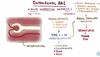
Results from some form of injury to the podocytes (often due to immune complex or complement deposition). This damages their capacity to prevent protein +/- blood from crossing the glomerulus (loss of anionic charge), allowing protein to leak into the urine. This results in loss of oncotic pressure within the plasma, causing fluid to leak into tissues.

Why do those with nephrotic syndrome tend to have hypoalbunaemia?
Due to urinary losses of protein, although a contribution from protein catabolism by tubular epithelial cells may also be relevant.
White bands in the nails (Muehrcke’s bands) are a characteristic sign of hypoalbuminaemia.

Why can hypercholesterolaemia and hypercoagulability occur in nephrotic syndrome?
In response to hypoalbuminaemia, there is a non-specific increase in liver protein synthesis in an attempt to compensate. However, by doing this, it also increases it’s synthesis of lipoproteins and clotting factors. As these are not lost in the urine, they accumulate, causing hyperlipidaemia and hypercoagulability
Why does oedema occur in nephrotic syndrome?
Underfill Model
- Reduced oncotic pressure leads to fluid shift from vascular to extracellular fluid compartment.
- Reduced PV stimulates activation of RAAS and release of ADH -> sodium and water reabsorption in the distal nephron.
- Capillary hydrostatic pressure is increased -> promotes further fluid shift into extravascular space
Overfill model
- Results from defect of sodium excretion in distal nephron.
- Sodium retention -> increased blood volume due to suppressed vasopressin and angiotensin/aldosterone levels.
- Increased capillary hydrostatic pressure and low oncotic pressure favours fluid movement into the extravascular space
What are clinical signs of nephrotic syndrome in children?
-
Oedema
- Periorbital (earliest sign)
- Scrotal, vulval, leg, ankle
- Ascites
- SOB - due to pleural effusions + abdominal distention
- Infections - loss of protective immunoglobulins in urine
- Frothy urine - due to albumin/lipurea
- May have haematuria
- GI disturbance
- Anorexia
What investigations would you do if you thought that a child had nephrotic syndrome?
- Bloods - FBC, ESR, U+Es, creatinine, albumin, complement C3/4
- Urine dipstick - Protein ++++
- Protein-Creatinine ratio
- 24 hr urine collection (GOLD STANDARD)
- Urine microscopy and culture
- Hep B/C screen
What is normal for protein/creatinine ratio?
<20mg/mmol
What is regarded as being in the nephrotic range when looking at protein creatinine ratio?
>200mg/mmol
For 24 hour urine collection, what is regarded as being a normal level of protein in the urine?
<60mg/m2/24 hrs
For 24 urine collection, what is regarded as being in the nephrotic range for protein in the sample?
>1g/m2/24hrs
What are the main causes of nephrotic syndrome in children?
-
Steroid sensitive
- Minimal change disease
-
Steroid resistant
- Focal segmental glomerulosclerosis
What is minimal change disease?
There is diffuse loss of podocyte foot processes (known as effacement). This is thought to occur due to abnormal secretion lymphokines by T cells, which leads to a reduction in electrostatic repulsion of negatively charged molecules (such as proteins) by the podocytes, leading to increased glomerular permeability to certain proteins -> selective proteinuria
https://www.youtube.com/watch?v=4Y4PWTXjyEc&t=219s

What findings would you see on electron microscopy with minimal change disease?
- Podocyte effacement
- Vacuolation
- Microvilli on visceral epithelium

What percentage of those with nephrotic syndrome have steroid sensitive disease?
85-90%
What age range does minimal change disease occur in?
1-10 yrs
What would suggest that it is not minimal change disease causing nephrotic syndrome?
- Evidence of autimmune disease
- Steroid resistance
What clinical features would indicate that the nephrotic syndrome may be steroid sensitive?
- No macroscopic haematuria
- Normal BP
- Normal complement
- Normal renal function
What is the definition of steroid resistant nephrotic syndrome?
Not responding to high dose steroids for 4 weeks
What is the definition of preoteinuria?
>3.5 g protein in 24 hrs
If you had confirmed the nephrotic syndrome as steroid sensitive (i.e. MCD), how would you manage the child?
-
Oral corticosteroids - 8-12 weeks
- 60mg/m2/day - reduce to 40mg/m2/day after 4 weeks
What proportion of steroid sensitive nephrotic syndromes resolves?
95% within 2-4 weeks
What proportion of those with steroid sensitive nephrotic syndrome relapse?
80%, of which 50% are frequent
What are complications of nephrotic syndrome at presentation/relapse?
- Hypovolaemia
- Thrombosis
- Infection
- Hypercholesterolaemia
Why does hypovolaemia occur in nephrotic syndrome?
During the initial phase of oedema, the intravascular compartment becomes depleted. This causes peripheral vasoconstriction and sodium retention. Low urinary sodium and high packed cell volume indicate hypovolaemia
What is a child at risk of in hypovolaemia caused by nephrotic syndrome?
- Shock
- Thrombosis
Why are children with nephrotic syndrome at risk of thrombosis?
Hypercoagulable state caused by:
- Urinary losses of antithrombin III
- Thrombocytosis which may be exacerbated by steroid therapy
- Increased clotting factor synthesis
- Increased blood viscocity
What is hypoalbuminaemia and what does it cause?
Reduced serum albumin concentration -> decreased intravascular oncotic pressure which leads to fluid shifts into ECF

What is the pathophysiology of FSGS?
Podocytes become damaged, allowing proteins and lipids to pass into the urine, resulting in proteinuria and lipiduria. Over time, the accumulation of lipids and proteins around the focally affected areas cause hyalinosis which leads to sclerosis
https://www.youtube.com/watch?v=l7ZyAmGA98w

What are examples of steroid resistant nephrotic syndromes?
- FSGS
- Membranoproliferative glomerulonephritis
- Membranous nephropathy
If treating a child for suspected steroid sensitive nephrotic syndrome, and it didn’t respond to treatment, what would be your next step?
Renal biopsy
What is haematuria?
Urine that is red in colour or tests positive for haemoglobin on urine sticks - should be examined under microscope
Defined as >10 RBCs per high-powered field
How can you distinguish glomerular haematuria from lower urinary tract haematuria?
Glomerular haematuria is more brown (due to deformed RBC’s and casts), and is often accompanied by proteinuria
Lower urinary tract haematuria is bright red, and occurs at the beginning of end of the urinary stream. This is not normally accmpanied by proteinuria
What is the most common cause of haematuria?
UTI - seldom as only symptom
What are causes of proteinuria?
- Orthostatic proteinuria
- Glomerular abnormalities
- Febrile illness - transient
- After exercise - transient
- Increased glomerular filtration pressure
- CKD
- HTN
- Tubular proteinuria
- UTI/STI
What are non glomerular causes of haematuria?
- Infection
- Trauma
- Stones
- Tumours
- Sickel cell disease
- Bleeding disorders
- Renal vein thrombosis
- Hypercalciuria
What are glomerular causes of haematuria?
- Acute glomerulonephritis
- Chronic glomerulonephritis
- IgA nephropathy
- Familial nephritis - alports syndrome
- Thin basement membrane disease
What is nephritic syndrome?
Clinical syndrome which shows signs of nephritis. Defined by haematuria, varying degrees of proteinuria, dysmorphic RBCs, and often RBC casts on microscopic examination of urinary sediment
What is the pathophysiology of nephritic syndrome?
Main driver is immune complex mediated inflammatory damage. Pathophysiology is dependent on where the immune complexes are depositied in the glomerulus. However, there are common pathophysiological processes seen in each cause:
- Proliferation of cells within glomerulus; mesangial cells , endothelial cells reducing capillary lumen diameter, and epithelial cells with Bowman’s space given rise to crescent formation.
- Infiltration of inflammatory cells; neutrophils, macrophages etc.
- Basement membrane thickening; caused by deposition of antibodies, immune complexes and complement.
https://www.youtube.com/watch?v=1nuSSsAnRVM

How does nephritic syndrome present clinically?
- Oedema - IV full as glomerulus stops functioning
- Oliguria - glomerulus stops functioning
- Hypertension / fluid overload
- Haematuria
- Variable Proteinuria

What is important to remember about nephrotic and nephritic syndromes?
THEY ARE NOT DIAGNOSIS - need to find the cause of the syndromes
Why does the glomerulus stop functioning in nephritic syndrome?
Increased glomerular cellularity restricts glomerular flow, leading do decreased glomerular filtration
What are causes of acute nephritis?
- Post infectious nephritis
- Vasculitis, SLE, Wegeners, microscopic polyarteritis
- IgA nephropathy
- Antiglomerular basement membrane disease (Goodpastures)
What is post infectious/streptococcal glomerulonephritis (PSGN)?
Nephritis which usually follows strep sore throat or skin infection. Infection initiates a type III hypersensitivity reaction which leads to immune complex formation. These complexes either become deposited in the GBM, lying in the subepithelial space, or form in the GBM when antigen is deposited in the GBM and antibodies bind to it. This deposition leads to an inflammatory response, which leads to nephritic syndrome.
https://www.youtube.com/watch?v=a24ZQLlRs9U

What sort of skin infection is most likely to lead to PSGN?
Impetigo
How long after a skin/throat infection does PSGN normally present?
5-21 days - shorter time period for throat infection
If a child had haematuria, what investigations would you initially perform?
- MSU microscopy and culture - RBC’s, WBC’s, hyaline, granular/red cell casts
- Protein and calcium excretion
- Kidney US
-
Bloods
- FBC, platelets, coag screen
- U+Es, creatinine, calcium, phosphate, albumin
- Sickle cell screen
- CXR - if fluid overloaded
If you suspected a glomerular cause of haematuria, what specific investigations could you perform?
- ESR, complement, ANA
- Throat swab/ASOT/AntiDNAaseB
- Hep B/C screen
- Renal biopsy
-
If alport’s suspected
- Mother’s urine for haematuria
- Hearing test
What would indicate that the cause of nephritic syndrome was PSGN?
- Bacterial culture - throat swab
- Positive Streptolysin assay (ASOT)
- Low C3 complement - by 6-8 weeks
How would you treat PSGN?
- Antibiotics
- Diuretics - fluid overload
What is IgA nephropathy?
This is a type III hypersensitivity reaction that occurs due to abnormal IgA structure (caused by abnormal glycosylation of serine and threonine -> galactose deficient) This leads to the IgA not being recognised and degraded as it normally would, thus leading to accumulation. In addition, IgG is also produced against the IgA and form immune complexes with it, which are deposited in the mesnagium of the kidneys. This activates the alernative complement pathway, which causes glomerular destruction.
https://www.youtube.com/watch?v=794YzMoIc28

When does IgA nephropathy typically occur?
Usually 1-2 days after URTI
How does IgA nephropathy typically present?
Episodes of macroscopic haematuria +/- chronic microscopic haematuria
What difference would you see in the investigation of IgA nephropathy compared to PSGN?
Normal ASOT and complement
PSGN - Low C3, Positive ASOT
What is Henoch-Schonlein Purpura?
HSP is a small-vessel leukocytoclastic vasculitis characterised by the tissue deposition of IgA and C3-containing immune complexes within affected organs. Its pathogenesis is similar to IgA nephropathy. The kidney damage occurs due to mesangial cell injury.
What can cause HSP to occur?
1-3 days Post infection - Viral URTI, Streptococcus
What are the clinical features of HSP?
- Fever
- Characteristic purpuric skin rash - symmetrically over buttocks , extensors of arms and legs, and the ankles
- Arthralgia - particularly knees and ankles
- Periarticular oedema
- Abdominal pain - colicky
- GN - macroscopic/microscopic haematuria. Can have proteinuria, which if severe enough can lead to nephrotic sundrome

What would you suspected if you saw the following?

Henoch Schoenlein Puprura
What is characteristic about the rash seen in Henoch Schoenlein Purpura?
Initially urticarial, rapidly becomeing maculopapular and purpuric
Characteristically palpable
How would you diagnose HSP?
Clinical Diagnosis
-
Palpable purpura, plus
- Abdominal Pain
- Renal involvement
- Arthritis/arthralgia
- Biopsy - IgA deposition
How would you manage a child with HSP?
- Glucocorticoid therapy - May help with gastrointestinal involvement
-
Immmunosuppresion - Severe renal disease (nephrotic / deranged creatinine / biopsy)
- Prednosolone / cyclophosphamde
-
Long term
- Hypertension and proteinuria screening
What is Alport’s syndrome?
https://www.youtube.com/watch?v=mJ6ULJrdW7I
X-linked dominant mutation of the COL4A5 gene, which leads to a mutation in type 4 collagen. This leads to deformed basement membrane formation, especially in the kidneys, ears ad eyes.

How does Alport’s syndrome affect the kidneys?
https://www.youtube.com/watch?v=mJ6ULJrdW7I
Defects in type IV collagen leads to a thin and overly porous GBM, which allows RBCs and, over time, protein to leak into the urine. Sclerosis also occurs, leading to renal insufficiency and Renovascular hypertension.

How does Alports syndrome affect the ear?
https://www.youtube.com/watch?v=mJ6ULJrdW7I
Not entirely known, however it is hypothesised that the collagen defects affect the basement membrane of the hair cells in the inner ear, leading to sensorineural hearing loss due to inability to generate nerve signals

How does alports syndrome affect the eye?
Differences in collagen structure lead to the lense capsule being unable to maintain the shape of the lense. Other proeblems include lense opacity, myopia, white/yellow flecks around the macula, and recurrent erosions of the cornea.
https://www.youtube.com/watch?v=mJ6ULJrdW7I

How does alport’s syndrome present?
- Haematuria - microscopic and macroscopic
- Proteinuria
- Hypertension
- Deafness - high tone sensori-neural loss
- Eye problems - anterior lenticonus, macular changes
How would you investigate suspected Alport’s syndrome?
- Clinical signs - Frank haematuria, vision or hearing problems, microscopic haematuria with no apparent cause
- Family History
- Kidney/Skin biopsy
- Genetic - COL4A mutation
How would you treat Alport’s syndrome?
- ACEi/ARBs - for proteinurea
- Lens replacement - if needed
- Dialysis/Transplantation - kidneys
What is an uncomplicated presentation of GN?
- Haematuria
- Oliguria
- Hypertension
- Periorbital oedema
- GI disturbance
- Loin Pain
What can complicate the presentation of glomerulonephritis?
- Hypertensive encephalopathy - Restless, drowsy, severe headache, fits, visual disturbance, vomiting, coma
- Uraemia - Acidosis, twitching, stupor, coma
- Cardiac - Gallop rhythm, cardiac failure +/- enlargement, pulmonary oedema
What is the definition of an acute kidney injury?
Abrupt loss of kidney function, resulting in reterntion of urea and other nitrogenous waste products, and dysregulation of extracellular volume and electrolytes. Due to kidney dysfunction, there is a rapid rise in serum creatinine, and anuria/oligura results.
What is oliguria classified as?
<0.5ml/kg/hr
What are the different types of of AKI?
- Prerenal
- Intra Renal
- Post renal

What are causes of pre-renal AKI?
-
Absolute loss of fluid
- Major haemorrhage
- Vomiting/Diarrhoa
- Severe burns
-
Relative loss of fluid
- Heart failure
- Distributive shock
-
Renal Hypoperfusion
- Drugs
- Renal artery stenosis/occlusion
- Hepato-renal syndrome

What happens physiologically when pre-renal AKI occurs?
Decreased blood flow -> decreased GFR. This results in accumulation of creatinine and urea in the blood (azotemia), and oliguria (<0.5 ml/kg/hr). RAAS is also activate, resulting in increased aldosterone release from the adrenal gland, resulting in increased Na+ reabsorption at the kidneys. Water and urea follow the Na+ reabsorptions. The urine produced has low Na+ and is more concentrated.
https://www.youtube.com/watch?v=6i6UKQjGJJs

What are intra-renal causes of AKI?
- Glomerular disease - GN
-
Tubular injury
- Acute tubular necrosis (ATN)
- Ischaemia
- Toxins/drugs
- Obstruction
-
Interstitial
- Acute interstitial nephritis - NSAIDs, Autoimmune
- Pyelonephritis
-
Vascular
- HUS
- Vasculitis
- Embolus
- Renal vein thrombosis
What is acute tubular necrosis?
Epithelial cells which line the tubules necrose due to events such as ischaemia or nephrotoxic drugs (aminoglycosides, lead, myoglobin, radiocontrast). NEcrotic cells slough off and plug the tubule, causing increased pressure in the tubule, lowering the GFR. Due to loss of epithelial cells, solute exchange is affected, leading to hyperkalaemia and metabolic acidosis
https://www.youtube.com/watch?v=ISFEgK8sfb8

What is acute interstitial nephritis?
This is thought to be a Type I or Type IV hypersensitivity reaction which involves inflammation of the interstitium caused by infiltration of immune cells such as neutrophils and eosinophils. Typically it’s caused by medications (NSAIDs, penicillins, diuretics), but can also be caused by autoimmune disease and vasculitis. Unless the cause is removed, it can lead to renal papillary necrosis
https://www.youtube.com/watch?v=ISFEgK8sfb8

What happens to the urine in intra-renal AKI?
Due to impaired reabsorption and secretion, the concentration of urea, sodium and water in the urine can all increase. This can differs from pre-renal AKI urine, which can be low in sodium, water and urea (depending on the cause)

What are post-renal causes of AKI?
-
Obstruction
- Compression - tumours
- Kidney stones
What happens phsiologically in post-renal AKI?
GFR decreases as the pressure gradient across the glomerulus decreases due to increased pressure in the tubule. This reduces the amount of waste products being filtered, and those that are filtered are acutally forced back into the blood by the high pressure within the tubule (except creatinine). As time progesses, pressure within the tubule damages the epithelial cells, resulting in subsequently less reabsorption of solutes over time.
https://www.youtube.com/watch?v=vnTR_y3Sf-k

How would you manage a child with a pre-renal AKI?
-
Most commonly fluid depletion/hypovlaemia
- Fluid replacement and circulatory support
How would you manage a child with renal AKI?
- Fluid restriction and diuretics - if fluid overload
- High calorie, normal protein diet
- Management of electrolyte disturbance
- REMOVE THE CAUSE - if possible
How would you manage post-renal AKI?
- Remove obstruction
- Nephrostomy/bladder catheterisation
When would dialysis be indicated in AKI?
Refractory
- A – Acidosis
- E – Electrolytes – refractory hyperkalemia
- I – Intoxications – SLIME: salicylates, lithium, isopropanol, methanol, ethylene glycol
- O – Overload
- U – Uremia
What can cause acute tubular necrosis?
- Crush injury
- Bruns
- Dehydration
- Shock
- Sepsis
What is haemolytic uraemic syndrome?
This is a clinical triad of AKI, microangiopathic haemolytic anaemia, and thrombocytopenia. It is typically secondary to a GI infection with verocytotoxin-producing E. coli 0157 or shigella, therefore has a prodrome of bloody diarrhoea.
What is the pathogenesis of HUS?
The toxin enters the system via the GI mucosa and preferentially localises to the endothelial cells of the kidney, where it causes intravascular thrombogenesis. Coagulation is activated, thus narrowing the microcirculation. RBCs become damaged passing through this circulation, causing the microangiopathic haemolytic anaemia seen in HUS.

How would you treat a child with HUS?
-
Based on symptoms
- Fluid balance, electrolytes, acidosis
- No antibiotics
- Hypertension
- Aware of other organs
- Renal replacement therapy
What are signs that HUS is developing?
Colitis -> haemoglobinurea -> oliguria +/- CNS signs -> encephalopathy - > coma
What are the stages of CKD in children?

What are clinical features of CKD?
Most asymptomatic, but stage 4 and stage 5:
- Anorexia/Lethargy
- Polydipsia/Polyuria
- Faltering growth
- Signs of renal osteodystrophy
- HTN
- Acute on chronic failure
- Unexplained normochromic, nocrmocytic anaemia
What are complications of CKD?
- Metabolic bone disease
- Metastatic calcification
- Accelerated atherosclerosis
- Hyperkalaemia
- Fluid overload
- Anaemia
How would you manage CKD in a Child?
- Diet - dietetic input
- Renal osteodystrophy prevention - phsophate restriction
- Anaemia - EPO
- Haemodialysis
What is renal osteodystrophy?
A disease of poor bone mineralisation due to renal failure. Renal failure causes increased calcium excretion and decreased phosphate ecretion, which leads to hypocalcaemia and hyperphosphataemia. This increases PTH in response to hypocalcaemia, causing bone resorption to increase in an attempt to correct hypocalcaemia. As the kidney is also not producing calcitriol, calcium absoprtion from the gut is deacreased.
The increased PTH leads to marrow fibrosis (osteitis fibrosis cystica)
