Neoplasia I- Histology and Cytology Flashcards

1
Q

Neoplasia

A

new growth

2
Q

Cancer

A

Latin for crab

tend to adhere to any part that they seize on in an obstinate manner

3
Q

Oncology

A

Study of tumors or neoplasms

4
Q

Tumor parenchyma

A

= neoplastic cells

5
Q

Reactive stroma

A

connective tissue, includes fibrous tissue, blood vessels, and inflammatory cells

in many malignant tumors connective tissue contains abundant and is firm: desmoplasia (desmoplastic stroma)

6
Q

Desmoplasia

A

in many malignant tumors connective tissue contains abundant collagen and is firm

7
Q

Nomenclature of benign mesenchymal neoplasms

A

cell of origin + oma

8
Q

Benign mesenchymal tumor of fibrous tissue

A

fibroma

9
Q

Benign mesenchymal tumor of adipose tissue (lipid)

A

lipoma

10
Q

Benign mesenchymal tumor of cartilage

A

chondroma

11
Q

Benign mesenchymal tumor of bone

A

osteoma

12
Q

Benign mesenchymal tumor of blood vessels

A

hemangioma

13
Q

Benign mesenchymal tumor of lymph vessels

A

lymphangioma

14
Q

Benign mesenchymal tumor of smooth muscle

A

leiomyoma

15
Q

Greek for smooth

A

leio

16
Q

Benign mesenchymal tumor of striated muscle

A

rhabomyoma

17
Q

Greek for rod

A

rhabdo

18
Q
A

Lipoma

Cells on slide are regular size = benign
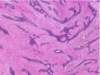
19
Q
A

Leiomyoma

Spindle cells resemble smooth muscle

same size and shape and color = benign

20
Q

Nomenclature of malignant mesenchymal neoplasms

A

cell of origin + ~sarcoma

sar =fleshy (greek)

21
Q

Malignant mesenchymal tumor of fibrous tissue

A

fibrosarcoma

22
Q

Malignant mesenchymal tumor of adipose tissue

A

liposarcoma

23
Q

Malignant mesenchymal tumor of cartilage

A

chondrosarcoma

24
Q

Malignant mesenchymal tumor of bone

A

osteosarcoma

25
Malignant mesenchymal tumor of blood vessels
angiosarcoma
26
Malignant mesenchymal tumor of lymph vessels
lymphangiosarcoma
27
Malignant mesenchymal tumor of smooth muscle
leiomyosarcoma
28
Malignant mesenchymal tumor of striated muscle
rhabdomyosarcoma
29
liposarcoma you can see that the sizes of the cells differ. very abnormal.
30
Leiomyosarcoma you can see mitotic figures. Color is nor regular, neither are cell shapes.
31
Leiomyoma vs leiomyosarcoma
32
Tumors derived from glands (epithelial)
adenoma ex: adenoma in the colon, adrenal gland, liver, kidney,salivary gland
33
Epithelial neoplasm producing fingerlike or warty projections
papilloma ex squamous papilloma upper respiratory tract, urinary bladder
34
benign epithelial neoplasm forming large cystic masses
cystadenoma ex ovarian cystadenoma, cystadenoma of pancreas
35
benign epiithelial neoplasm forming visible projection above mucosal surface
polyp ex adenomatous polyp in the colon
36
Tubular adenoma of the colon adenomatous = polyp projection into lumen crypts look a little darker because nuclei are enlarged
37
Normal uterus, fallopian tibes, and ovaries vs overian cystadenoma you can see that it is benign bc growth is smooth and regular
38
Malignant epithelial neoplasm tumor derived from glands
adenocarcinoma ex colon, breast, prostate, lung, stomach
39
Malignant epithelial neoplasm derived from squamous epithelium
squamous cell carcinoma ex skin, cervix, esophagus
40
Malignant epithelial neoplasm derived from kidney (renal) cells
Renal cell carcinoma
41
Malignant epithelial neoplasm derived from liver cells
hepatocellular carcinoma
42
Fibroadenoma
benign neoplasm of the breast biphasic neoplasm derived from fibroblasts and glandular epithelium
43
Fibroadenoma ## Footnote benign neoplasm of the breast biphasic neoplasm derived from fibroblasts and glandular epithelium
44
fibroadenoma
45
fibroadenoma
46
fibroadenoma
47
Teratoma
Teratoma is a germ cell tumor arising from totipotent germ cells in testis or ovaries terato- teras- wonder monster Can form somatic tissues derived from all 3 germ layers endodermal- enteric type glands, respiratory eithelium mesodermal- cartilage, smooth muscle, ect ectodermal- squamous epithelium, neuronal tissue
48
Ovarian teratoma
49
ovarian teratoma
50
Tumors that are exceptions to nomenclature
melanoma of the skin seminoma of the testes both actually malignant
51
melanoma of the skin
52
Seminoma of the testes
53
Criteria that destinguish benign from malignant tumors
local invasion degree of differentiation rate of growth and proliferation distant spreas (metastasis)
54
Squamous cell carcinoma- well differentiated can see squamous epithelium that are not supposed to be there. Have breached the basement membrane
55
Squamous cell carcinoma well differentiated can see quamous epithelium that have moved. lots of pink cytoplasm
56
Squamous cvell carcinoma poorly differentiated cant really tell where it came from
57
squmous cell carcinoma poorly differentiated you can see some mitotic bodies
58
malignant tumor- anapalsia complete lack of differentiation cant tell where it is from can see mitotic figures
59
Differentiation of a tumor Close resemblance to normal cells of original tissue
tumor is well differentiated
60
Differentiation of a tumor little or no resemblance
tumor is poorly differentiated
61
Differentiation of a tumor no resemblance to original tissue
tumor is anaplastic (ANAPLASIA)
62
local invasion invasice breast carcinoma with infiltrative borders/ invasive edges
63
Benign Fibroadenoma Circumscribed
64
Malignant Invasive breast carcinoma infiltrative, locally invasive
65
Benignvs malignant Benign fibroadenoma, circumscribed invasive breast carcinoma, infiltrative, locally invasive
66
Invasive adenocarcinoma of colon on L it is normal but on R side you see glands that should not be there. Invasion.
67
Invasive adenocarcinoma of the breast On the L you see normal breast globular structure on the R you see tumor nest. Irregulat, uncontrolled.
68
Normal squamuos Mucosa of cervix
69
Dysplasia
pre cancerous lesion
70
Squamous dysplasia of cervix We see abnormal really large nuclei. See some mitotic figures. Not yet invasive bc it hasnt breached the basement membrane
71
Dysplasia
disordered growth Commonly found in epithelial organs dysplasic can be a precursor lesion to invasive cancer
72
Dysplasia vs invasive cancer
dyspasia - intact basement membrane invasive cancer- breach of basement membrane
73
Histologic features of dysplasia
Loss of architectural orientation Loss in uniformity of individual cells Nuclear pleomorphism Large hyperchromatic nuclei with high nuclear-to-cytoplasmic ratio
74
Normal vs squamous dysplasia of uterine cervix
75
Benign tumors typically grow
slowly
76
Malignant tumors typically grow
faster
77
Tumor necrosis is a feature of what type of tumor
malignant
78
Rapidly growing cancers may outgrow their..
blood supplu results in reduced blood supply, ischemia, and tumor necrosis
79
Leiomyoma vs leiomyosarcoma
80
Viable fleshy tumor L Necrotic tumor R Necrosis typically looks yellow
81
Metastasis