Mod XI - M&M25 - Miller66 - Thoracic: Flashcards
ANESTHESIA FOR THORACIC SURGERY
Don’t forget to go over the reading assignment for additional procedures not covered in this lecture
M&M25 - Miller66
ANESTHESIA FOR THORACIC SURGERY
OBJECTIVES
- Describe indications, components, and preoperative assessment for thoracic surgeries
- Identify potential anesthetic and surgical complications
- State contraindications for OLV/DLT.
- Be able to identify mispositioning of DLT based on case scenarios
- Describe basic principles of postoperative pain management for thoracic surgery patients
ANESTHESIA FOR THORACIC SURGERY
lung isolation techniques have been in existence for as long as ET intubation itself - “Closed endobronchial anesthesia”, aka lung isolation technique, first performed in
1928
ANESTHESIA FOR THORACIC SURGERY
Closed endobronchial intubation, with the use of a bronchial blocker was 1st performed in
1936
ANESTHESIA FOR THORACIC SURGERY
First use of a double-lumen endotracheal tube (DLT) in
1950
DLT technology continuously evolving
ANESTHESIA FOR THORACIC SURGERY
DLT technology continuously evolving. However, what continues to be a its main concern?
Maintaining effective gas exchange in the face of ventilation perfusion mismatches
ANESTHESIA FOR THORACIC SURGERY
Two important anesthetic techniques for thoracic surgery
Lung isolation to facilitate surgical access within the thorax
Management of one-lung ventilation (OLV)
ANESTHESIA FOR THORACIC SURGERY
Benefits of OLV
Provides quiet surgical field
(This is very important in thoracoscopic surgeries)
Thoracic surgeons consider lung separation an absolute requirement for pulmonary surgery
Surgery can be performed on a lung while it’s being ventilated
Thoracic surgery alone is not an absolute indication for OLV
ABSOLUTE AND RELATIVE INDICATIONS FOR OLV
ABSOLUTE INDICATIONS FOR OLV
Lung isolation to prevent contamination/infection of health lung
Regulate distribution of ventilation to one lung
Unilateral lung lavage
Most common thoracic surgeries create relative indication for lung separation
ABSOLUTE AND RELATIVE INDICATIONS FOR OLV
RELATIVE INDICATIONS FOR OLV
Most common thoracic surgeries create relative indication for lung separation, in that they can safely accomplished without it
Surgical exposure for thoracic procedures- high Priority
•TAA
•Pneumonectomy
•Thoracoscopy
•Upper lobectomy
•Mediastinal exposure
Surgical exposure-medium (lower) priority
•Middle and lower lobectomies & segmental resections
•Esophageal resection
•Procedures on the thoracic spine
Severe hypoxemia r/t unilateral lung disease
METHODS OF LUNG ISOLATION
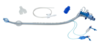
1.DOUBLE-LUMEN TUBES
•Bifurcated tube with both an endotracheal and an endobronchial lumen
•Can be used to achieve isolation of either right or left lung
2.SINGLE-LUMEN TUBES
•Tube is advanced into the contralateral mainstem bronchus for ventilation while the surgical side is collapsed
3.BRONCHIAL BLOCKERS
•Blockade of a mainstem bronchus to allow lung collapse distal to the occlusion
METHODS OF LUNG ISOLATION
What the most common type of lung isolation?
DOUBLE-LUMEN TUBES
- Bifurcated tube with both an endotracheal and an endobronchial lumen
- Can be used to achieve isolation of either right or left lung

METHODS OF LUNG ISOLATION
Placement options for Double-lumen tube (DLT)
- Direct laryngoscopy
- Via tube exchanger
- Fiberoptically
METHODS OF LUNG ISOLATION
Advantages of Double-lumen tube (DLT)
Easy to place successfully
Repositioning rarely required
Bronchoscopy to isolated lung
Suction to isolated lung
CPAP easily added
Can alternate one-lung ventilation to either lung easily
Placement still possible if bronchoscopy not available
Best device for absolute lung isolation
METHODS OF LUNG ISOLATION
Disadvantages of Double-lumen tube (DLT)
Size selection more difficult
Difficult to place in patients with difficult airways or abnormal tracheas
Not optimal for postoperative ventilation
Potential laryngeal trauma
Potential bronchial trauma
METHODS OF LUNG ISOLATION
Placement options for Bronchial Blockers (BB)
- Arndt
- Cohen
- Fuji
- EZ Blocker
METHODS OF LUNG ISOLATION
Advantages of Bronchial Blockers (BB)
Size selection rarely an issue
Easily added to regular ETT
Allows ventilation during placement
Easier placement in patients with difficult airways and in children
Postoperative two-lung ventilation by withdrawing blocker
Selective lobar lung isolation possible
CPAP to isolated lung possible
(Often used when lung isolation requirements were not anticipated at the begining of the case; so rather than switching out for a DLT, the decision was made to place a BB)
METHODS OF LUNG ISOLATION
Disadvantages of Bronchial Blockers (BB)
More time needed for positioning
Repositioning needed more often
Bronchoscope essential for positioning
Limited right lung isolation due to RUL anatomy
Bronchoscopy to isolated lung impossible
Minimal suction to isolated lung
Difficult to alternate one-lung ventilation to either lung
(Also have a higher incidence for being dislodged when compared to DLT)
METHODS OF LUNG ISOLATION
Which lung isolation technique is used when a DLT is not an option?
A. BB
B. SLT
A. BB
(These are not commonly used in the clinical setting, but are used moe often than single lumen tubes (SLT) when a DLT is not an option)
METHODS OF LUNG ISOLATION
The final option for lung isolation is to use either an SLT or an endobronchial tube that is advanced into the contralateral mainstem bronchus, protecting this lung while allowing collapse of the lung on the side of surgery
Why is this technique rarely used today in adult practice (except in some cases of difficult airways, carinal resection, or after a pneumonectomy),
Limited access to the surgical lung for bronchoscopy, suctioning or CPAP
owing to the limited access to the nonventilated lung and the difficulty in positioning a standard SLT in the bronchus
METHODS OF LUNG ISOLATION
Advantages of Endobrochial tube
Like regular ETTs, easier placement in patients with difficult airways
Longer than regular ETT
Short cuff designed for lung isolation
METHODS OF LUNG ISOLATION
Disadvantages of Endobrochial tube
Bronchoscopy necessary for placement
Does not allow for bronchoscopy, suctioning, or CPAP to isolated lung
Difficult one-lung ventilation (right lung)
METHODS OF LUNG ISOLATION
Advantages of Endotracheal tube advanced into bronchus
Easier placement in patients with difficult airways
METHODS OF LUNG ISOLATION
Disadvantages of Endotracheal tube advanced into bronchus
Does not allow for bronchoscopy, suctioning, or CPAP to isolated lung
Cuff not designed for lung isolation
Extremely difficult right one-lung ventilation
DOUBLE-LUMEN ENDOTRACHEAL TUBES
The DLT Consists of:
a single tube with two lumens
Bronchial lumen (blue cuff)
Tracheal lumen

DOUBLE-LUMEN ENDOTRACHEAL TUBES
Bronchial lumen (blue cuff)
Longer than the tracheal lumen
Designed to be inserted into the Right of Left main bronchus
Used to ventilate the lung that it is inserted into
DOUBLE-LUMEN ENDOTRACHEAL TUBES
Tracheal lumen
Terminates in the trachea (mid-trachea)
Corresponding port will ventilate the opposite lung
DOUBLE-LUMEN ENDOTRACHEAL TUBES
Why are Right and left DLT are manufactured?
DLT can be used to achieve isolation of either right or left lung
DOUBLE-LUMEN ENDOTRACHEAL TUBES
What’s the most widely used DLT?
Left DLT
DOUBLE-LUMEN ENDOTRACHEAL TUBES
Sizing of DLT is determined by:
Patient’s gender and height
DOUBLE-LUMEN ENDOTRACHEAL TUBES
Which DLT size is used for females? which size is used for males?
Females: 35-37
Males: 39-41
DOUBLE-LUMEN ENDOTRACHEAL TUBES
What are the charateristics of internal vs external diameters of DLTs?
Internal diameter of DLT are reduced b/c of the double-lumen
External diameters of DLT are large
Size 37 Fr = 11 mm external diameter
Smallest DLT 25 Fr = 7.5 mm external diameter
DOUBLE-LUMEN ENDOTRACHEAL TUBES
Why are DLTs are not typically used in children?
Large external diameters
DOUBLE-LUMEN ENDOTRACHEAL TUBES
Which method is suggested by some books to determine proper positioning/size of the DLT? Typically DLT size is simply based on:
CT-scan with a superimposed DLT to predict proper positioning/size
This is actually never done in practice
Typically DLT size is simply based on gender and height
Comparative Diameters of Single- and Double-Lumen Tubes
How do external diameters of SLTs and DLTs compared? Why should DLTs never be advanced againts resistance?
Compared with SLTs, DLTs have a large external diameter
This is why DLTs should not be advanced against significant resistance
Selection of Double-Lumen Tube Size Based on Adult Patient—s Sex and Height
What the appropriate Size of Double-Lumen Tube (Fr) for Female <160 (63 in)* vs Female >160 cm in height?
35 Fr. vs 37 Fr
Selection of Double-Lumen Tube Size Based on Adult Patient—s Sex and Height
What the appropriate Size of Double-Lumen Tube (Fr) for Male <170 (67 in)* vs Male >170 cm in height?
39 Fr. vs 41 Fr
Comparative Diameters of Single- and Double-Lumen Tubes
Photograph of the cut cross sections of several SLTs and DLTs.

Note that:
The external diameter of a 35-Fr DLT is larger than that of an 8.0-mm (internal diameter) SLT, and a 41-Fr DLT is larger than a 10-mm SLT.

ANATOMIC CONSIDERATIONS
Why are Right and left-sided DLTs designed differently?
To accommodate differences in bronchial anatomy between left and right side must be taken into acount
Right side main bronchus is shorter than the left side
Distance from carinal bifurcation to right upper lobe takeoff = 1.5-2 cm
Distance from carinal bifurcation to left upper lobe takeoff = 4.5-5 cm
ANATOMIC CONSIDERATIONS
Which main bronchus is shorter?
Right side main bronchus is shorter than the left side
Distance from carinal bifurcation to right upper lobe takeoff = 1.5-2 cm
Distance from carinal bifurcation to left upper lobe takeoff = 4.5-5 cm

ANATOMIC CONSIDERATIONS
Right-sided DLTs are made to reduce the chance of obstruction of the orifice of the right upper lobe - Such differences include:
Modifications in the R DLT => slot in the endobronchial cuff for ventilation of right upper lobe
Note view If FO scope is passed at the end of the endobrochial lumen
Note view if FO scope is passed at the tracheal lumen

RIGHT VERSUS LEFT SIDED DLT
PROS of the RIGHT SIDED DLT
Distorted anatomy of the entrance of left mainstem bronchus (tumor or TAA)
Site of surgery involving left mainstem bronchus
Left lung transplant, L-pneumonectomy
RIGHT VERSUS LEFT SIDED DLT
CONS of the RIGHT SIDED DLT
More difficult to use
More easily mispositioned
Not stocked by many places to reduce the cost of purcahsing an item that is rarely used
RIGHT VERSUS LEFT SIDED DLT
PROS of the LEFT SIDED DLT
•Most commonly used
- Quickest/most successfully placed
- Repositioning rarely required
- Best for absolute lung separation
- Large lumen easy for suctioning
- Placement still possible w/o bronchoscopy
RIGHT VERSUS LEFT SIDED DLT
CONS of the LEFT SIDED DLT
- Can be difficult to select proper size
- Tear in tracheal cuff during intubation
- Potential for laryngeal, tracheal, and bronchial injuries
RIGHT VERSUS LEFT SIDED DLT
It is also typical to manage some cases that would require a Right side DLT w/ a Left side DLT - (e.g.
Left pneumonectomy
In this case a Left DLT can be used, but it would to be removed prior to stappling the left maintsem bronchus
LEFT SIDED DOUBLE-LUMEN TUBES
Blue cuff = ? lumen going where?
Blue cuff volume? (mL)
Blue = Bronchial = B
Bronchial cuff volume = ( 3 mL)

LEFT SIDED DOUBLE-LUMEN TUBES
White cuff = ? lumen going where?
White cuff volume ?(mL)
White cuff = portion of the tube that will sit in the trachea, just above the carina
White cuff volume = 5 mL

LEFT SIDED DOUBLE-LUMEN TUBES
Why is it important to never over inflate either of the cuffs (Bronchial vs tracheal cuffs?
Cuff can rupture, which reduces chances of maintaining OLV
Tissue corrosion to surrounding the tube can occur

LEFT SIDED DOUBLE-LUMEN TUBES
Suction can be placed in either of the lumens - for what purpose?

To help facilitate collapse of the lung during surgery
Suction that can be placed in either lumen to help facilitate complete lung collapse

LEFT SIDED DOUBLE-LUMEN TUBES
What the purpose of the Connector?

Connector that allows for ventilation of both lungs together

PLACEMENT OF A DOUBLE-LUMEN TUBE
Describe inertion of the the DLT:
DLT is passed by direct laryngoscopy, with the stylet in place, just beyond the vocal cords
DLT is advanced until endobronchial cuff has passed beyond the vocal cords
Remove stylet and turn DLT 90 degrees counterclockwise (for a left-sided DLT placement)
Finally, tube is advanced to appropriate depth or until resistance is met, which is usually around 27 to 29 cm at the lip

PLACEMENT OF A DOUBLE-LUMEN TUBE
Representation of when the Remove stylet and turn DLT 90 degrees counterclockwise (for a left-sided DLT placement)
the Remove stylet and turn DLT 90 degrees counterclockwise (for a left-sided DLT placement)

PLACEMENT OF A DOUBLE-LUMEN TUBE
Finally, tube is advanced to appropriate depth or until resistance is met, which is usually around 27 to 29 cm at the lip

PLACEMENT OF A DOUBLE-LUMEN TUBE
Based on the Average anatomy of 170 cm patient, optimal depth strongly correlates with
Patient’s height

PLACEMENT OF A DOUBLE-LUMEN TUBE
As previously noted, the tube is typically inserted to 27 to 29 cm at the teeth/lip - Optional technique - The correct depth can also be calculated by:
Depth = (patient’s height/10) cm + 12

PLACEMENT OF A DOUBLE-LUMEN TUBE
What should you do if you meet resistance before the tube had reached the calculated depath?
If you ever meet resistance, you wanna stop advancing the tube
You may not actually reach the correctly calculated depth before you meet Resistance

PLACEMENT OF A DOUBLE-LUMEN TUBE
Once the tube is in place you must check correct placement - During bilateral lung ventilation, the tracheal cuff is inflated, and auscultation should occur to
Confirm bilateral ventilation

PLACEMENT OF A DOUBLE-LUMEN TUBE
Once bilateral breath sounds and chest rise are confirmed, the tracheal lumen is clamped, and the port distal to that clamp is opened
The Bronchial cuff is then inflated and auscultation should occur

over the side of the lung being ventilated by the bronchial lumen
In the case of this picture, no chest rise would be expected over the right side
Chest rise and lung sounds would be expected over the left side

PLACEMENT OF A DOUBLE-LUMEN TUBE
Auscultation alone should not be used to confirm placement of a DLT - What’s more?
A fiber optic scope should be inserted into the tracheal lumen for inittial positioning, and for any subsequent time the pt’s position changes

PLACEMENT OF A DOUBLE-LUMEN TUBE
Through the tracheal view, the blue or bronchial cuff should not herniated out of the left bronchus, and it should be seen at
approximately 5 mm below the tracheal carina in the left bronchus

PROBLEMS RELATED TO DOUBLE-LUMEN TUBES
The most commonly occuring problem with positioning of the of a DLT is
Mispositioning, whereby the tube is either
Tip of tube
Too proximal,
Too distal, or
Placed in the incorrect bronchus
Any of these could be the reason why the intended lung is not collapsing
Fiberoptic broncoscopy should be used to reconfirm and repostition the tube

PROBLEMS RELATED TO DOUBLE-LUMEN TUBES
Any of the causes of malpositioning could be the reason why the intended lung is not collapsing - What should be used to reconfirm and repostition the tube?
Fiberoptic broncoscopy

PROBLEMS RELATED TO DOUBLE-LUMEN TUBES
Tracheal and bronchial injuries can occur, and is typically seen when
DLT is too small
Undersized DLT migrates too distally than anticipated causing the main body of the DLT to be positioned into a bronchus

PROBLEMS RELATED TO DOUBLE-LUMEN TUBES
Hoarseness can occur with
Traumatic intubations
Too large DLT causing pressure on surrounding tissue
Movement of DLT with inflated cuffs

BRONCHIAL BLOCKER PLACEMENT
OLV w/ a BB may be desired if DLT cannot be placed, eg. :
Previous oral or neck surgery
Difficult intubation
Anticipated need for mechanical ventilation post op
May be used preferentially in patients with previous oral or neck surgery
Used for patients with anticipated postoperative mechanical ventilation

BRONCHIAL BLOCKER PLACEMENT
How are BB placed
Through a SLT, with the assistance of a bronchoscopy
Blockade of a mainstem bronchus
Collapse distal to the occlusion
Requires the use of a bronchoscopy for correct placement

VENTILATORY SETTINGS FOR OLV
What are the GOALS of mechanical ventilation for OLV?
Maintain adequate arterial oxygen saturation,
Protect the lung, and
Optimize surgical field for adequate visualization and manipulation
VENTILATORY SETTINGS FOR OLV
•Tidal volumes, •Historically
Large TV used
Thought behing 10-15 mL/kg of TV was that this will prevent atelectasis
VENTILATORY SETTINGS FOR OLV
Now understood that high volume predispose the dependent lung to
Volutrauma
Which causes the release of local mediators (cytokine inflammatory mediators)
Could lead to Acute Lung Injury (ALI)
It is now Recommended to keep TV 5-6 mL/kg
VENTILATORY SETTINGS FOR OLV
It also recommended to keep Peak Inspiratory Pressures (PIP) < 25 cm H2O - Peak airway pressures > 40 cmH2O may contribute to:
“Hyper-inflation injury”
VENTILATORY SETTINGS FOR OLV
It’s important to determine An appropriate air-oxygen mixture - This value will vary from pt to pt - Be sure to leave room for “reserve” - why?
Some facilities will keep the pt at an FiO2 of 1.0 the entire procedure
This can cause atelectasis, and eliminate the reserve intervention in the face of a declining oxygenation
If FiO2 less than 1.0 is used, and the pt experiences hypoxemia, increasing the FiO2 to 1.0 would buy the practitionner time to plan another intervention
However, if the FiO2 starts out at 1.0, there is no “reserve”
Also, on 1.0 FiO2, by the time the SpO2 is falling, the insult that’s causing the hypoxiawould be more advanced and the saturation will continue to decline during diagnosis and management of the issue
VENTILATORY SETTINGS FOR OLV
Volume Control versus Pressure control
Pressure control will curtail sudden increases in PAP,
however must watch for rapid changes in tidal volumes
PHYSIOLOGY OF TWO-LUNG VENTILATION - AFFECT OF POSITION ON GAS EXCHANGE
Effect of positioning on ventilation - Pts are typically placed in which position for thoracic surgery?
Lateral decubitus position
Gravitational effects on gas and blood flow => Result of differences in ventilation and perfusion in different lung segments

PHYSIOLOGY OF TWO-LUNG VENTILATION - AFFECT OF POSITION ON GAS EXCHANGE
Normal V/Q ratio =
0.8 (ventilation 4L/min; perfusion 5L/min)
PHYSIOLOGY OF TWO-LUNG VENTILATION - AFFECT OF POSITION ON GAS EXCHANGE
Position changes can result in changes in ventilation/perfusion, especially in the anesthetized and paralyzed pt
When the anesthetized pt is placed in the lateral decubitus position, the ventilation/perfusion ration is
DECREASED V/Q ratio
So there is physiologic shunting increased to 20-30%
vs normal shunt = 3-5%
PHYSIOLOGY OF TWO-LUNG VENTILATION - AFFECT OF POSITION ON GAS EXCHANGE
Why is V/Q ratio DECREASED in the lateral decubitus position?
Increased of the compliance of the Nondependent lung, It becomes easier to ventilate
Aslo noted is decreased perfusion of the Nondependent lung
The displacement of the relaxed diaphragm by abdominal viscera, and the downward gravity of the mediastinum result in DECREASED compliance of the Dependent lung
Perfusion the Dependent lung is increased
PHYSIOLOGY OF TWO-LUNG VENTILATION - AFFECT OF POSITION ON GAS EXCHANGE
Further reduction in movement of dependent hemithorax in lateral decubitus position d/t:
Utilization of bean bag or axillary roll

PHYSIOLOGY OF TWO-LUNG VENTILATION - AFFECT OF POSITION ON GAS EXCHANGE
During Two-lung ventilation, when the pt is in the lateral decubitus position, the Distribution of blood flow between nondependent and dependent lung is:
40%:60%

PHYSIOLOGY OF TWO-LUNG VENTILATION - AFFECT OF POSITION ON GAS EXCHANGE
When One-lung ventilation is initiated, lateral decubitus, the Distribution of blood flow between nondependent and dependent becomes:
20%:80%

PHYSIOLOGY OF TWO-LUNG VENTILATION - AFFECT OF POSITION ON GAS EXCHANGE
During Two-lung ventilation, when the pt is in the lateral decubitus position, the Distribution of blood flow between nondependent and dependent lung is 40%:60%
When One-lung ventilation is initiated, lateral decubitus, the Distribution of blood flow between nondependent and dependent is reduced to 20%:80%
Why?
Hypoxic pulmonary vasoconstriction (HPV)
HPV reduces blood flow to the non-dependent lung by 50% in an effort to reduce the physiologic shunting
Without autoregulation of pulmonary blood flow, (aka without HPV), a large physiologic shunt of 40% would occur

The Reflex intrapulmonary feedback mechanism that improves gas exchange and arterial oxygenation is known as:
HYPOXIC PULMONARY VASOCONSTRICTION (HPV)
It is Present in most mammals

HYPOXIC PULMONARY VASOCONSTRICTION (HPV)
In response to Hypoxia, normal tissue will vasodilate - With HPV, Alveolar hypoxia leads to:
Increased pulmonary vascular resistance (PVR) and vasoconstriction to reduce blood flow to nonventilated lung (non-dependent lung)
This reduces perfusion during OLV
Blood is then redirected towards the dependent, more perfused lung

HYPOXIC PULMONARY VASOCONSTRICTION (HPV)
When is HPV initiated? When does its maximum effect occur? when does it return to baseline?
Within second of hypoxia
Maximum effect after ~15 minutes
May not return to baseline for several hours following prolonged OLV

INHIBITION OF HYPOXIC PULMONARY VASOCONSTRICTION
Can be caused by:
•Volatile anesthetics inhibit HPV in dose-dependent fashion
•Higher doses = more inhibition
•Halothane > Enflurane > Isoflurane
•Doses < 1 MAC = weak inhibition of HPV
•Nonventilated lung à reduced delivery of inhalational agents to alveoli àless inhibition of HPV
•Vasodilators
•Nitroglycerin, Nitroprusside, Phosphodiesterase inhibitors, beta blockers, calcium channel blockers
•Nitrous Oxide
Very high/very low pulmonary artery pressures
Hypocapnia = Vasodilation
High mixed venous PaO2
Also low PaO2, why?
Excessive PEEP
Pulmonary infection
Increased cardiac output
INHIBITION OF HYPOXIC PULMONARY VASOCONSTRICTION
How do volatile anesthetics inhibit HPV
in dose-dependent fashion
•Higher doses = more inhibition
INHIBITION OF HYPOXIC PULMONARY VASOCONSTRICTION
Which volatile anesthetics inhibit HPV the most?
Older agents
•Halothane > Enflurane > Isoflurane
INHIBITION OF HYPOXIC PULMONARY VASOCONSTRICTION
How do Modern volatile anesthetics inhibit HPV?
At Doses < 1 MAC = modern agents are weak inhibition of HPV
Cause only a 4% increase in the shunt that would be expected with a normal HPV response
•Nonventilated lung => reduced delivery of inhalational agents to alveoli àless inhibition of HPV
INHIBITION OF HYPOXIC PULMONARY VASOCONSTRICTION
How do Vasodilators such as Nitroglycerin, Nitroprusside, etc affect HPV
(Phosphodiesterase inhibitors, beta blockers, calcium channel blockers)
Can reduce HPV by causing the opposite effect
Which is vasodilation in the non ventilated pulomonary vasculature
INHIBITION OF HYPOXIC PULMONARY VASOCONSTRICTION
Why is Nitrous Oxide generally avoided?
Nitrous Oxide can cause an increase in PVR
Which can either cause or exacerbate Pulm HTN, thus opposing HPV
INHIBITION OF HYPOXIC PULMONARY VASOCONSTRICTION
Why does low PaO2 inhinbits HPV?
…
INHIBITION OF HYPOXIC PULMONARY VASOCONSTRICTION
How to Very high/very low pulmonary artery pressures affect HPV?
Low PAP as in those seen with hypovolemic pts can trigger an adrenergic vasoconstriction, which reduces blood flow to the well ventilated lung
Increase PAP will increase PVR, which will vasoconstrict the pulmonary vasculature, potentially reducing flow to ventilated lung and exacerbating hypoxemia
INHIBITION OF HYPOXIC PULMONARY VASOCONSTRICTION
How does Hypocapnia affect HPV?
Hypocapnia causes vasodilation
Hypocapnia causes reduced pulmonary vascular resistance and vasodilation, which oppose HPV
INHIBITION OF HYPOXIC PULMONARY VASOCONSTRICTION
How does High PaO2 affect HPV?
High PaO2 can cause atelectasis which further induces hypoxemia
INHIBITION OF HYPOXIC PULMONARY VASOCONSTRICTION
How does Excessive PEEP affect HPV?
Excessive PEEP can potentially reduce perfusion into well ventilated areas
This can cause zone 1 ventilation/perfusion ratio, where more of the lung enters into a death space ventilatory pattern
INHIBITION OF HYPOXIC PULMONARY VASOCONSTRICTION
How does Increased cardiac output inhibits HPV?
Increased cardiac output is an interesting inhibitor of HPV
Thought to potentially recruit constricted vessels, thereby increasing the shunt
HYPOXEMIA DURNG ONE-LUNG VENTILATION
Hypoxemia during OLV is defined as:
PaO2 < 90%
Acceptable saturation = >90%
Brief periods of saturation in high 80% acceptable in patients without significant comorbidities
Lower acceptable saturations will be higher in pts with organs that are highly susceptible to hypoxemia, including those with CV or cerbrovascular disease
HYPOXEMIA DURING ONE-LUNG VENTILATION
Hypoxemia is set to now occur in about what % of pts under OLV?
5-10%
HYPOXEMIA DURING ONE-LUNG VENTILATION
Previously, hypoxemia occurred frequently, 20-30% of cases. Now 5% - Improvement is the result of:
Improvement in lung isolation
Anesthetic agents
Better understanding of the physiology behind OLV
HYPOXEMIA DURING ONE-LUNG VENTILATION
What are anesthetic goals of OLV?
Maximize pulmonary vascular resistance in nonventilated lung (or non-dependent lung) while minimize PVR in ventilated or dependent lung
Thinking about the physiology behind this will help you remember
HYPOXEMIA DURING ONE-LUNG VENTILATION
First step if hypoxemia occur during OLV is to
Check for tube malpositioning
A large portion of hypoxemic episodes are remedied by tube repositioning
HYPOXEMIA DURING ONE-LUNG VENTILATION
Beside tube malpositioning, other causes of hypoxemia during OLV include:
Bronchospasm
Decreased cardiac output
Hypoventilation
Low FiO2
Pneumothorax of dependent lung
PREDICTION OF HYPOXEMIA DURING OLV
Factors That Correlate with an Increased Risk of Desaturation and hypoxemia During One-Lung Ventilation include:
- High percentage of ventilation or perfusion to the operative lung on preoperative scan
- Poor PaO2 during two-lung ventilation, particularly in the lateral position intraoperatively
- Right-sided thoracotomy
- Normal preoperative spirometry (FEV1 or FVC) or restrictive lung disease
- Supine position during one-lung ventilation
In these pts, it could be anticipated that prophylactic measures would need to be employed
PREDICTION OF HYPOXEMIA DURING OLV
The most important predictor of hypoxemia during OLV is:
the PaO2 during Two-Lung Ventilation, specifically in the lateral position
PREDICTION OF HYPOXEMIA DURING OLV
Normal preoperative spirometry (FEV1 or FVC) or restrictive lung disease
Perfusion scans and spirometry can be used to help predict the potential for hypoxemia during OLV
Perfusion to the operative lung is inversely proportional to the physiologic shunting
Pts with a lower FEV1 such as those with obstructive lung disease have be shown to experience less hypoxemia than pts with normal spirometry or pts with restrictive lung disease
This is thought the be the result of intrincic or auto-peep that helps keep the pt airway patent during OLV
PREDICTION OF HYPOXEMIA DURING OLV
Right sided thoracotomy are also a/w higher incidences of hypoxemia - why?
This is b/c the right lung is larger than the left, so perfusion is higher to the right
PREDICTION OF HYPOXEMIA DURING OLV
Supine position with OLV has also been show to have less favorable outcomes than the lateral position - why?
This is d/t to the gravitational changes that occur with ventilation and perfusion
Therapies for Desaturation during One-Lung Ventilation
Severe or precipitous desaturation: resume two-lung ventilation (if possible).
Gradual desaturation
Ensure that delivered FiO2 is 1.0.
Check position of double-lumen tube or blocker with fiberoptic bronchoscopy.
Ensure that cardiac output is optimal; decrease volatile anesthetics to <1 MAC.
Apply a recruitment maneuver to the ventilated lung (this will transiently make the hypoxemia worse).
Apply PEEP 5 cm H2O to the ventilated lung (except in patients with emphysematous pathology).
Apply CPAP 1-2 cm H2O to the nonventilated lung (apply a recruitment maneuver to this lung immediately before CPAP).
Use intermittent reinflation of the nonventilated lung.
Partial ventilation techniques of the nonventilated lung:
Lung oxygen insufflation
Lobar insufflation
Lobar collapse (using a bronchial blocker)
Use mechanical restriction of the blood flow to the nonventilated lung
PEEP, positive end-expiratory pressure; CPAP, continuous positive airway pressure.
Therapies for Desaturation during One-Lung Ventilation
Re-inflation of the non-ventilated lung can cause HPV to
be more effective
HPV has a pre-conditionning effect
HPV response to a second hypoxic challenge after re-expansion will be greater than the first