Mod VI: Congenital Heart Disease Part2 Flashcards
(77 cards)
Cyanotic Heart Defects
Predominantly Right-to-Left Shunts/Mixing lesions include:
Tetralogy of Fallot
Transposition of the Great Arteries
Hypoplastic Left Heart Syndrome (HLHS)
Tricuspid valve abnormalities (Ebstein’s anomaly)
Truncus arteriosus
Total anomalous pulmonary venous connection

Cyanotic Heart Defects
Pathophysiologic changes a/w predominantly Right-to-Left Shunts/Mixing Lesions include:
Decreased pulmonary blood flow
leading to:
Hypoxemia
LV volume overload
LV dysfunction

Cyanotic Heart Defects
Hemodynamic goals for predominantly Right-to-Left Shunts/Mixing Lesions include:
Maintain SVR
(Squatting)
Decrease PVR
(via Hyperoxia - Hyperventilation - Avoiding lung hyperinflation)

Cyanotic Heart Defects
Cyanotic Heart Defects are complex lesions that produce
Ventricular outflow obstruction
Obstruction favors shunt towards unobstructed side
Intracardiac shunting
Affected by ratio SVR:PVR with mild obstruction
Direction and magnitude fixed with large obstructions
Atresia extreme form obstruction
Shunting occurs proximal to atretic valve
Survival depends on distal shunt (PDA, PFO, VSD) where blood flows in opposite direction
Cyanotic Heart Defects - Predominantly Right-to-Left Shunts/Mixing Lesions
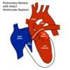
Which Cyanotic Heart Defects are a/w decreased pulmonary blood flow?
TOF
Pulmonary atresia
Tricuspid atresia
Cyanotic Heart Defects - Predominantly Right-to-Left Shunts/Mixing Lesions
Which Cyanotic Heart Defects are a/w increased pulmonary blood flow?
Transposition of the great vessels
Truncus arteriosus
Hypoplastic left heart
Cyanotic (R→L) Heart Defects
The congenital heart condition that involves four abnormalities occurring together, including a defective septum between the ventricles and narrowing of the pulmonary artery, leading to cyanosis is also known as:
Tetralogy of Fallot

Cyanotic (R→L) Heart Defects
Most common CHD producing R to L shunt
Tetralogy of Fallot

Cyanotic (R→L) Heart Defects - Tetralogy of Fallot
What’s the prevalence of Tetralogy of Fallot in neonate?
3rd most prevalent CHD in the neonate

Cyanotic (R→L) Heart Defects - Tetralogy of Fallot
Anatomic defects associated with Tetralogy of Fallot:
VSD (R-to-L)
Aorta that overrides the pulmonary tract
Obstruction of pulmonary outflow tract
Right ventricular hypertrophy

Cyanotic (R→L) Heart Defects - Tetralogy of Fallot
Pathophysiologic characteristics seen with Tetralogy of Fallot:
R-to-L shunting
↓ Pulmonary blood flow
Polycythemia (>65%)
D/t body attempt to compensate for lack of O2 by producing more RBCs
Ductal dependent pulmonary blood flow (L-R shunt) in neonate with severe obstruction (PGE1)

Cyanotic (R→L) Heart Defects - Tetralogy of Fallot
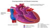
Blood flow in Tetralogy of Fallot
Note:
VSD in TOF is with R=>L shunt
Misplaced aorta that overrides pulmonary tract
Stenosis of the pulmonary valve and pulm artery out of the RV
Thickening of the RV wall

Cyanotic (R→L) Heart Defects - Tetralogy of Fallot
Blood flow in Tetralogy of Fallot
See picture
Note “pulmonary atresia”

Cyanotic (R→L) Heart Defects - Tetralogy of Fallot
Manifestations Tetralogy of Fallot:
Hypoxemia/cyanosis
Clubbing
Squatting
(↑ SVR by reducing blood flow to femoral arteries)
Ejection murmur
Hypercyanotic attacks (“tet spells”), as evidenced by:
Infundibular “spasm” => worsen RV outflow tract obstruction
↓ SVR
Can occur w/o provocation but often associated with crying or exercise
Accompanied by hyperventilation & syncope
Cyanotic (R→L) Heart Defects - Tetralogy of Fallot
Treatment of Tetralogy of Fallot includes:
IV fluids
Knee-to-chest
Phenylephrine (↑ SVR)
Esmolol
MSO4
(caution w/ ↓ venous return and CO a/w MSO4 )
Cyanotic (R→L) Heart Defects - Tetralogy of Fallot
The surgical palliation to Tetralogy of Fallot in which the Left subclavian artery is shunted to the left pulmonary artery to increase pulmonary blood flow is known as:
Blalock-Taussig shunt

Cyanotic (R→L) Heart Defects - Tetralogy of Fallot
What’s the difference btw the Traditional and the Modified Blalock-Taussig shunts?
The traditional Blalock-Taussig shunt uses the actual subclavian artery, whereas
The Modified Blalock-Taussig shunt uses a graft to to divert some of the subclavian artery blood flow to the PA
Schematic drawing of original Blalock-Taussig shunt on patient’s right side and modified Blalock-Taussig shunt on left side

Cyanotic (R→L) Heart Defects - Tetralogy of Fallot
Complete correction of Tetralogy of Fallot involves:
Closure VSD
Removal obstructing infundibular muscle
Pulmonic valvulotomy
Cyanotic (R→L) Heart Defects - Tetralogy of Fallot
The main goal of Anesthetic management of Tetralogy of Fallot is to lessen the R-to-L shunt. How can this be accomplished?
Maintain intravascular volume
Maintain SVR/Avoid decreasing
Avoid ↑ PVR
(By avoiding acidosis, hypoxemia, excessive PIP)
Maintain higher FiO2 and lower ETCO2 to prevent PVR increase
Cyanotic (R→L) Heart Defects - Tetralogy of Fallot
Options for inducing Anesthesia in Tetralogy of Fallot
Inhalation with pink patients
Ketamine IV/IM with cyanotic patients
Cyanotic (R→L) Heart Defects - Tetralogy of Fallot
How does R-to-L shunting in Tetralogy of Fallot effect on rate of inhalational induction?
Slows inhalational induction
Slows uptake
(D/t to less blood flow to the lungs in general; Opposite of L-to-R shunts)
Dilutional effect
Cyanotic (R→L) Heart Defects - Tetralogy of Fallot
How does R-to-L shunting in Tetralogy of Fallot effect on rate of IV induction?
Accelerates onset in IV agents
Cyanotic (R→L) Heart Defects
A form of congenital heart disease whereby there is a complete absence of the tricuspid valve. Therefore, there is an absence of right atrioventricular connection. This leads to a hypoplastic (undersized) or absent right ventricle. This condition is also known as:
Tricuspid Atresia

Cyanotic (R→L) Heart Defects - Tricuspid Atresia
Anatomical defects/physiologic characteristics of Tricuspid Atresia:
Complete absence of right atrioventricular connection
Severe hypoplasia or absent RV
Pulmonary blood flow dependent on PDA (L-R shunting)
LA & LV handle both systemic and pulmonary circulations
Systemic venous return shunted from RA => LA via ASD or PFO
Mixing of O2 and deO2 in LA → LV → Aorta = CYANOSIS
90% associated with VSD allowing some blood to enter RV
Normal related great arteries or with transposition