MCBG Session 9 - Post-translational Modification Flashcards
Outline the basis for post-translational modification.
- All proteins adopt a unique 3-dimensional structure to become active
- Some proteins may need additional processing after translation
I. Proteolytic cleavage – breaking peptide bonds to remove part of the protein
II. Chemical modification – addition of functional groups to amino acid residues
Outline protein sorting.
- Protein destined for the cytosol, or posttranslational import into organelles are synthesised on free ribosomes
- Protein destined for a membrane or secretory pathway via co-translational insertion are synthesised by ribosomes and on the rER

Requirements for protein sorting:
- A signal (address), intrinsic to the protein
- A receptor that recognises the signal and which directs it to the correct membrane
- A translocation machinery energy to transfer the protein to its new place
Identify the types of secretion from cells.
- Constitutive secretion – constant flow of extracellular proteins out of the cell e.g. collagen – example of secreted and modified protein
- Regulated secretion:
I. Endocrine cells – secreting hormones
II. Exocrine cells – secreting digestive juices
III. Neurocrine cells – secreting neurotransmitters
Explain how proteins are targeted to the ER/secretory pathway (co-translational transport)
- Protein synthesis on bound ribosomes; cotranslational transport of proteins into or across ER membrane
- Budding and fusion of ER-to-Golgi vesicles to form cis-Golgi
- Retrograde Golgi-to-ER transport
- Cisternal progression
- Retrograde transport from later to earier Golgi cisternae
- Consitutive secretion / Regulated secretion
- Sorting into Lysosomes
- Endocytosis

What are the functions of the ER?
- Insertion of proteins into membranes
- Specific proteolytic cleavage
- Glycosylation
- Formation of S-S bonds
- Proper folding of proteins
- Assembly of multi-subunit proteins
- Hydroxylation of selected Lys and Pro residues
What does glycolisation of proteins do?
- Correct protein folding
- Protein stability
- Facilitates interactions with other molecules
- Deficiencies in N-linked glycosylation lead to severe inherited human disease: Congenital disorders of glycosylation (CDG)
What is misfolding?
- Protein may be trapped in mis-folded conformation
- Protein contains mutation resulting in mis-folding
- Protein may be incorrectly associated with other sub-units
How do ER chaperone proteins attempt to correct misfolding?
- BiP: “Binding Immunoglobulin Protein”
- Calnexin and Calreticulin
- Retain unfolded proteins in the ER
- Act as sensors to “monitor” extent of protein mis-folding
- Mediate increased transcription of chaperones
- Mediate reduction in translation
What happens if protein misfolding cannot be corrected?
- Protein may be returned to cytosol for degradation
- Protein may accumulate to toxic levels in the ER resulting in disease
- This may arise due to single mutation
What is O-linked glycolisation?
O-linked glycosylation: attachment of sugar to -OH group
I. Occurs in Golgi apparatus
II. Attachment of sugar to hydroxyl group of serine, threonine
III. Important in proteoglycans
IV. Component of extracellular matrix and mucus secretions

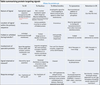
Summarise protein target signals to the ER, nucleus, mitochondria, lysosomes and retention in the ER respectively in terms of:
- Nature of signal
- Location of signal within the primary sequence
- Folded or unfolded during transfer?
- Involvement of specialist proteins
- Signal retained or cleaved?
- Requires energy?