MCBG Session 16 - Case Studies Flashcards
What is cytogenetics?
Cytogenetics: the study of the genetic constitution of cells through the visualisation and analysis of chromosomes.
What are the benefits of cytogenetic analysis?
- Accurate diagnosis/prognosis of clinical problems
I. Identify the syndrome associated with abnormality
II. Account for phenotype
III. Account for pregnancy loss
- Better clinical management – E.g. hormone treatment for Klinefelter syndrome
- Prenatal diagnosis – TOP of affected pregnancy/planning management at birth
- Assess future reproductive risks
I. Risk of live born abnormal child
II. Previous Down’s pregnancy, approx. 1% increase above pop risk of another
The referral reasons for cytogenetics are due to constitutional abnormalities and acquired abnormalities. Outline the former.
- Prenatal diagnosis – Chorionic villus sampling and Amniocentesis
- Birth defects – dysmorphism, congenital malformations, mental retardation, developmental delay (abnormal behaviour, learning difficulties), specific syndromes (Down syndrome, Williams syndrome, DiGeorge syndrome)
- Abnormal sexual development
- Infertility
- Recurrent foetal loss

The referral reasons for cytogenetics are due to constitutional abnormalities and acquired abnormalities. Outline the latter.
- Leukaemia’s
I. Acute diseases – AML/ALL
II. Chronic diseases – CML
III. Myelodysplasia/ Myeloproliferative disorders
- Solid tumours
- Specific translocations/abnormalities can give prognostic information
Outline the chromosome analysis. (incl define karyotyping)
- Karyotyping: the systematic sorting of chromosomes
- Whole genome screen 5-10Mb resolution
- Metaphase chromosome stained, paired up and grouped together
- Abnormalities described using standard nomenclature ISCN
Outline the steps involved in chromosome analysis.
- Count the number of chromosomes
- Identify each chromosome pair
- Assess if there is any missing or extra material – Are the bands in the right place?
- All pairs must be seen at the correct resolution twice
- All chromosomes independently rechecked once

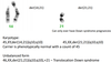
Provide examples of standard nomeclature:
- Normal female
- Normale male
- Female with trisomy 21
- Male with chromosome 7 inversion
- 46,XX – normal female
- 46,XY – normal male
- 47,XX,+21 – female with trisomy 21
- 46,XY,inv(7)(p11.2q11.23) – male with chromosome 7 inversion
Identify some numerical cytogenetic abnormalities (incl. define aneuploidy)
- Aneuploidy – loss and gain of whole chromosomes
- Arise due to errors at cell division in meiosis
I. Trisomies – Down syndrome +21, Patau syndrome +13 and Edwards syndrome +18
II. Monosomies – Turner syndrome 45,X (X inactivation, only full monosomy to be viable)
What is polyploidy?
- Gain of a whole haploid set of chromosomes
- Triploid 3n
- 69, XXX

What are the causes of polyploidy?
- The most common cause of polyploidy is polyspermy: fertilisation of an egg by more than one sperm.
- Triploidy occurs in 2-3% of all pregnancies and ~15% of all miscarriages: term deliveries die shortly after birth
- Tetraploidy is rarer (1-2%) but tetraploid cells are often found at prenatal diagnosis as a cultural artefact
- Diploid/triploid mosaicism seen in livebirths
What are the causes of aneuploidy?
- Originates from non-disjunction at one of the meiotic cell divisions
- Forms gametes with a missing chromosome and an extra chromosome – which chromosomes involved will influence viability.
- Can occur during mitotic cell division – causes mosaicism i.e. two cell populations in an individual
What is anaphase lag?
- Chromosomes can be ‘left behind’ at cell division because of defects in spindle function or attachment to chromosomes
- The lagging chromosomes may be lost entirely in mitosis or meiosis

What is Down syndrome?
- Trisomy 21
- Frequency 1:650-1000
- Hypotonia
- Manifestations: characteristic facial features, intellectual disability, heart defects
- Increased prevalence of leukaemia
- Increased incidence of early Alzheimers
What is Edwards syndrome?
- Incidence 1:6000; female predominance
- Maternal meiosis II error
- Modal lifespan 5-15 days
- Nearly all diagnoses made prenatally
- Visual features: Small lower jaw, prominent occiput, low-set ears, rocker bottom feet, overlapping fingers

What is Patau syndrome?
- Trisomy 13
- Incidence 1:12 000
- Majority die in neonatal period
- Holoprosencephaly
- Polydactyly
- Multiple congenital abnormalities

What is X chromosome inactivation?
- Only one X chromosome is ever active in a human cell
- Males only have one X chromosome
- Females have two X chromosomes
- X inactivation ensures individuals have same X chromosome complement that is active
Why is X chromosome inactivation problematic?
- Males only have a single X chromosome.
- However, the X and Y chromosomes have short regions in common at the tips of the long and short arms, allows for pairing during cell division
- Two pseudo-autosomal regions (PAR1 and PAR2)
- Turner syndrome patients will be monosomic for genes in the PARs
- SHOX gene (within PAR) associated with short stature

What is Turner Syndrome?
- Incidence 1:2500
- Majority cases absent paternal X; phenotypic differences depending on parental origin of X
- Visual features: puffy feet, redundant skin at back of the neck
- Manifestations: short stature, heart defects, mild learning difficulties, neck webbing, infertility

Outline what is meant by mosaicism.
- Mosaicism: the presence of 2/more cell lines in an individual. Usually caused by mitotic non-disjunction. Occurs throughout the body or tissue limited
- Degree of mosaicism depends on when the error occurred
I. First post zygotic division – no mosaicism looks like a meiotic event
II. Subsequent divisions – 3 cell lines, monosomy cell line usually lost
- Trisomic conceptus ‘rescued’ to give mosaicism – anaphase lag
- Non-disjunction in later cell division

Identify some cytogenetic structural abnormalities.
- Translocations –Reciprocal & Robertsonian
- Inversions – Paracentric & pericentric
- Deletions – incl. microdeletions
- Duplications
- Insertions
- Rings
- Marker chromosomes
- Isochromosomes
Outline recipocial translocations.
- Two break rearrangements
- Usually unique to a family – t(11;22) is an exception
- Carriers produce balanced and unbalanced gametes
- If unbalanced offspring will have an abnormal phenotype dependant on regions of trisomy and monosomy
- Segregation analysis using pachytene diagram to assess this imbalance

Outline robertsonian translocations.
- Two acrocentric chromosomes fused together – 13,14,15 ,21,22
- Mono or dicentric – 13;14 most common
- Chromosome count of 45 in balanced carriers
- Trivalent formed at meiosis – Not very stable
- Aneuploidy risk
I. Females have higher risk than males
II. Homologous carriers can’t have normal pregnancy
Outline the types of segregation in meiosis I.
- Alternate – balanced
- Adjacent I – non homologous centromeres, most common form to give imbalance
- Adjacent 2 – homologous centromeres
- 3:1 non disjunction
- 4:0 non disjunction – all balanced
Outline Alternate segregation.
Alternate – alternate centromeres segregate together

Outline Adjacent segregation.

What is FISH?
- Fluorescent in situ hybridisation (FISH)
- Molecular cytogenetic technique
- Allows us to answer specific questions – Need to know what to look for
- Probes used for specific chromosomes or loci
- ISCN for FISH probes

What are centromere probes?
- Large probes, easy to see Metaphase and interphase
- Used for: copy number analysis and identifying derivative chromosomes and markers

What is the function of whole chromosome paints?
Can identify individual chromosomes even when they are rearranged

What are locus specific probes?
- Microdeletion/duplication syndromes
- Too small to see on G-banded chromosomes

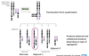
Outline prenatal aneuploidy screening as a form of interphase analysis
- PND - up to 14 days in culture- causes anxiety
- Uncultured cells
- FISH probes for 13,18,21,X &Y – Common aneuploidies
- Result in 24-48hrs
- Full karyotype -14 days
- 99+% concordance with full karyotype – Many patients TOP after FISH

Outline Leukaemia FISH as a form of interphase analysis.
- Look for different types of abnormalities
- Translocations – fusion probes for the genes involved
- Gene rearrangements – breakapart probes
- Amplifications – locus specific probes (E.g. her2, c-myc oncogenes)

What is microarray comparative genomic hybridisation (aCGH)?
- Examines the whole genome at high resolution
- Copy number changes
I. Can’t detect balanced rearrangements
II. Not used for mutation detection
- Uses patient DNA not chromosomes
- Compare normal control DNA to patient DNA
- 15-20% abnormality rate in developmental delay cohort.

What is CGH slide scanning?
- Scan slide with 3 μm laser scanner
- Expect 1:1 ratio for test and reference – giving a yellow colour
- Excess red = more reference DNA and deletion of test DNA
- Excess green = more test DNA and duplication of test DNA
What are the aCGH referral groups?
- Learning difficulties/ developmental delay/ multiple congenital abnormalities
- Normal karyotype
- Balanced de novo karyotype – is it really balanced ?
- Unbalanced karyotypes to assess gene content
Read the advantages and disadvantages of Array CGH?
- Advantages
I. Examines the entire genome at a high resolution
II. Targeted against known genetic conditions and sub telomere regions
III. 1 array is the equivalent of many thousands of FISH investigations & can be automated
IV. Detailed information on genes in del/dup region
V. Better phenotype/genotype correlation
- Disadvantages
I. Arrays are more expensive than karyotyping
II. Will not detect balanced rearrangements – not suitable for referrals
III. Copy number variation (CNV) – what is genuine abnormality ?
IV. Mosaicism may be missed
Outline array analysis.
- Scan array slides
- Input scans into software – Aligent Cytogenomics
- Run appropriate algorithm for array type
- Results file produced Quality criteria must be reached
- Interpret any copy number calls (CNV? Pathogenic?)
- Follow up using FISH/MLPA/array
Outline the results in accordance to:
I. Normal result
II. Pathogenic (causative) change
III. Uncertain change
IV. Benign finding
- Normal result
- Pathogenic (causative) change
I. Clear pathogenic finding
II. Specific gene(s) or Syndromic region
- Uncertain change
I. Possibly pathogenic – novel copy number change with relevant gene content
II. Likely benign – novel, large copy number change with no genes or those unlikely to be relevant to phenotype
- Benign finding
I. Polymorphic finding (use Database of Genomic Variants to assess)
II. Generally not reported
