MCBG Session 11 - Protein Function and Regulation Flashcards
(20 cards)
Briefly, introduce the concept of short term regulation.
- Substrate availability will affect the rate of enzyme activity
- Isoenzymes are different forms of the same enzyme that have different kinetic properties
- Some coenzymes will have limited availability e.g. NAD/NADH
- Product inhibition – accumulation of the product of a reaction inhibits the forward reaction e.g. Glucose-6-phosphate inhibits hexokinase activity

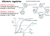
Outline allosteric regulation.
- Allosteric enzymes show a sigmoid relationship between rate and substrate concentration, instead of the rectangular hyperbola seen for simple enzymes.
- Multi subunit enzymes can exist in 2 different conformations:
I. T state – low affinity
II. R state –high affinity
- The substrate binding to one subunit makes subsequent binding to other subunits progressively easier.

What are allosteric activators and inhibitors?
- Allosteric activators - increase the proportion of enzyme in the R state.
- Allosteric inhibitors - increase the proportion of enzyme in the T state
To supplement allosteric regulation, outline the example of Allosteric regulation of phosphofructokinase

Identify some common covalent modifications of protein activity.
- Modification: Phosphorylation
I. Donor molecule - ATP
II. Example of modified protein - Glycogen phosphorylase
III. Protein function - glucose homeostasis; energy transduction
- Modification: Acetylation
I. Donor molecule - Acetyl CoA
II. Example of modified protein - Src
III. Protein function - DNA packing; transcription
- Modification: ADP ribosylation
I. Donor molecule - NAD+
iI. Example of modified protein: RNA polymerase
III. Protein function: Transcription
- Modification: Ubiquitination
I. Donor molecule - Ubiquitin
II. Example of modified protein - Cyclin
III. Protein function - Control of the cell cycle
Outline phosphorylation and explain why it is so effective.
- Protein kinases: transfer the terminal phosphate from ATP to -OH group of Ser, Thr, Tyr
- Protein phosphatases: reverse the effects of kinases by catalysing the hydrolytic removal of phosphoryl groups from proteins.
- Why is protein phosphorylation so effective?
I. Adds 2 negative charges
II. A phosphoryl group can make H-bonds
III. Rate of phosphorylation/dephosphorylation can be adjusted
IV. Links energy status of the cell to metabolism through ATP
V. Allow for amplification effects

Explain the amplification of enzyme cascades by proteolytic cleavage.
- When enzymes activate enzymes, the number of affected molecules increases geometrically in an enzyme cascade.
- Amplification of signals by kinase cascades allows amplification of the initial signal by several orders of magnitude within a few milliseconds
- E.g. Glycogen breakdown and synthesis are reciprocally regulated.

Provide examples of enzyme in biological systems being activated by specific proteolytic cleavage.
- Digestive enzymes are synthesised by zymogens (inactive precusors) in the stomach and pancreas.
- Some protein hormones are synthesised as inactive precursors
- Blood clotting is mediated by a cascade of proteolytic activations that ensures a rapid and amplified response.
- Many developmental processes are controlled by the activation of zymogens to contribute to tissue remodeling.
- Programmed cell death (apoptosis) is mediated by proteolytic enzymes, caspases, which are synthesised in inactive (procaspase) form.

Provide an example of how endogenous inhibitors regulate protease activity.

Outline long term regulation.
- Change in rate of protein synthesis – Enzyme induction/repression
- Change in rate of protein degradation – Ubiquitin-proteasome pathway

Outline the blood clotting cascade.
- Intrinsic pathway: Damaged endothelial lining of the blood cells promotes the binding of factor XII
- Extrinsic pathway: Trauma releases tissue factor (factor III)
- Both pathways activate Factor X (common endpoint)
- Thrombin is subsequently activated
- Thereafter, a fibrin clot is formed.
- Cascade allows the formation of a clot from the activation of very small amounts of the initial factor.

Outline the extrinsic pathway.
- Membrane damage exposes extracellular domain of tissue (factor III)
- Autocatalytic activation of Factor VII

Outline the intrinsic pathway.
- Membrane damage plays a role in activation of the intrinsic pathway
- Factor IX and X are targeted to membrane by Gla domains.
- Ca2+ plays a role
- Required for sustained thrombin activation

Describe the modular structure of prothrombin.
- The protease function (thrombin part) is contained in the C-terminal domain
- The two kringle domains help keep prothrombin in the inactive form
- Gla domains target it to appropriate sites for its activation.

Describe the calcium-binding region of prothrombin.
- Prothrombin binds calcium ions via Gla residues.
- Only prothrombin next to the site of damage will be activated.
- Clots will be localised to the site of damage.

Outine the formation of a blood clot.
- Thrombin cleaves fibrinopeptides A and B from the central globular domain of fibrinogen.
- Globular domains at the C-terminal ends of the b and g chains interact with exposed sequences at the N-termini of the cleaved b and a chains to form a fibrin mesh or clot.
- The newly formed clot is stabilised by the formation of amide bonds between the side chains of lysine and glutamine residues in different monomers.
- This cross-linking reaction is catalysed by transglutaminase, which is activated from protransglutaminase by thrombin.

Outline the structure of fibrinogen.
- 340 kDa protein
- 2 sets of tripeptides, α, β, γ, joined at N-termini by disulphide bonds
- 3 globular domains linked by rods
- N-termini regions of α and β chains are highly negatively charged and prevent aggregation of fibrinogen

Outline the steps involved in the regulation of the clotting process.
- Localisation of (pro)thrombin Dilution of clotting factors by blood flow, and removal by liver
- Digestion by proteases
I. For example, factors Va and VIIIa are degraded by protein C
II. Protein C is activated by thrombin binding to endothelial receptor, thrombomodulin
III. Defects in protein C can cause thrombotic disease
- Specific inhibitors Antithrombin III (AT3) Enhanced by heparin binding AT3-heparin does not act on thrombomodulin-bound thrombin.

Outline classic haemophilia as a defect in factor VIII.
- Factor VIII (‘antihaemophilic factor’) is not a protease, but markedly stimulates the activity of factor IXa , a serine protease.
- The activity of factor VIII is markedly increased by limited proteolysis by thrombin and factor Xa. This positive feedback amplifies the clotting signal and accelerates clot formation.
- Treatment with recombinant factor VIII

State the key control points in blood clotting
- Inactive zymogens present at low concentration.
- Proteolytic activation.
- Amplification of initial signal by cascade mechanism.
- Clustering of clotting factors at site of damage.
- Feedback activation by thrombin ensures continuation of clotting.
- Termination of clotting by multiple mechanisms.
- Clot breakdown controlled by proteolytic activation.
