Male and female reproductive tract histology Flashcards
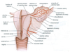
What is the ovary attached to?
What is anchored by?
Posterior face of broad ligament
Anchored by ovarian ligament (to uterus) and suspensory ligament (to pelvic wall)

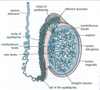
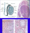
How is the internal structure of the ovary divided?
Inner medulla (loose connective tissue and blood vessels)
Outer cortex (ovarian follicles)
In which part of the ovary are the follicles found?
Outer cortex
Describe the cortical stroma of the ovary?
Highly cellular connective tissue
Scattered with smooth muscle cells
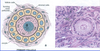
Which organ is this?

Ovary
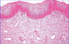
Describe the surface epithelium of the ovary?
Simple squamous or cuboidal
Continuous with mesothelium
Tunica albuginea beneath it
70% ovarian tumours arise here

Describe the tunica albuginea of the ovary?
Beneath the surface epithelium
Dense connective tissue
Oocytes deep to it

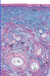
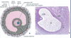
What are primordial oocytes?
Smallest oocytes
Arrested in prophase of meiosis 1
Squamous follicle cells on outside, surrounded by common basal lamina

What are primary oocytes?
Oocyte surrounded by zona pellucida (within follicle cell layer)
Enlarged
Follicular cells become cuboidal and multilayered granulosa cells
Stromal cells start to form theca interna and externa

Which cells form the stratum granulosum in the primary oocyte?
Follicular cells
Which cells form the theca interna and externa in the primary oocyte?
Stromal cells
What is a secondary follicle?
Stratum granulosum thickened
Antrum appears
Cumulus oophorus: stalk of granulosa cells that suspend oocyte
Corona radiata formed by granulosa cells around oocyte after release

What is the cumulus oophorus?
Stalk of granulosa cells that suspend oocyte in secondary follicle

What is the corona radiata?
Granulosa cells around oocyte form corona radiata after release

What is a Graafian follicle?
Mature follicle
When does the oocyte complete its first meiotic division?
Under LH surge
When does the primary oocyte become a secondary oocyte?
When it completes its first meiotic division after the LH surge
What happens after the secondary oocyte is formed?
Follicle ruptures > oocyte released into body cavity > uterine tubes > corpus luteum formed
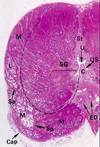
What is the corpus luteum formed from?
Follicle that has lost its oocyte
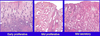
Describe the corpus luteum?
Stromal, granulosa and thecal cells invade cavity to differentitate into luteal cells
Contain lipid and become vascularised

What is the function of the corpus luteum?
Produces progesterone and oestrogen to prepare endometrium for implantation
How long does the corpus luteum last for?
14 days without fertilisation
No fertilisation > regresses to form corpus albicans
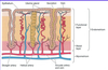
Which important processes occur in the uterine tubes?
Collects released oocytes
Fertilisation and initial development
Describe the structure of the uterine tubes?
Serosa: mesothelium plus thin connective tissue
Muscularis: smooth muscle
Mucosa: connective tissue plus epithelium
How does the structure of the uterine tubes facilitate the movement of oocytes?
Cilia move oocyte towards isthmus
Smooth muscle peristalsis
Where does fertilisation usually occur?
Ampulla of uterine tubes
What are the layers of the uterine wall?
Endometrium (mucosa)
Myometrium (muscularis)

Describe the external surface of the uterine wall?
Covered by perimetrium, which consists of mesothelium and underlying elastic connective tissue
Describe the myometrium?
Three layers of smooth muscle
Inner and outer layers are longitudinal, middle layer is circular
Middle layer is very vascular

How does the uterus accommodate pregnancy?
Enlarges individual smooth muscle cells
Generates more smooth muscle cell
Increases connective tissue
Describe the structure of the endometrium?
EPITHELIUM
Mix of ciliated and secretory columnar cells
Simple epithelium
SECRETORY GLANDS
Penetrate into lamina propria
HELICAL ARTERIES
BASAL LAYER
Regenerates functional layer after sloughing off
Stem cell resevoir

How does the endometrium chnage throughout the menstrual cycle?
What controls this?
Changes thickness
Growth driven by oestrogen
Breakdown due to decrease of oestrogen and progesterone

Describe the structure of the proliferative endometrium?
Abundant coiled glands, which are relatively small and undeveloped

Describe the structure of the secretory epithelium?
Large, well developed glands that contain secretion

Describe the difference between the endocervix and the ectocervix structure?
Endocervix: simple columnar epithelium, glandular, secretory
Ectocervix: simple stratified epithelium, non-glandular

Where are cells shed from in the cervix?
Ectocervix
Stratified epithelium sheds cells
Describe the epithelium of the vagina?
Stratified squamous epithelium
Non-keratinised

Describe the muscular structure of the vagina?
Smooth muscle
Thin inner and thick outer
Continuous with muscle of uterus
Describe the glands of the vagina?
No glands
Lubricated by cervical glands or glands in vestibule
Which factors may cause the structure of the breast to vary?
Age
Stage of menstrual cycle
Reproductive status
At which stages of life are the male and female mammary glands similar?
Until puberty
Describe how puberty affects the male and female mammary glands?
What are the stimuli?
Male: regression due to testosterone
Female: growth due to oestrogen and progesterone
What accounts for most of the growth in female breasts afetr puberty?
Extra adipose tissue
When do mammary glands become active?
Pregnancy
How are female mammary glands kept inactive until pregnancy?
Negative influence of stromal cells
Describe the structure of mammary glands?
Modified sweat glands
15-20 lobes
Lactiferous ducts form branching network and in in terminal duct lobular units
Surrounding intralobular stromal tissue

What are terminal ductal lobular units?
Functional metabolic unit that sits at the head of the mammary gland
Made up of interlobular collecting ducts and acini

What happens to the mammary glands just before and during menstruation?
Gland involutes, some cells apoptose
Describe the appearrance of inactive mammary glands?
Sparse
Cuboidal/columnar epithelial cells
Surrounding myoepithelial cells
Extensive dense connective tissue

Describe the appearrance of active mammary glands?
Terminal ductules elongate and branch
Epithelial and myoepithelial cells proliferate
Large cuboidal epithelial cells that contain lipid and secretory product in lumen

Describe the contents of breast milk?
Protein, lipid and carbohydrate mixture
High in IgA
What stimulates milk production?
Prolactin
Describe the first breast milk that is released?
Colostrum
High protein, low lipid and carbohydrate
Describe the lactation process?
Suckling > inhibits prolactin release-inhibiting hormone > increased prolactin and oxytocin released
Prolactin stimulates milk production
Oxytocin acts on myoepithelial cells > milk ejection
Describe the changes that occur in the mammary glands after menopause?
Mammary glands involute
Secretory cells disappear
Connective tissue loses elastic and collagen fibres and fibroblasts
Describe the structure of the testis?
Surrounded by tunica albuginea
Divided into 250 compartments by septa from tunica albuginea
Seminiferous tubules located in compartments

Describe how the seminiferous tubules connect to the vas deferens?
Seminiferous tubules > rete testis > efferent ductules > epididymis > vas deferens

Describe the structure of the seminiferous tubules?
Stratified epithelium
Surrounded by tunica propria, which contains myoid cells and Leydig cells

Describe the function of the seminiferous tubules?
Spermatogenesis site
Tunica propria contracts to move sperm down tubules
Leydig cells produce testosterone

What is the function of Sertoli cells?
Support cells for spermatogenesis
Promote cell division and privide correct environment
When are Leydig cells active?
First 5 months of foetal life
Puberty onwards
Describe the tubuli recti?
Last, straight part of seminiferous tubule
Lined with Sertoli cells only and finally simple cuboidal epithelium
Connect to rete testis

Describe the structure and function of the rete testis?
Channels lined with ciliated cuboidal cells
Allox mixing of spermatozoa from different tubules
How is the rete testis connected to the epididymis?
Efferent ductules

Describe the structure of the epididymis?
Ciliated pseudostratified columnar epithelium
Epithelium surrounded by smooth muscle
Highly coiled (4-6m)
Head, body and tail
Spermatozoa in lumen

Describe the function of the epididymis?
Site of sperm maturation
Decapacitation (inability of sperm to fertilise egg)
Absorbs most testicular fluid around sperm, clean up debris
Describe the pathway of the vas deferens?
Epididymis > vas deferens > abdomen via inguinal canal > over lip of pelvis > descends to prostate > through prostate as ejaculatory duct
Describe the structure of the vas deferens?
Pseudostratified ciliated columnar epithelium
Thick smooth muscle coat (not in ejaculatory duct)
Describe the structure of the seminal vesicles?
Thick mucosa
Thin smooth muscle
Describe the function of the seminal vesicles?
Secrete fructose and prostaglandin rich secretion
Modify acid of vagina (alkaline)
Provide 50% volume of semen
Describe the structure of the prostate?
Lobular
Urethra located centrally
Inferior paired ejaculatory ducts
Branching prostatic (urethral) sinuses
Main prostatic glands (located peripherally), drain into urethra via long ducts
Submucosal glands (located medially), drain via prostatic sinuses

Describe the zones of the prostate?
Central zone: surrounds ejaculatory ducts
Peripheral zone: surrounds central zone
Transitional zone: surrounds urethra
Periurethral zone: immediatley adjacent to urethra

Describe the histology of the prostate?
Heterogenous epithelium: columnar or cuboidal, stratified
Separated by connective tissue
Abundant smooth muscle

What may be found within the lumen of prostate glands?
Prostatic concretions
Contain protein, calcium phosphate, cholesterol and cellular debris
More common with age

Describe the function of the prostate gland?
Produces a complex secretion that intially clots the ejaculate
Fibrolysin (in secretion) then dissolves clot
Describe the structure of the penis?
Three separate masses of erectile tissue: 2 corpora cavernosa and 1 corpus spongiosum
Surrounded by tunica albuginea
Urethra within corpus spongiosum

Describe how penile erection occurs?
Hydraulic means
Depends on blood and blood pressure
Describe the histology of the penile tissue?
Cavernous tissue contains many potential vascular spaces
These spaces are surrounded by smooth muscle and connective tissue

Describe how the nervous system controls penile function?
Parasympathetic stimulation: relaxes cavernosal smooth muscle and dilates arteries supplying blood > swelling of the cavernous bodies compresses veins in supercial penis, restricting outflow > erection
Sympathetic stimulation: restricts inflow of blood and contracts cavernosal smooth muscle > detumescence
Describe the histology of the clitoris?
Body: bilateral cavernous tissue surrounded by tunica albuginea
