Lungs and Pleura Flashcards

1
Q

What does the right pleural cavity and the left pleural cavity contain?

A

RPC - Right lung LPC - Left lung

2
Q

What are the 3 spaces outlined in pink, blue and yellow?

A

Pink - Right pleural cavity Blue - Mediastinum Yellow - Left pleural cavity

3
Q

What is the mediastinum and what does it contain?

A

Space between the 2 pleural cavities

Contains trachea, heart, oesophagus, blood vessels and nerves

4
Q

What are the 3 spaces outlined in yellow, blue and pink?

A

Yellow - Right pleural cavity

Blue - Mediastinum

Pink - Left pleural cavity

5
Q

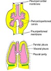
Complete the diagram of devloping lungs and pleura

A
6
Q

At how many weeks does the respiratory system start to develop?

A

4-6 weeks

7
Q

Does the lungs and pleura arise from the ectoderm, mesoderm or endooderm layer?

A

Endoderm

8
Q

How does the lungs and pleura develop?

A
  1. Endodermal out pouches form lung buds and bronchial buds
  2. Rapid division of the airways occurs
  3. Hence the lungs push their way out into the primitive thoracic cavity.
  4. As they do they take the lining with them (visceral pleura)
9
Q

Is this the visceral or parietal pleura?

Closely adheres to the lungs including the fissures

A

Visceral pleura

10
Q

What is the function of visceral pleura?

A

Creates a smooth and slippery surface

11
Q

Which 2 structures does the visceral pleura connect to?

A

The parietal pleura and the hilum

12
Q

Complete the diagram

A
13
Q

Complete the diagram of the segments of parietal pleura

A
14
Q

Is this the visceral or parietal pleura?

Lines the internal aspect of the thoracic wall

A

Parietal pleura

15
Q

What are the 4 parts of the paritetal pleura and where do they line?

A
  1. Costal (Internal rib cage)
  2. Mediastinal (Lateral wall of mediastinum)
  3. Diaphragmatic (Superior diaphragm)
  4. Cervical (Cervical region)
16
Q

What is the pleural cavity?

A

Space between the 2 layers of pleura

17
Q

What does the pleural cavity contain?

A

A thin layer of serous fluid

18
Q

How is the pleural cavity important for breathing?

A

Surface tension of the serous fluid hold lungs against thoracic wall

Lungs expand as the thoracic cavity expands

19
Q

Which 2 pathologies cause the surface tension between the pleural layers to be lost?

A

Pneumothorax

Haemothorax

20
Q

What is this arrow pointing to?

A

Pleural cavity

21
Q

Label the pleural cavity recesses

A
22
Q

Why is the pleural cavity not symmetrical?

A

Because of the heart

23
Q

When can the pleural cavity recesses be occupied by lung?

A

During forced inspiration

24
Q

Name the innervation for the 4 parts of the parietal pleura

Cervical

Costal

Mediastinal

Diaphragmatic

A
  • Cervical – 1st intercostal nerve
  • Costal – intercostal nerves
  • Mediastinal – phrenic nerve
  • Diaphragmatic – lower intercostals and phrenic nerves
25
How is the visceral pleura innervated? What type of innervation and where from.
Autonomic (sympathetic and parasympathetic) from pulmonary plexuses
26
Label the nerves on the diagram
27
Label the lobes and fissures on each lung.
RIGHT LUNG 1) Superior 2) Middle 3) Inferior Green line - Oblique fissure Blue line - Horizontal fissure LEFT LUNG 1) Superior 2) Inferior Green line - Oblique fissure
28
What are the 2 anatomical features the left lung has that the right lung does not?
1. Lingula 2. Cardiac notch
29
Label the 3 surfaces highlighted on the lung
Yellow - Costal Green - Mediastinal Blue - Diaphragmatic
30
What is the top and bottom of the lung referred to as?
The apex and base
31
What are the 3 borders of the lung?
1. Anterior 2. Inferior 3. Posterior
32
Complete the diagram of the pleural and hilum structures
33
Name the 6 structures which make up the hilum
1. Bronchi 2. Pulmonary arteries 3. Pulmonary veins 4. Bronchial arteries and veins 5. Pulmonary plexus 6. Lymph vessels and nodes
34
Label the hilum
35
Where does deoxygenated blood leave the heart from?
Right ventricle via left or right pulmonary artery
36
Where does oxygenated blood enter the heart?
Left atrium via left or right superior or inferior pulmonary veins
37
Which 2 structures do the bronchial arteries supply oxygen and nutrients to?
Lung and visceral pleura
38
Where do the left and right bronchial arteries originate from?
Left arteries branch from descending aorta. Right arteries branch from intercostal arteries.
39
Where do bronchial veins terminate?
Azygous system of veins
40
Complete the diagram of bronchial arteries and veins.
41
Which nervous system innervates all viscera in the body?
Autonomic nervous system (sympathetic and parasympathetic)
42
Where does the sympathetic and parasympathetic innervation of the lungs originate from?
Sympathetic – from sympathetic trunk Parasympathetic – from vagus
43
What is the effect of the sympathetic and parasympathetic nervous system on the - Bronchial musculature Pulmonary vasculature
Sympathetic * Bronchial musculature – dilates * Pulmonary vasculature – constricts Parasympathetic * Bronchial musculature – constricts * Pulmonary vasculature - dilates
44
What 3 nodes does lymph drain via on its way to the hilum?
1. Bronchopulmonary (Hilar) nodes 2. then Tracheobronchial nodes 3. then Paratracheal nodes
45
Complete the diagram of the lymph nodes
46
Name the 7 levels of the respiratory tree
1. Trachea 2. Primary bronchi (main) 3. Secondary bronchi (Lobar) 4. Tertiary bronchi (Segmental) 5. Bronchioles (Terminal/Respiratory) 6. Alveolar sacs 7. Alveoli
47
Complete the diagram of the bification of the trachea
48
Where does the trachea run?
Distal to larynx Runs from C6 to T4/5
49
What 2 tissues makeup the trachea? Do they sit anteriorly or posteriorly?
Anteriorly c-shaped cartilage rings Posteriorly Trachealis muscle
50
What is the name of the angle where the trachea bifurcates? What spinal level is this?
The sternal angle T4/5
51
What 2 types of membrane trac is the trachea lined by?
Mucous membrane and cilia
52
Complete the diagram of the respiratory tree
53
Which main bronchus is wider, shorter and runs more vertically?
Right main bronchus
54
Where do the main bronchi enter the lung?
Hilum
55
How many secondary bronchi are in each lung?
3 in the right lung 2 in the left lung
56
What does each tertiary bronchus supply?
A bronchopulmonary segment
57
How many bronchopulmonary segments does the right and left lung have?
Usually, 10 in the right lung vs. 8-10 in the left lung
58
Which section of the respiratory tree contains cartilage?
Primary bronchi
59
Where does the blood supply for bronchopulmonary segments come from?
A single branch of the pulmonary artery
60
What seperates the bronchopulmonary segments?
Connective tissue
61
Why are bronchopulmonary segements surgically resectable?
They're removable in isolation
62
How many generations of conducting bronchioles are there?
20-25
63
Which part of the respiatory tree does not contain cartilage?
Bronchioles
64
What is the difference between terminal bronchioles and respiratory bronchioles?
* Terminal bronchiole gives rise to several generations of respiratory bronchioles * Respiratory bronchioles characterized by out pockets of alveoli
65
What are the different zones called?
Conducting zone Respiratory zone
66
What are the black stars?
Alveolar sacks
67
What are alveolar ducts?
Eelongated airways lined by alveoli which lead to common spaces called alveolar sacs
68
What is each alveolus surrounded by?
Capillaries
69
What are the 2 cells types in alveoli and what percentage do they make up?
Type 1 pneumocytes (95%) Type 2 pneumocytes (5%) – secrete PULMONARY SURFACTANT
70
Complete the diagram of the respiratory tree