Lung Tumors Flashcards

1
Q

What is the reasoning as to why adenocarcinomas have increased in prevalence in more recent years?

A

• Filters on cigarettes may make us inhale deeper and pull the smoke into alveolar portion of the lungs

2
Q

Differentiate a driver and passenger mutation.

A

Driver mutations are the mutations that allow the cell to become immortal in the first place.

Passenger mutations occur as a result of the rapid growth of the cell with a relatively unchecked genomic structure

3
Q

What are the 2 common signal transduction pathways used by RTKs (receptor tyrosine kinases)?

A
4
Q

T or F: as tumors proliferate they become a heterogenous mass of cells, all of which may have different mutagenic properties.

A

True, as cancer proliferates passenger mutations may accumulate and confer advantages to certain cells.

5
Q

What are oncogenes?

A

Viral or Cellular (proto-oncogenes) genes that encode proteins needed to subvert normal growth control mechanisms

6
Q

T or F: proteins driving cancer are mutated versions of proteins normally present in the cell

A

FALSE, these genes may just be amplified causing overexpression of a normal gene product

7
Q

What is lung cancer screeing a good idea?

A

Because Prognosis of cancer is related to the stage of cancer at the time of diagnosis

8
Q

With what two key substances do we see cigarette smoke having a synergistic effect with in causing lung cancer?

A

Asbestos (construction workers, mechanics, miners, etc)

Radon (uranium miners, people in houses on top of radon heavy ground)

9
Q

What is suggested by the fact that only 10% of smokers get lung cancer?

A

10% get lung cancer but the others don’t so there must be a predisposition to getting lung cancer

10
Q

What are some reasons you might see a nodule in your lung on CXR?

A

• Hamartoma
• Granuloma
• Infection (particularly fungal, mycobacterial)
• Rheumatoid lung
• Silicosis

11
Q

If you see a nodule on a CT scan of a smoker, what factors should you consider?

A

Is the patient high risk?
- do they smoke, how old, other exposures, hemoptysis, other malignancies

Is the nodule high risk?
- Size (more than 3cm, typically malignant, less than 0.8 typically not), location (upper lung - favors malignancy), Appearance (spiculated or calcified), Growth

12
Q

What physical characterisitics of a lung tumor would make you think it is maligant?

A

A mass greater than 3cm, that appeared as a spiculated lesion in the upper lobe, that was growing

13
Q

What physical characteristics of a tumor would make you think its benign?

A

A mass smaller than 0.8cm that is not in the upper lobe and appears calcified without evidence of growth

14
Q

What is a hamartoma?
• describe what they are?
• What tissue are they typically composed of when in the lung?

A

Hamartoma is **normal tissue that is abnormally arranged

•**
Typically this just looks like a rounded COIN LESION on x-ray, that is PERIPHERALLY located, SOLITARY, and WELL CIRCUMSCRIBED.

• these are nodules of MATURE connective tissue that show evidence that they may be neoplastic

15
Q

What is a choristoma?

A

Choristoma - normal tissue in an abnormal location

16
Q

What is this lesion and what would you expect it to look like on CXR?
• describe the key gross characteristics.

A

Key gross characteristics: Lobulated, grey-white glistening nodule

Hamartoma - typically present as well circumscribed solitary coin lesions near the periphery.

17
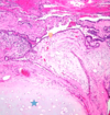
Q

This tissue was biopsied from someone’s lung.
• is this mass benign or maligant?
• how do you know?

A

Benign, this is a haratoma containing Cartilage, Fat, and respiratory Epithelial cells. None of the components of this specimen appear to be neoplastic (all tissue is neatly formed).
18
Q

What are the 4 histologic type of primary lung cancer?

A
  • *1. Adenocarcinoma
    2. Squamous cell carcinoma
    3. Large cell carcinoma
    4. Small cell carcinoma**
19
Q

Note: 87% of people with lung cancer are smokers or ex-smokers. People who smoke 1 ppd have a 10x risk of getting lung cancer. People who smoke 2 ppd have a 60x risk of getting lung cancer

A

Note: 87% of people with lung cancer are smokers or ex-smokers. People who smoke 1 ppd have a 10x risk of getting lung cancer. People who smoke 2 ppd have a 60x risk of getting lung cancer

20
Q

Do you expect this mass to be cancerous when a biopsy is done?

A

Yes this is a PERIPHERALLY located spiculated mass that appears to be large

• Peripheral location implies that this is an adenocarcinoma or Bronchoalveolar carcinoma

(note: Gupta does not seem to distinguish between these two, instead uses the term lepidic growth pattern of adenocarcinoma)

21
Q

What is the most common histological type of cancer among non-smokers and female smokers?
• what are some key features of this cancer?

A

Most common histologic type:
• Lung Adenocarcinoma, these tumors are glandular and often produce mucin

22
Q

What is lepidic growth?
• what type of lung cancer is this associated with?

A

Lepidic growth - tumor that creeps along that alveoli to thicken them and sometimes gives a pneumonia-like consolidation appearance

23
Q

What’s in your differential when you see a tumor as shown here?

A

This tumor appears as a single mass and is peripherally located. this indicates two things:

  1. This is probably a primary lung cancer b/c its a single lesion
  2. Peripheral location means it could be Adenocarcinoma (or bronchoalveolar) OR Large Cell carcinoma
24
Q

What is this showing?

A

Adenocarcinoma that has caused a pneumonia-like consolidation in an entire lobe

25
Q

What tumor-type is likely shown here?
• how would you confirm this by histological means?

A

Lung Adenocarcinoma, a mucicarmine stain is extremely useful and should be positive since most of these tumors are mucin secreting. It stains mucin pink

26
Q

What is cancer is this?

A

Adenocarcinoma of the lung

27
Q

What cancer is this?

A

Lepidic pattern of growth in a lung adenocarcinoma (bronchoalveolar carcinoma)

28
Q

What cancer(s) come into your differential when you see this on CT?

A

Squamous cell carcinoma or Small cell carcinoma (remember that squamous lung is due to respiratory to sqaumous metaplasia from the stress of smoke and that small cells are derived from neuroendocrine cells that are typically found centrally)

29
Q

Who is most likely to get squamous cell carcinoma?
• what are 3 three things to think about when you hear squamous cell carcinoma?

A

Males that Smoke are the most likely people to get squamous cell carcinoma of the lung

  1. Squamous cell tumors have a tendency to cavitate
  2. You will see Keritin Pearls or intercellular bridges on histo.
  3. PTHrP may be produced leading to hypercalcemia in these people
30
Q

Flip to see a quick review of how Calcitriol whose renal sythesis is upregulated in squamous cell carcinoma as a result of PTHrP affects osteoclasts

A
31
Q

What type of lung cancer is shown here?
• what characteristics give this away?

A

This looks like squamous cell carcinoma of the lungs which is typically located centrally as shown here and is associated with cavitation (this is why we can’t use most VEGF drugs here)

32
Q

What cancer is this histology indicative of?

A

Squamous Cell Lung Carcinoma
• You can clearly see keratin pearls here

Below we can see the intracellular bridging

33
Q

What type of lung cancer is this?
• key features?
• where was this tumor likely found?

A

Large Cell Lung Carcinoma
• Large Cells with prominent nucleoli with vesicular chromatin. No glandular or squamous differentiation is evident

• Large Cell carcinomas are often found peripherally

34
Q

T or F: large cell carcinoma has a good prognosis.

A

FALSE, px. for large cell carcinoma is poor

35
Q

What primary lung cancer is this most likely to be?
• why?

A

Small Cell carcinoma of the lung. These are typically centrally located with infiltrative growth into surrounding tissues

36
Q

What lung cancer has the strongest correlation to smoking?

A

Small Cell Lung Carcinoma

37
Q

What are some key histologic features to look for in small cell carcinoma?
• what are some paraneoplastic syndroms to associate with this cancer?

A
  • Small cells with scant cytoplasm (high N/C) and Granular Chromatin
  • Nuclear molding, abundant mitoses, crush artifact

Paraneoplastic Syndroms:

  • *• SIADH** - (excessive resoption via aquaporins of H2O causing hyponatremia, vaptans maybe could be used to treat in severe case)
  • *• excessive ACTH secretion** - could cause cushings like effects with moonface and gynecomastia
  • *• Lambert-Eaton Syndrome** - mimics myasthenia gravis by blocking communication at neuromuscular junction via Abs to Ca2+ volted channels
38
Q

What does this lung histology show?

A

Small cells with a high N/C ratio along with Crush Artifact, nuclear molding and abundant mitoses

Shown below is the Azzopardi phenomenon that sometimes happens when a bunch of DNA from these cells deposits around a vessel

39
Q

What two stains are very useful in determining if something is a small cell carcinoma?
• whay do these stains work?

A

Chromogranin and synaptophysin - are used for NEUROENDOCRINE tumors. Since small cell carcinoma of the lung is a neuroendocrine tumor, this stuff works

40
Q

What type of lung cancer is shown here?
• where in the lung was this biopsy most likely taken?

A

Carcinoid Tumor shown here. This tumor is normally Centrally Located
Notice that these are bland cells with granular chromatin

Notice on the picture below how the tumor forms nests of little island in the stroma

41
Q

Carcinoid Tumor
• who is usually affected?
• How common is this?
• Px?

A

Younger people (50’s) are typically affected and the prognosis is much better than most lung cancer. Not associated with smoking and accounts for only 1-5% of primary lung tumors.

42
Q

What primary lung tumor is this?

A

Carcinoid tumor. You can tell this tumor is centrally located (often endobronchial) by its close proximity to the bronchus seen in the picture.

• Sometimes you even get a collar button appearance inside the airway (seen below)

43
Q

What paraneoplastic syndrome is this guy suffering from?
• what are some symptoms of this syndrome?
• what chemical is most responsible for the manifestation of this syndrome?

A

Carcinoid syndrome is caused predominantly by serotonin as well as other mediators like neuron-specific enolase, bombesin, calcitonin, and others. Symptoms include diarrhea, flushing, and cyanosis. Only about 10% of bronchial carcinoids give rise to this.

44
Q

How is lung cancer staged?

A

TMN

T category
• tumor size and how close is it to invading other structures into and out of the lung

N category
• How many nodes have been invaded

M category
• Distant Metastases (liver, brain, bone)

45
Q

What is the most prevalent driver mutation in adenocarcinomas?
• what is the most prevalent in adenocarcinomas of non-smokers?
• what non-smokers often get adenocarcinoma of the lung?

A

Across all Adenocarcinomas of the lung K-ras mutations are the most prevalent. In non-smokers the EGFR mutation is the most common.
Non-smoking, Asian Females often get adenocarcinoma associated with EGFR mutations

46
Q

what causes SVC sydrome in someone with lung cancer?

A

SVC sydrome results from things pressing on the SVC resulting in a build of pressure in the upper extremities and head. If this condition develops slowly enough anastomoses will help with venous return

Notably: the azygos vein, the hemiazygos vein, and the connecting intercostal veins. The second pathway is the internal mammary venous system plus tributaries and secondary communications to the superior and inferior epigastric veins. The long thoracic venous system, with its connections to the femoral veins and vertebral veins, provides the third and fourth collateral routes, respectively.

47
Q

What is the etiology of these tumors in the lung?

A

These tumors are most likely from METASTATIC cancer because it is multiple lesions. Often these are called Cannonball nodules

48
Q
A
49
Q

What is the most common cause of pleural tumors?
• what is the typicall pattern of involvment?

A

Lung and Breast tumors that metastasize into the pleura are the most common causes of pleural tumors. Typically these appear as nodules or masses in the pleura and effusions. Rarely is there diffuse growth.

50
Q

Malignant Mesothelioma:
• where does it arise from?
• Latency?
• Association with smoking?
• Prognosis

A

Malignant Mesothelioma typically arises from either parietal or visceral pleura. Typically it takes 20 years for this disease to manifest after heavy exposure to asbestos (an is only seen in 7-10% of these ppl.) As opposed to primary lung cancers, mesothelioma risk is not increased by smoking. Patients typically live only a year after diagnosis.

51
Q

What chromosomal deletions are tied to mesothelioma?
• viruses?

A

Chromosomal deletions 1p, 3p, 6q, 9p, 22q are common

Simian Virus 40 (SV40) is often present in people with mesothelioma

52
Q

Note how this histologic section looks a lot like adenocarcinoma that has metastasized. A good clinical hx. is very important here

A
53
Q

Differentiate Mesothelioma and Adenocarcinoma of the lung on the basis of:
• Histochemical Stains
• Immunohistochemical stains
• Electron Microscopy

A

Adenocarcinoma typically will stain with a mucin-specific histochemical stain and a Carcinoembryonic antigen immunohistochemical stain. On electron microscopy they often have stubby microvillus rootlets.

Mesothelioma typically stains with a hyaluronic acid histochemical stain and a calretinin IHC stain. Microvilli in these cells are long and slinder.

54
Q

Why is the calretinin stain not that useful?

A

Calretinin can really only assure that what you’re looking at is mesothelial cells, but it doesn’t tell you if the cells are cancerous

Shown below is a calretinin stain

55
Q
A