Lung Path 3 - Interstitial through Diseases of Vascular Origin (Singh) Flashcards

1
Q

List some of the Interstitial lung diseases

A

UIP: ususal interstitial pneumonia

NSIP: non-specific interstitial pneumonia

COP: Cryptogenic Organizing Pneumonia
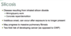
Sarcoidosis
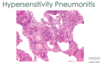
Hypersensitivity Pneumonia
DSIP: Desquamative Interstitial Pneumonia
RB-ILD: Respiratory Bronchiolitis-Interstitial Lung Disease

  • *LCH:** Langerhans Cell Histiocytosis
  • *PAP:** PulmonaryAlveolarProteinosis
2
Q

What are the clinical sign/sx’s of chronic interstitial pulmonary diseases?

A
  • Dyspnea and Tachypnea
  • End-respiratory crackles “velcro-like”
  • Restrictive patters on PFT (decreased DLCO, FVC)
  • Basilar infiltrates on CXR
  • Cyanosis (late)
  • NO wheezing
3
Q

What is characteristically seen on CXR of someone w/ chronic interstitial pulmonary diseases?

A
  • Bilateral lesions that take form of small nodules, irregular lines, or
  • Ground-glass shadows
4
Q

Grossly, what is the end-stage lung seen in chronic interstitial pulmonary diseases referred to as?

A

Honeycomb lung

5
Q

Which 2 chronic interstitial lung diseases are categorized as granulomatous?

A
  • Sarcoidosis
  • Hypersensitivity pneumonitis
6
Q

What are some of the enviornmental and occupational factors which may be associated with Idiopathic Pulmonary Fibrosis?

A
  • Cigarette smoking
  • Expsoure to metal fumes and wood dust (industrial)
  • Occupations such as: farming, hair-dressing, and stone-polishing
  • GERD has also been implicated
7
Q

Loss-of-function mutations in which 2 genes has been implicated in Idiopathic Pulmonary Fibrosis?

A
  • TERT
  • TERC

*Encode components of telomerase

8
Q

What genetic factors have been assoicated with Idiopathyic Pulmonary Fibrosis?

A

Telomerase mutations

Surfactant mutations

MUC5B variant

9
Q

Some cases of Idiopathic Pulmonary Fibrosis are associated with a genetic variant leading to ↑ secretion of which mucin?

A

MUC5B

10
Q

Idiopathic pulmonary fibrosis is a disease most common in which age group?

A

>50 yo

11
Q

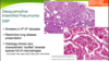
The histologic pattern of fibrosis seen in Idiopathic Pulmonary Fibrosis is referred to as what?

What is the imaging modality to diagnosed this condition based on its characteristic appearance?

A

- Cystic space surrounded by thick excessive fibrosis

- On CT

Usual interstitial pneumonia (UIP)

12
Q

Microscopically, what is the hallmark of Idiopathic Pulmonary Fibrosis; there is coexistence of what?

A

Patchy interstitial fibrosis*** (normal mixed with fibroblastic foci)

–> varies in intensity and age

Coexistence of early (fibroblastic foci) and late (collagenous) lesions

13
Q

In Idiopathic Pulmonary Fibrosis the formation of cystic spaces lined by hyperplastic type II pneumocytes or bronchiolar epithelium is known as what?

A

Honeycomb fibrosis

14
Q

The earliest lesions in Idiopathic Pulmonary Fibrosis contain exuberant what?

A

Fibroblastic proliferation (fibroblastic foci)

15
Q

Diagnosis of Idiopathic Pulmonary Fibrosis requires what?

A

Classic findings on high-resolution CT or pulmonary biopsy

16
Q

What are the initial sx’s of Idiopathic Pulmonary Fibrosis and the later sx’s with progression?

A
  • Early —> ↑ dyspnea on exertion and dry cough
  • Later –> hypoxemia, cyanosis and clubbing
17
Q

What is the prognosis and only definitive therapy for Idiopathic Pulmonary Fibrosis?

A

Median survival 3-5 years after Dx

Lung transplantation

Medication to arrest fibrosis inlcude Tyr kinase inhibitors and TGF-B inhibitors

18
Q

Why is it important to recognize pt’s with Nonspecific Interstitial Pneumonia?

A

Have much better prognosis than those w/ UIP

19
Q

What is the histomorphology of the cellular and fibrosing patterns of Nonspecific Interstitial Pneumonia?

A

Cellular = mild to moderate chronic interstitial inflammation; in UNIFORM parenchyma (can be patchy, but she emphaisized uniform)

Fibrosing = diffuse or patchy fibrotic lesions of roughly the SAME age

Resident cells include lyphocytes and eosinophils surrounding mild to moderate alveolar thickening

–> can be treated at this point.

20
Q

What is a major morphological difference between the lesions of Nonspecific Interstitial Pneumonia and Idiopathic Pulmonary Fibrosis?

A

In NSIP the lesions are at roughly SAME stage of development

21
Q

What 4 morphologial findings are absent in Nonspecific Interstitial Pneumonia?

A
  • NO fibroblastic foci
  • NO honeycombing
  • NO hyaline membranes
  • NO granulomas
22
Q

Which population is most likely to be affected by Nonspecific Interstitial Pneumonia?

A

Female NON-smokers in their 6th decade of life

23
Q

What are the key features of Nonspecific Interstitial Pneumonia seen on high-resolution CT?

A

Bilateral, SYMMETRIC, predominantly lower lobe reticular opacities

24
Q

How do patients with Cryprogenic Organizing Pneumonia (formerly BOOP) present and what is seen radiographically?

A
  • Cough and dyspnea
  • Patchy SUBpleural or

- peribronchial areas of airspace consolidation

25
Q

What is the hallmark histology seen with Cryprogenic Organizing Pneumonia?

A
  • Polypoid plugs of loose organizing CT (Masson bodies) within alveolar ducts, alveoli, and often bronchioles
  • Balls of Fibroblasts
  • superimposed on prior infection or fibrosis of chronic inflmmatory process
26
Q

Prognosis and Tx of Cryprogenic Organizing Pneumonia?

A
  • Some recover spontaneously
  • Most need oral steroids x 6 months + for complete recovery
27
Q

Cryprogenic Organizing Pneumonia is most often caused by what?

A

As a response to infection or inflammatory injury to the lungs

28
Q

Which 3 autoimmune/CT disease can manifest as interstital lung disease?

A
  • Rheumatoid arthritis
  • Systmic sclerosis (scleroderma)
  • SLE

Each of these can present as IPF, NSIP, or Organiznig pneumonia.

29
Q

Inhalation of particles, pneumoconiosis, stimulate what inside the lung which lead to progression of disease?

A

Resident INNATE immune cells

30
Q

Inhalation of coal dust may be a part of what 4 diseases?

A
  • Anthracosis
  • Coal macules/nodules
  • Progressive massive fibrosis –> “Black lung”
  • Caplan syndrome
31
Q

What occurs to the inhaled carbon pigment from coal, leading to Anthracosis?

A

Engulfed by alveolar or interstitial macrophages, which then accumulate in the CT along lymphatics in lungs or in hilus

32
Q

Where are lesions (macules and nodules) of simple coal workers’ pneumoconiosis most often seen in the lungs?

A

Upper lobes and upper zones of lower lobes

33
Q

Indoor use of smoky coal (bituminous) for cooking and heating is associated with what complication?↑

A

↑ risk of lung cancer

34
Q

What is the most prevalent occupational disease worldwide?

A

Silicosis

35
Q

Which race has a higher risk for Silicosis?

A

Blacks

36
Q

Which form of silica (crystalline/amorphous) is the most fibrogenic?

A

Crystalline (i.e., quartz, cristobalite, and tridymite)

37
Q

What occurs after inhalation of silica particles; what is activated and what is released?

A
  • Particles phagocytosed by macrophages
  • Activate the inflammasome
  • Leads to release of inflammtory mediators, IL-1 and IL-18
38
Q

Where in the lungs and in which LN’s are the nodules of silicosis seen?

A
  • Hilar LN’s
  • Upper zones of lungs
39
Q

Histologically, what is the hallmark lesion seen with silicosis?

A

Central area of WHORLED COLLAGEN fibers w/ a peripheral zone of dust-laden macrophages

These dense collagenous nodules manifest as “eggshell calcification” of lymph nodes on radiography

40
Q

Thin sheets of calcification may occur in the LN’s of pt with silicosis and are seen radiographically as what?

A

Eggshell calcification

41
Q

Silicosis is associated with an increased susceptibility to what infection; why?

A

TB; impaired ability of pulm. macrophages to kill phagocytosed mycobacteria

42
Q

The onset of Silicosis may occur in what 3 ways (classification/duration)?

A
  • Slow and insidious = 10-30 yrs after exposure
  • Accelerated = within 10 yrs
  • Rapid = week to months after intense exposure to fine dust high in silica
43
Q

Pt’s with Silicosis have double the risk for developing?

A

Lung cancer (as opposed to CWP)

44
Q

Which asbestos fiber (serpentine or amphibole) is the most dangerous and pathogenic?

A

Amphibole = more aerodynamic, smaller (easy to inhale to deeper pats of the lungs) and less soluble

45
Q

Which property of asbestos fibers accounts for the remarkable synergy btw tobacco smoking and development of lung carcinoma in asbestos workers?

A

Toxic chemicals ca be adsorbed onto the fibers

46
Q

Histologically, asbestosis is marked by diffuse pulmonary interstital fibrosis, indistinguishable from other causes, except for the presence of?

A

Asbestos bodies –> Golden-brown, fusiform or beaded rods (covered in macrophage spit)

47
Q

In asbestosis, inorganic particulates may become coated with iron-protein complexes and are called what?

A

Ferruginous bodies

48
Q

Most common manifestation of asbestos exposure seen on the anterior and posterolateral aspects of the parietal pleura and over the dome of the diaphragm?

Known as?

A

-Well-circumscribed pleural plaques formation (dense collagen, often calcified)

“Candlewax drippings

49
Q

In contrast to other types of pneumoconiosis, where in the lungs does asbestosis typically begin?

A

Lower lobes and subpleurally

50
Q

What type of neoplasm is assoicated with asbestosis?

A

mesothelioma

May occur decades after expsure

Lifetime exposure risk is 10%

51
Q

Which cytotoxic drug used in cancer therapy may lead to pulmonary damage and fibrosis?

A

Bleomycin

52
Q

What is the most common clinical pattern/organs of involvement seen with Sarcoidosis?

A
  • Bilateral HILAR lymphadenopathy and Lung involvement
  • Eye and skin lesions are 2nd in frequency
53
Q

What is the CD4/CD8 T-cell like in pt’s with Sarcoidosis?

A
  • CD4/CD8 T-cell ratios range from 5:1 to 15:1
  • Suggests pathogenic involvement of CD4+ helper T cells
54
Q

Which cytokine found in the bronchoalveolar fluid of pt with Sarcoidosis is a marker of disease activity?

A

TNF

55
Q

Pt’s with Sarcoidosis have abnormal immune responses including anergy to what?

A

Common skin test Ags such as Candida or tuberculosis PPD

56
Q

Which 2 HLA genotypes are associated with Sarcoidosis?

A
  • HLA-A1
  • HLA-B8
57
Q

What is the characterisic histological finding in the tissues of pt with Sarcoidosis?

A

Non-caseating (Non-necrotizing) granulomas composed of clustered epitheloid macrophages, often w/ giant cells

58
Q

What are 2 characteristic morphological findings within giant cells of pt with Sarcoidosis?

A

inclusions known as asteroid bodies

Schaumann bodies –> Laminated concentrations of Ca2+ and proteins

59
Q

Why is there a high diagnostic yield of bronchoscopic biopsies for Sarcoidosis?

A

Relatively high frequency of granulomas in the bronchial submucosa

60
Q

Which LN’s are most frequently affected by Sarcoidosis?

A

Hilar and mediastinal

61
Q

Which 3 sites other than lungs and LN’s are also commonly affected by Sarcoidosis and what is seen in each?

A
  • Spleen - may be enlarged; granulomas often form small nodules
  • Liver - may be enlarged; scattered granulomas often in portal triad
  • Bone marrow - lesions w/ tendency for phalangal bones of hands and feet; small areas of bone resorption within marrow cavity w/ widening of the bony shafts
62
Q

What 3 types of skin lesions may be encountered in Sarcoidosis?

A

Subcutaneous nodules

Erythema nodosum

Flat, slightly red and scaling, resembling those of SLE

63
Q

Scattered granulomas associated with Sarcoidosis may be found in the liver, most often where?

A

Portal triads

64
Q

What is the ocular involvement in some cases of Sarcoidosis?

A
  • Iritis or uveitis, either uni- or bilaterally
  • Corneal opacities, glaucoma, and total vision loss may occur
  • Often accompanied by inflammation of the lacrimal glands w/ suppression of lacrimation
65
Q

Bilateral sarcoidosis of the parotid, submaxillary, and sublingual glands constitutes a combined uveoparotid involvement designated as what?

A

Mikulicz syndrome

66
Q

Muscle involvement in Sarcoidosis may be asymptomatic, but weakness, tenderness, aches, and fatigue should prompt consideration of what?

A

Occult Sarcoid Myositis

67
Q

Majority of pt’s with Sarcoidosis will present due to what signs/sx’s?

A
  • Insidious onset of respiratory abnormalities (i.e., dyspnea, SOB, cough, chest pain, hemoptysis)
  • Constitutional sx’s –> fever, fatigue, weight loss, night sweats

–> FEQUENTLY INCIDENTAL FINDING

68
Q

Elevated serum levels of what enzyme and ion may be seen in Sarcoidosis?

A
  • ↑↑↑ ACE
  • ↑ 1ᾳ-hydroxylase –> hypercalcemia
69
Q

What is the prognosis of Sarcoidosis; how does death most often occur?

A
  • 60-70% recover w/ minimal or no residual manifestations
  • 20% have permanent loss of lung function or vision
  • 10-15% die of cardiac or CNS damage
70
Q

Describe a “typical” sarcoidosis pt

A

<40

Blacks are at an increased risk

Lung complains

ACE elevation

Alpha hydroxylase in liver also elevated –> hyperCa2+

71
Q

Staging of Sarcoidosis

A
72
Q

Hypersensitivity pneumonitis primarily involves what structures in the lungs in constrast to asthma?

A

Alveolar walls; granulomatous rxn to inhaled Ags

Airway-centered granulomata with assoicated lymphocytes

Asthma = conducting airways

73
Q

Why is history so important in the diagnosis of Hypersensitivity Pneumonitis?

A
  • Disorders such as Pigeon breeder’s lung (protein from bird feces)

Farmer’s lung (spores in hey)

Hot tub lung - MAC

Humidifier and air-conditioner lung are in this class as well

–> Pet birds and moldy basements are easily missed unless asked about specifically

74
Q

Presence of noncaseating granulomas in 2/3’s of pt’s with hypersensitivity pneumonitis suggests what type of hypersensitivity rxn?

A

T-cell mediated (type IV)

75
Q

Farmers lung is due to exposure to dusts generated from humid, warm, newly harvested hay that permits rapid proliferation of which spores?

A

Thermophilic actinomycetes (hey)

76
Q

The histologic changes of hypersensitivity pneumonitis are characteristically centered around which lung structures?

A

Histologic changes are primiarly centered on bronchioles

1) Lymphocytes, plasma and macrophages in interstitium

(no eosiionophils)

2) noncaseating gramulomas in 2/3 of pts
3) interstitial fibrosis and obliterative bronchiolitis later
4) intra-alveolar infiltrate in 50% + of pts

77
Q

What are 3 histomorphological changes seen with hypersensitivity pneumonitis?

A
  1. Interstitial pneumonitis w/ lymphocytes, plasma cells, and macrophages
  2. Noncaseating granulomas
  3. Interstital fibrosis w/ fibroblastic foci, honeycombing, and obliterative bronchiolitis (late stages)
78
Q

How soon after exposure to antigenic dust do sx’s of hypersensitivity pneumonitis appear; last how long; what are the sign’s/sx’s?

A
  • Appear 4-6 hours after exposure; lasting 12 hours to days
  • Sx’s = recurring fever, dyspnea, cough, and leukocytosis
79
Q

What is seen on CXR of pt with hypersensitivity pneumonitis and what will PFT’s show?

A
  • CXR = micronodular interstitial infiltrates
  • Pulmonary Function Test = acute restrictive disorder pattern
80
Q

Why is it important to recognize the diseases of hypersensitivity pneumonitis as far as prognosis goes?

A
  • If unresolved —> chronic fibrotic lung disease, respiratory failure w/ dyspnea and cyanosis and a ↓ TLC and compliance
  • If exposure removed = sx’s and disease resolve
81
Q

What is the most striking morphological finding in Desquamative Interstitial Pneumonia?

A

Large # of macrophages w/ abundant cytoplasm containing dusty brown pigment (smoker’s macrophages) in the airspaces

82
Q

Some of the macrophages in desquamative interstitial pneumonia contain lamellar bodies which are what?

A

Vacuoles of surfactant from necrotic type II pneumocytes

83
Q

What is the morphology of the alveolar septa in desquamative interstitial pneumonia and they are lined by what?

A

Thickened septa lined by plump, cuboidal pneumocytes

84
Q

Desquamative interstitial pneumonia most often presents in which age group associated w/ what risk factor; what are the sign’s/sx’s?

A
  • Men or women in the 4th or 5th decade; virtually all of whom smoke
  • Insidious onset of dyspnea + dry cough over weeks to months, often with clubbing of digits = restrictive lung disease presentation
85
Q

Patients with desquamative interstitial pneumonia often have an excellent response to what; what is the prognosis like?

A
  • Steroids and cessation of smoking
  • Excellent prognosis
86
Q

Respiratory bronchiolitis-interstitial lung disease is part of a spectrum with desquamative interstitial pneumonia, except when does it present and what are the sx’s like?

A
  • Earlier presentation (3rd-4th decades)

- Still smoking related

  • Less symptomatic; still has restrictive pattern w/ similar sx’s
87
Q

Hallmark of respiratory bronchiolitis-interstitial lung disease is presence of pigmented intraluminal macrophages where?

A

First- and second-order respiratory bronchioles

Fewer macrophages

Peribronchiolar metaplasia (fewer cilliated cells)

Fibrosis is only seen in advanced cases

88
Q

Patchy submucosal and peribronchiolar infiltrate of lymphocytes and histiocytes in a smoker is characteristic of what disease?

A

Respiratory bronchiolitis-interstitial lung disease

89
Q

Pulmonary Langerhans Cell Histiocytosis is most commonly seen in whom?

A

Young smokers; who improve after smoking cessation

90
Q

Imaging of the chest in pt w/ Pulmonary Langerhans Cell Histiocytosis will show what?

A

Cystic and nodular abnormalities

91
Q

Peripheral cysts of Pulmonary Langerhans Cell Histiocytosis may rupture and cause what?

A

Pneumothorax

92
Q

What is a characteristic histiological finding in Langerhans Cell Histiocytosis (LCH)?

The langerhans cells of Pulmonary Langerhans Cell Histiocytosis will stain positive for what and negative for?

A

Stellate lung lesions *

  • Positive for S100, CD1a and CD207 (langerin)
  • Negative for CD68
93
Q

In some cases of Pulmonary Langerhans Cell Histiocytosis, the Langerhans cells show neoplastic change due to activating mutation in what?

A

Serine/threonine kinase BRAF

94
Q

Major histology and cell types of Pulmonary Langerhans Cell Histiocytosis?

A
  • Eosinophils
  • Langerhans cells (immature dendritic cells)
  • Varying fibrosis and cysts
95
Q

Pulmonary Alveolar Proteinosis (PAP) is a rare disorder related to which defects?

A
  • Defects in granulocyte-macrophage CSF (GM-CSF)

or

  • Pulmonary macrophage dysfunction = accumulation of surfactant in the intra-alveolar and bronchiolar spaces
96
Q

Pulmonary Alveolar Proteinosis (PAP) is characterized by what radiologic findings?

A

Bilateral PATCHY asymmetric pulmonary opacifications

97
Q

Autoimmune Pulmonary Alveolar Proteinosis (PAP) occurs primarily in whom and is due to antibodies against what?

A
  • Primarily adults; represents 90% of PAP cases
  • Antibodies against GM-CSF
98
Q

Loss of GM-CSF signaling seen in Pulmonary Alveolar Proteinosis (PAP) causes what?

A

Blocks terminal differentiation of alveolar macrophages impairing their ability to catabolize surfactant

99
Q

Pulmonary Alveolar Proteinosis (PAP) can be treated how if autoimmune vs. secondary?

A
  • Autoimmune = SQ injections of GM-CSF
  • Secondary = Tx the underlying disorder
100
Q

Secondary Pulmonary Alveolar Proteinosis (PAP) is uncommon and associated with what other disorders?

A
  • Hematopoietic disorders (d/2 GM-CSF)
  • Malignancies
  • Immunodeficiency disorders
  • Lysinuric protein intolerance (inborn error of metabolism)
  • Acute silicosis
101
Q

Hereditary Pulmonary Alveolar Proteinosis (PAP) is extremely rare and occurs in whom?

A

Neonates

may be autoimmune - most common

secondary, or

hereditary - very rare

102
Q

Pulmonary Alveolar Proteinosis (PAP) is characterized by what morphological changes within the alveoli?

A

Homogenous, granular precipitate containing surfactant proteins that fill the avleoli and airspaces

Causing focal-to-confluent consolidation of large areas of the lungs w/ minimal inflammation

103
Q

What is found in the alveolar precipitate of Pulmonary Alveolar Proteinosis (PAP) and what does it stain positive for?

A
  • Cholesterol clefts and surfactant proteins
  • Stains (+) for PAS
104
Q

What is the standard of care and provides underlying benefit for pt’s with Pulmonary Alveolar Proteinosis (PAP) regardless of cause?

A

Whole-lung lavage

105
Q

How do adult pt’s with Pulmonary Alveolar Proteinosis (PAP) present (signs/sx’s)?

A

Cough + andundant sputum w/ chunks of gelatinous material

106
Q

What is the most frequently mutated gene in surfactant disorders and via what inheritance pattern?

A

Autosomal recessive disorder assoc. w/ ABCA3

107
Q

Small lamellar bodies with electron dense cores are diagnostic for which mutation causing a surfactant dysfunction disorder?

A

ABCA3 mutation

108
Q

Fractures of which bone cause a particularly high risk for PE?

A

Hip fractures

109
Q

What are some primary and secondary hypercoagulable states that are risk factors for PE?

A
  • Primary = factor V Leiden, prothrombin mutations, and antiphospholipid syndrome
  • Secondary = obesity, recent surgery, cancer, OC use, and pregnancy
110
Q

Which type of emboli are often seen in pt’s who die after chest compressions performed during CPR?

A

Small bone marrow emboli

111
Q

Indwelling central venous lines can act as a nidus for formation of what type of thrombi, with what complication?

A

Right atrial thrombi, which can embolize to lungs

112
Q

PE have what 2 deleterious pathophysiologic consequences?

A
  • Respiratory compromise due to the non-perfused, although ventilated, segment
  • Hemodynamic compromise due to ↑ resistance to pulmonary blood flow caused by the embolic obstruction
113
Q

In pt’s w/ adequate CV function, which arterial supply sustains the lung parenchyma following PE; what is seen (hemorrhage/infarction)?

A
  • Bronchial arterial supply
  • Hemorrhage may occur WITHOUT infarction
114
Q

Majority of infarcts associated w/ PE affect which lobes and occur as (single/multiple) lesions?

A
  • Lower lobes
  • Multiple lesions
115
Q

How can a PE be distinguished from a postmortem clot?

A

Presence of the lines of Zahn in the thrombus

116
Q

Infarction associated with PE is most often seen in whom?

A

Pt’s w/ inadequate CV function

117
Q

How does pulmonary infarction appear morphologically in the early stages and what is seen on the apposed pleural surface?

A
  • Hemorrhagic as raised, red-blue area
  • Pleural surface covered by fibrinous exudate
118
Q

What occurs 48 hours after pulmonary infarct and what are the morphological changes as time progresses?

A
  • Red cells begin to lyse –> infarct becomes paler and eventually red-brown as hemosiderin is produced
  • Over time, fibrous replacement begins at margins as a gray-white peripheral zone —> contracted scar
119
Q

If a pulmonary infarct is caused by an infected embolus, there may be intense neutrophilic inflammatory rxn, and these lesions are knowna as what?

A

Septic infarcts

120
Q

What is electromechanical dissociation in regards to the clinical presentation of a large pulmonary embolus?

A

ECG has a rhythm, but no pulses are palpated due to no blood entering pulmonary aterial circulation

121
Q

If pt survives after a sizable pulmonary embolus the clinical syndrome may mimic what other condition, with what associated signs/sx’s?

A
  • May mimic MI
  • Severe chest pain + dyspnea + shock
122
Q

What are the associated signs/sx’s of a pulmonary infarct?

A
  • Dyspnea
  • Tachypnea
  • Fever
  • Chest pain
  • Cough + hemoptysis
123
Q

Fibrinous pleuritis associated with pulmonary infarcts may produce what Physical Exam findings on the lung exam?

A

Pleural friction rub

124
Q

How soon can pulmonary infarct be seen on CXR and what is seen?

A
  • 12-36 hours
  • Wedge-shaped infiltrate
125
Q

Via which imaging modality is the diagosis of PE usually made?

A

Spiral CT angiography

126
Q

What are complications which may develop due to small pulmonary which are unresolved; greater risk for developing what?

A
  • Pulmonary HTN
  • Chronic cor pulmonale
  • Pt’s have 30% chance of suffering 2nd embolus
127
Q

Talc embolisms are common in whom?

A

IV drug users

128
Q

Pulmonary HTN is defined as mean pulmonary artery pressure ≥______mmHg at rest

A

≥25 mmHg at rest

129
Q

What are the 5 classification of pulmonary HTN as classified by the WHO?

A
  1. Pulmonary arterial HTN - primarily vascular disease
  2. 2’ to left-heart failure
  3. 2’ to chronic pulmonary parenchymal disease or hypoxemia
  4. 2’ to thromboembolic pulmonary HTN
  5. Multifactorial
130
Q

List 5 common causes of pulmonary HTN

A
  1. Chronic obstructive or interstitial lung disease (group 3)
  2. Congenital or acquired heart disease (group 2)
  3. Recurrent thromboemboli (group 4)
  4. Autoimmune disease (group 1)
  5. Obstructive sleep apnea (also group 3)
131
Q

Idiopathic pulmonary HTN has what type of inheritance pattern?

A

Autosomal Dominant w/ incomplete penetrance

132
Q

Inactivating germline mutations in which gene has been implicated in familial pulmonary HTN?

A

BMPR2 gene

133
Q

Haploinsufficiency for BMPR2 leads to what pathologic changes causing pulmonary vascular thickening and occlusion?

A

Proliferation of endothelial cells AND vascular smooth m. cells

134
Q

What are the hallmark morphological changes of pulmonary muscular and elastic arteries and right ventricle seen with all forms of pulmonary HTN (list 3)?

A
  • MEDIAL hypertrophy of the pulmonary and elastic arteries
  • Pulmonary arterial ATHEROSCLEROSIS
  • RV hypertrophy
135
Q

Which vessels are most prominently affected by pulmonary HTN and what’s seen morphologically?

A

Arterioles and small arteries = striking MEDIAL hypertrophy and INTIMAL fibrosis

136
Q

What type of lesions are small, vascular channels (tufts of capillary) that may be seen with severe, long standing pulmonary HTN?

A

Plexiform lesions

137
Q

Idiopathic pulmonary HTN is most commonly seen in whom?

A
  • Women who are 20-40 yo
  • Occasionally young children
138
Q

What is the usual cause of death that ensues within 2-5 years of symptomatic idiopathic pulmonary HTN?

A

Decompensated cor pulmonale often w/ superimposed thromboembolism and pneumonia

139
Q

In Goodpasture Syndrome autoantibodies destroy the glomerular BM in renal glomeruli and pulmonary alveoli giving rise to what disease in each system?

A
  • Rapidly progressive glomerulonephritis (RPGN)
  • Necrotizing hemorrhagic interstitial pneumonitis
140
Q

Which sex and age group is most often affected by Goodpasture Syndrome?

A

Males in the teens or 20’s; majority are active smokers

141
Q

Which 2 HLA subtypes are associated with Goopasture Syndrome?

A

HLA-DRB1*1501 and *1502

142
Q

What is the histology of the damage to alveolar walls in Goodpasture Syndrome?

A

Focal necrosis w/ hemosiderin-laden macrophages

143
Q

What is the most common cause of death in pt with Goodpasture Syndrome?

A

Uremia

144
Q

Most cases of Idiopathic Pulmonary Hemosiderosis occur in whom and how does it present?

A
  • Most often young children; but some cases in adults
  • Insidious onset of productive cough, hemoptysis, and anemia assoc. w/ diffuse pulmonary infiltrations
145
Q

Tx for Idiopathic Pulmonary Hemosiderosis?

A

Long-term immunosuppression w/ prednison or azathioprine

146
Q

What are the diagnostically important features of Polyangiitis w/ Granulomatosis (Wegener), especially in contrast to Sarcoidosis?

A
  • Capillaritis and scattered, POORLY formed granulomas
  • Granulomas of sarcoidosis are rounded and well-defined
147
Q

Which biopsy technique may provide the only tissue necessary for diagnosis of Polyangiitis w/ Granulomatosis (Wegener)?

A

Transbronchial biopsy

148
Q

What is Pneumoconiosis?

A

Occupational exposure to inhaled pollutants (dust or vapor)

The smaller the particle, the worse the response is.

149
Q

What is the cuase of Pneumoconiosis?

What makes it worse?

A

Repeated exposure to high level of pollutants

Exaggerated response is most likely d/2 a genetic component

SMOKING makes it worse d/2 impaired cilliary clearance

150
Q

What are the variants of coal workers’ pneumoconiosis?

A

Anthracosis

Coal macules/nodules

Progressive massive fibrosis –> worst outcome

Most Pt will have one of the milder forms, and will not have a progressive disease