Lectures 4-6 (Q1) Flashcards

1
Q

What is the radiopharmaceutical used in Scintigraphy?

A

Technetium

(99mTc)

2
Q

What are the main clinical applications of Scintigraphy (5)?

A
  • Bone scans → lameness evals
  • Thyroid scans
  • Parathyroid scans
  • Renal scans
  • PSS studies
3
Q

What agent is used for bone scans?

A

MDP

(Methyline Di-Phosphate)

4
Q

What does MDP bind to in bones?

A

hydroxyapatite crystals

5
Q

What is MDP localization in bone dependent on?

A

Depends on the rate & extent of bone remodelling and blood perfusion

6
Q

What is the drawback of scintigraphy?

A
  • Not very specific (but is highly sensitive)
  • Lesions can be ID easily but can’t DX w/o using further techniques
7
Q

List the safety concerns you must take into account when using MDP.

A
  • Protective gear (i.e. gloves, etc)
  • Remains in the urine for 48 hrs.
  • Need to keep the animals in controlled areas to prevent exposure to non-protected individuals
  • Will have contaminated bedding & kennels
8
Q

List the 3 Imaging Phases of a Bone Scan.

A
  • Phase I: Vascular or pool phase
  • Phase II: Soft tissue phase
  • Phase III: Bone phase
9
Q

Characteristics of the Vascular phase of a bone scan?

A
  • Area imaged = vasculature & extravascular fluid
  • Very short phase
  • Only 1 area can be imaged
  • Best real-time
10
Q

Characteristics of the Soft tissue phase of a bone scan.

A
  • Distributed in the ECF of all body tissues
  • Occurs 1- 15 min post injection
11
Q

Characteristics of the Bone phase of a bone scan.

A
  • IDs pathological increases in blood flow & bone pathology
  • Occurs 2-4 hrs. post injection
12
Q

How is the image formed in Scintigraphy?

A
  • Gamma camera records radiation emitted from P
  • Photomultiplier tube inside the camera transforms the light into an electric signal
  • Intensity of radiation = pixel brightness
13
Q

What is proportional to Tc uptake in Scintigraphy?

A

Tc uptake is proportional to the intensity of radiation

14
Q

What is a “hot spot” on Scintigraphy?

A

An area of increased uptake =

increased bone/soft tissue activity

15
Q

What does 99mTcO4 allows you to visualize?

A

Thyroid morphology

16
Q

What does 131I or 123I allow you to visualize?

A

Thyroid fxn

17
Q

Which organ should show similar

uptake to the thyroid?

A

the salivary glands

18
Q

What is Fxnal Renal Scintigraphy used for?

What is the radiopharmaceutical used?

A
  • GFR calculation
  • 99mTc-DTPA
    • Diethylene triamine pentaacetic acid
19
Q

What % of 99mTc-DTPA is boung to plasma proteins?

A

5-10%

20
Q

What radiopharmaceutical is used for

Scintigraphy of PSS?

A
  • Sodium 99mTc-pertechnetate → trancolonic/transsplenic scintigraphy
  • <strong>99m</strong>Tc-mebrofenin → transsplenic scintigraphy + liver fxn assay
21
Q

Where will the radiopharmaceutical accumulate in a normal patient in a PSS Scintigraphy?

A

In the liver

22
Q

Where does the radiopharmaceutical accumulate in PSS patients?

A
  • Predominately in the cardiac chambers
  • Minimal to no uptake in the liver
23
Q

How is Leukoctye Scintigraphy used in Equines?

A

to ID foci of inflammation

24
Q

How are Scintigraphic Ventilation-Perfusion studies utilized?

A

To evaluate Ventilation/Perfusion matching

25
Q

How is MMA (macro-aggregated albumin) used?

A

To visualize the perfused lung →

gets stuck in pulmonary capillaries

26
Q

How is DTPA (Diethylene triamine pentaacetic acid) used?

A

Aersolized and visualizes the lung paranchyma

27
Q

What is the advantage of Scintigraphy?

A

More sensitive & can pick up bony changes before they are visible with radiographic methods (Rads, CT)

28
Q

What does SPECT stand for?

A

Single Proton Emission Computed Tomography

29
Q

How is SPECT different from Scintigraphy?

A

2 gamma cameras rotate around the P & takes images @ multiple positions along the 360° circle

30
Q

How are SPECT images often studied?

Why?

A
  • DUAL studies = scintigraphy overlayed on CT or MRI
  • to improve anatomic accuracy & improve spatial resolution
31
Q

What does PET scan stand for?

A

Positron Emission Tomography

32
Q

What type of energy is recorded by

PET scan detectors?

A

anihilation radiation

(sounds dangerous!)

33
Q

Anihilation radiation produces ____ photons that are emitted in _______ direction.

A
  • TWO
  • exactly opposite directions (180° from each other)
34
Q

What is the advantage of PET scans?

A
  • PET detectors are more sensitive & thus can detect very subtle pathologies
  • Functional imaging → agent accumulates in areas w/ high glucose metabolism (1° tumor & their metastasis)
  • Can study brain fxn
35
Q

Essential Radiographic Techniques?

A
  • Appropriate exposure
  • High definition film/screen combo
  • Use standard positioning techinques
  • Take at least 2 views
  • Collimate to the area of interest
  • Use sedation or GA if needed
  • Label appropriately
  • Use the same projections & exposure for follow up studies
36
Q

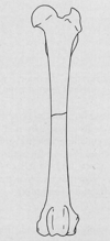
Label the parts of a long bone

A
37
Q

What supplies blood to normal, healthy bone?

(3)

A
  • Centrifugal from the medullary artery (~ 2/3 of blood supply)
  • Periostal vessels
  • Epiphyseal vessels → immature animals
38
Q

Characteristics of Woven Bone?

A
  • Unorganized structure
  • Formed in fetal life & during skeletal repair
  • “Fracture callus”
  • Replaced by lamellar bone during remodelling
  • Makes up the skeleten of lower vertebrates
39
Q

Characteristics of Lamellar Bone?

A
  • Orderly structure
  • Slower rate of formation
  • Occurs later in fracture healing
40
Q

Which type of bone lacks radiographic density?

A

Cancellous/Trabecular/Spongy bone

41
Q

Which form of bone is more dense radiographically?

A

Compact or Cortical bone

42
Q

What leads to the development of flat bones?

A

Intramembranous ossification

43
Q

Periosteal membrane around __1__ bone lays down __2__ laminae.

A
  1. Tubular (diaphyseal)
  2. Concentric
44
Q

How do the cells differentiate in

Endochondral ossificaiton?

A

along a Chondrogenic pathway

45
Q

How do long bones gain length?

A

Endochonral bone growth along growth plates

46
Q

Where is the physeal plate located?

A

Btwn the epiphysis & metaphysis

47
Q

Where is the Epiphyseal Plate located?

A

Beneath the surface of Articular cartilage

48
Q

Where are the 1° Ossification Centers located?

A

Diaphyses

(middle of bone)

49
Q

Where are the 2° Ossification Centers located?

A
  • Epiphyses (ends of bone)
  • Metaphysis
50
Q

Where are the Traction Centers (3° Ossification center) located?

A

Apophyses

51
Q

The metaphyseal width __1__ immediately to the width of the __2__ as it grows shaftward.

A
  1. decreases
  2. diaphysis
52
Q

What is the “cut back” zone?

A
  • Site of marked modeling in width in the Metaphysis→ cortical margin is irregular
  • Has a roughened cortical/periosteal border
  • In young, growing animals
53
Q

How do growth plates appear on rads?

A

appear poorly organized

54
Q

Give some examples of 3° Ossification Centers.

A
  • Supraglenoid tubercle scapula
  • Olecranon
  • Medial epicondyle of the humerus
  • Trochanter tertius femoris
  • Tibial apophysis
  • Tuber calcis
  • Tuber coxae
55
Q

What are the weakest parts of developing bone?

Consequence?

A
  • Physes
  • Physeal FXs are common in young P
56
Q

What does the shape of the physis partially determine?

(In regards to FXs)

A

Type of Salter-Harris FX

57
Q

What commonly occurs after injury to the physis?

A
  • Malformations of bone growth
    • abnormal length, shape, contour
58
Q

Damage to the Ulnar Physeal plate

(btwn the epiphysis & metaphysis) can cause what?

A

Premature closure of the growth plate → shortened ulna →cranial bowing → angular limb deformities

59
Q

Describe the different types of Salter Harris FXs

(Type I - V)

A
60
Q

List the 6 different types of Periosteal Rxns.

A
  • Smooth, well defined
  • Parallel/Lamellar → “onion skin”
  • Brush border
  • Palisading
  • Irregular, ill defined
  • Radiating → “sunburst”
61
Q

What type of Periosteal Rxn is this?

3 Rule outs?

A
  • Lamellar or “Onion skin” periosteal rxn
  • Ewing’s, osteomyelitis, or osteosarcoma
62
Q

What type of Periosteal Rxn is show here?

(lateral aspect of 5th metacarpal & digit)

A

Palisading Periosteal rxn

(ex. hypertrophic osteopathy)

63
Q

What type of Periosteal Rxn is seen here?

A

Sunburst Periosteal Rxn

(often highly aggressive)

64
Q

What type of Periosteal Rxn is seen here (arrows)?

A
  • Codman Triangle
  • Osteosarcoma, Ewing sarcoma, osteomyelitis, metastasis, chondrosarcoma ormalignant fibrous histiocytoma
65
Q

Characteristics of Benign Bone Lesions?

A
  • Short zone of transition
    • distinct margins, sclerotic border
  • Intact cortex
  • Intact, smooth periosteal new bone
  • Slow changes in appearance
66
Q

Characteristics of Malignant Bone Lesions?

A
  • Long zone of transition
    • Indistinct margins, less dense border
  • Interuppted cortex
  • Interuppted, irregular periosteal new bone
  • Changes appearance rapidly
67
Q

Is this lesion benign or malignant?

A

Benign

68
Q

Is this lesion Benign or Malignant?

A

Malignant

69
Q

What should you always do in conjunction when assess bone lesions?

A

Assess the surrounding soft tissue

70
Q

When do Pathological FXs often occur?

A

spontaneous or following minor trauma to a weakend bone

71
Q

Which animals get Fatigue FXs?

Where do these fxs typically occur?

A
  • Racing animals → horses & greyhounds
  • Metacarpals & metatarsals
72
Q

What can mimic FX lines?

(7)

A
  • Nutrient foramina
  • Overlying fascial plane fat
  • Gas in the fascial plane in open FXs
  • Normal growth plates
  • Skull sutures
  • Mach lines
  • Artefact from a damaged grid
73
Q

What is the only way that FX lines can be visualized radiographically?

A

if they are aligned PARALLEL to the X-ray beam

(it passes directly through them)

74
Q

Increased opacity of the cortex and medulla can be indicative of what 3 things?

A
  • Folding FX
  • Impaction
  • Overriding FX ends
75
Q

What is the best way to confirm a Chip FX?

A

Radiograph the opposite limb & compare

76
Q

List the 4 different criteria used to classify FXs.

A
  • Morphology
  • Cortical involvement
  • Fragment shape
  • Articular/physeal involvement
77
Q

What type of FX line is demonstrated here?

What typically causes this type in animals?

A
  • Comminuted FX w/ a Transverse FX line
  • High energy trauma → HBC
78
Q

What type of FX line is seen here?

A

Oblique FX line

79
Q

What type of FX line is seen here?

A

Spiral FX line

80
Q

What are Comminuted FXs?

A
  • Have at least three fracture fragments & the fracture lines interconnect.
  • The individual fracture lines that form the comminuted fracture may be transverse, oblique, or spiral.
81
Q

What type of FX is seen here?

A

Comminuted FX

82
Q

What type of fracture is seen here?

A

Butterfly FX (fragment)→ type of Communited

a bone break in which the center fragment is contained by two cracks & forms a triangle.

83
Q

What type of FX is shown here?

A

Multiple segmental FX

(type of Comminuted FX)

84
Q

How do Avulsion FXs typically occur?

A

when a fragment of bone tears away from the main mass of bone as a result of physical trauma

(often at the site of tendon & ligament attachement)

85
Q

What type of FX is seen here?

A

Avulsion FX

86
Q

What type of FX is seen here?

A
  • Greenstick FX
    • occurs when a young, soft bone
    • One side bends and the other side breaks
87
Q

Which Salter-Harris Type is shown here?

A

Salter-Harris Type I

(seperation @ the level of the physis)

88
Q

What Salter-Harris Type is shown here?

A

Salter-Harris Type II

(involves the physis & metaphysis)

89
Q

Which Salter-Harris Type is seen here?

A

Salter-Harris Type III

(involves the physis & epiphysis)

90
Q

Which Salter-Harris Type is seen here?

A

Salter-Harris Type IV

(involves physis, metaphysis & epiphysis)

91
Q

Which Salter-Harris Type is seen here?

A

Salter-Harris Type V

(crushed/compressed physis)

(May not be visisble @ time of injury)

(May lead to abnormal growth/deformity)

92
Q

What type of physeal FX is seen here?

A

Salter-Harris Type VI

93
Q

What type of Physeal FX is seen here?

A

Salter-Harris Type VII (AKA Ogden VII)

  • Isolated to the epiphyseal plate→ does NOT involve the physis
  • May be an avulsion fx
94
Q

What are the features of a Pathological FX?

A
  • Cortical thinning or destruction
  • Medullary bone cystic/destruction
  • Periosteal new bone is present
  • Shape/pattern/direction of FX line is unusual
  • Bone shape is often abnormal
95
Q

What is the likely cause of this fracture?

A

Pathologic FX

96
Q

Etiology of Sesamoid Bone Dz?

A
  • Congenital → bipartite sesamoid bones
  • Traumatic → nonunion FX
  • 2° changes → Modeling following arthrosis
97
Q

How do you initally assess a FX?

(8 questions you should be asking yourself)

A
  1. Location
    • which bones are involved? where on the bone?
  2. Age of FX
    • sharpness of FX ends? signs of bone healing or callus formation?
  3. Type of FX
  4. Displacement of the Fragments
    • Distracted? Impacted? or Overriding?
  5. Bone radiolucency or loss of normal architecture?
  6. Joint involvement?
  7. Foreign material present?
  8. Soft tissue changes?
  9. Other injuries elsewhere in the body?
98
Q

What can help you determine the age of a FX?

A
  • Margin of FX fragment
  • Fragment density
  • Fracture gap
  • Callus formation
  • Periosteal rxn?
99
Q

Signs of an ACUTE FX.

A
  • Sharply, delineated fx margins
  • Persistent “normal” bone density
  • Wide fracture gap
  • No callus formation
100
Q

Signs of a Chronic FX?

A
  • Indistinct, rounded fx margins
  • Loss of bone density
  • Bridged fx gap
  • Callus formation
101
Q

Describe the steps of FX healing in chronological order.

A
  1. Blurring of FX lines → bone resorption
  2. Widening of FX gap → bone resorption
  3. Formation of endosteal & periosteal callus
  4. Progressive bridging of FX gap & decreased luceny
  5. Remodelling of the callus → becomes smoother & smaller
  6. FX gap no longer visible
102
Q

What is the relationship btwn calluls formation and bone stability?

A

More stability = less callus formation

Less stability = more callus formation

103
Q

How can plates potentially complicate FX healing?

A

Plate provides stress protection → reduces the load on the bone → weakens the bone (instead of strengthening it) → may lead to reFX

104
Q

Periosteal tearing can lead to a _________ and complicate FX healing.

A

Bucket handle callus

105
Q

How can non-anatomic reduction complicate FX healing?

A
  • Malunion
  • Non-union
    • Hypertrophic
    • Atrophic
106
Q

How does an Atrophic Non-Union appear on a radiograph?

A
  • Pointed FX ends
  • No callus formation visisble
  • No osseous bridging of FX gap
107
Q

What is the 1/2 life & carrier molecule of Technetium?

A

Half life = 6 hrs

Carrier = MDP