Lecture I: Sensory Physiology Flashcards

1
Q

What are the two classification schemes by which peripheral nerves are classified?

A

1) Their contribution to a compound action potential (A, B, and C waves)
2) Based on fiber diameter, myelin thickness, and conduction velocity (classes I, II, III, IV)

2
Q

What are the features of the group Ia or Aα peripheral axons (i.e., fiber diameter, myelination conduction velocity, and receptor supplied)?

A
  • 13-20 μm (large)
  • 80-120 m/s (fast)
  • Heavily myelinated
  • Receptor supplied: primary muscle spindles, Golgi tendon organs

*These are the alpha-motorneurons

3
Q

What are the features of the group IV or C peripheral axons (i.e., fiber diameter, myelination, conduction velocity, and receptor supplied)?

A
  • 0.2-1.5 μm (small)
  • 0.5-2 m/s (slow)
  • Unmyelinated
  • Receptor supplied: skin mechanoreceptors, thermal receptors, and nociceptors (slow pain)
4
Q

An appropriate stimulus applied to a somatosensory receptor produces a ________ that, when large enough, leads to action potentials that can be carried over a considerable distance into the CNS.

A

An appropriate stimulus applied to a somatosensory receptor produces a generator potential that, when large enough, leads to action potentials that can be carried over a considerable distance into the CNS.

5
Q

What is the Weber-Fechner Law; how much stimulus required for noticeable difference?

A
  • There exists a logarithmic relationship between stimulus and perception
  • A 10% difference is usually required for conscious perception of change
6
Q

How does perceived intensity differ amongst muscles and cutaneous stimuli?

A
  • Muscle perceived intensity matches the actual intensity very closely
  • Cutaneous perceived intensity may diverge from the actual intensity substantially
7
Q

When a stimulus persists unchanged for several minutes without a change in position or amplitude, what occurs to the neural response and sensation; this is called what?

A
  • The neural response diminishes and sensation is lost
  • This is receptor adaptation
8
Q

Receptors that respond to prolonged and constant stimulation are classified as?

A

Slowly adapting receptors

9
Q

Receptors that respond only at the beginning or end of a stimulus are classified how; what activates them?

A
  • Rapidly adapting receptors
  • Only active when the stimulus intensity increases or decreases
10
Q

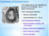
What are the 4 mechanoreceptors?

A
  1. Meissner corpuscle
  2. Pacinian corpuscle
  3. Merkel disk
  4. Ruffini ending
11
Q

What is a receptive field?

A

Individual mechanoreceptor fibers convey information from a limited area of skin

12
Q

Tactile acuity is highest and lowest where and how does this related to the size of the receptive field?

A
  • Tactile acuity is highest in fingertips and lips (smallest receptive field)
  • Tactile acuity is lowest in calf, back and thigh (largest receptive field)
13
Q

What is an afterdischarge?

A
  • In some cases of receptor adaption, the removal of the stimulus triggers AP’s as the ending “reforms.”
  • The persistance of a sensation after the stimulus eliciting the discharge has been removed
14
Q

What is Pre-synaptic inhibition; what kind of synapse; what is the end result?

A
  • Axo-axonal synapse
  • The post-synaptic cell is a pre-synaptic terminal
  • The end result of pre-synaptic transmission is reduced NT released from the inhibited pre-synaptic terminal
15
Q

What are the 4 steps in pre-synaptic inhibiton (what’s released and the end result)?

A
  1. Pre-synaptic terminal of neuron C synspases on the pre-synaptic terminal of neuron A, when acitvated neuron C releases GABA causing influx of Cl- into neuron A
  2. Results in hyperpolarization of pre-synaptic terminal of neuron A
  3. Less Ca2+ enters cytosol
  4. Leads to less NT released and reduced probability of AP’s in neuron B
16
Q

Pre-synaptic inhibition occurs where in the pathways involved in central processing of the senses; what impact does it have on the brain?

A
  • Occurs between neighboring receptors at the first synapse in their pathway
  • Every synapse along the way represents a chance to modify or respond to the stimulus
  • Increases the brain’s ability to localize the signal
17
Q

What is the most powerful form of inhibitory control in all primary afferent fibers?

A

Pre-synaptic inhibition

18
Q

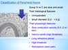
The somatosensory cortex has how many layers, how are the neurons arranged, and what is the importance of this arrangement?

A
  • Has 6 layers
  • Arranged in columns
  • Each column deals with one sensory modality in one part of the body
19
Q

Which layers of the primary sensory cortex houses the columns which are the main site of termination of axons from the thalamus?

A

Layer IV

20
Q

Which are the main output neurons from the primary sensory cortex?

A

Pyramidal cells

21
Q

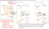
Where is the primary somatosensory cortex (S1) located, which Brodmann’s areas, and what is this part of the brain involved in?

A
  • Located in post-central gyrus
  • Brodmann’s 1, 2, and 3
  • First stop for most cutaneous senses
  • Involved in the integration of information for postion sense as well as size, shape discrimination
22
Q

Where is the secondary somatosensory cortex (S2) located, whre does it receive input from and what is this part of the brain involved in?

A
  • Wall of the sylvian fissure
  • Receives input from S1
  • Cognitive touch
  • Comparisons between objects, different tactile sensations and determining whether something becomes a memory
23
Q

Do the primary and secondary somatosensory areas (S1 and S2) have somatotopic representation?

A
  • S1 has detailed somatotopic representation
  • S2 has somatopic representation, BUT is NOT as detailed as S1
24
Q

What is the function of the parieto-temporal-occipital (PTO) association cortex?

A
  • High level interpretation of sensory inputs
  • Receives input from multiple sensory areas, including S1 and S2
  • Analyzes spatial coordinates of self in enviornment, names objects, and has many functions
25
Q

What are Corticofugal signals, where are they transmitted between and their function?

A
  • Transmitted back from cortex to lower relay stations in the thalamus, medulla, or SC
  • Controls the intensity of sensory sensitivity
  • Typically inhibitory and suppresses sensory input
26
Q

What is the Doctrine of specific nerve energies, tells you what?

A
  • No matter where along the afferent pathway is stimulated, the sensation that will occur is determined by the nature of the sensory receptor in the periphery connected to that pathway
  • Tells you the modality (i.e., what kind of receptor stimulated)
27
Q

What is the Law of Projections, tells you what?

A
  • No matter where along the afferent path we stimulate, the perceived sensation is always referred back to the area of the body in which the receptor is located
  • Tells you where you feel it (location)
28
Q

What are the 4 ways pain can be characterized by location?

A

1) Deep pain (i.e., bone pain)
2) Muscle pain
3) Visceral pain
4) Somatic/cutaneous pain

29
Q

Nociceptors are bare nerve endings used for sensing noxious stimuli, what are the 2 types of fibers involved and characteristics of each?

A

Aδ fibers: small, sparsely myelinated. Fast, sharp pain

C fibers: unmyelinated fibers associated with dull pain (slow pain)

30
Q

What are the different types of nociceptors (i.e., what are they sensitive to)?

A
  • Sensitive to both thermal and mechanical stimuli (majority)
  • Sensitive only to thermal or only to mechanical
  • Silent/sleeping: not active during initial injury, but can be “woken up.”
31
Q

Many mixed modality nociceptors express one of the ______family of receptors along with a mechanosensitive _____ channel.

A

Many mixed modality nociceptors express one of the TRP family of receptors along with a mechanosensitive Na+ channel.

32
Q

Unlike other receptors, nociceptors express a number of ligand-gated receptors (in addition to the stimulus-gated channels), this includes receptors for what ligands?

A
  • Substance P
  • The Kinins (i.e., bradykinin)
  • ATP
  • H+
33
Q

Mutations in which channel may lead to an absence of pain sensation or the production of a paroxysmal pain syndrome?

A

Nav1.7 mechanosensitive Na+

34
Q

What are the TRP family of receptors?

A
  • Transient receptor potential family of receptors (TRPV1, TRPA1, TRPM8)
  • Ligand-gated non-selective cation channel permeable to Ca2+, Na+, and/or K+
35
Q

When chemicals such as Substance P, Bradykinin, ATP, or H+ bind to a nociceptor how is sensitivity affected?

A
  • They change the sensitivity of nociceptors (usually increasing)
  • Activate silent nociceptors
36
Q

What is the source of the Substance P, the Kinins, ATP, and H+ which act to bind nociceptors and alter the sensitivity?

A
  • Activated nociceptors, the damaged tissue, and recruited WBC release these into the periphery as well as in the SC
37
Q

What is the NT released by Aδ fibers (for pain) in the SC and what receptor does it act on?

A

EAA which act on non-NMDA receptors

38
Q

What is the NT released by C fibers (for pain) in the SC and what receptor does it act on?

A
  • Substance P and EAA’s
  • Acts on both NMDA and non-NMDA receptors
39
Q

Nociceptors that travel with the spinoreticulothalamic pathway (slow pain) synapse where and then what?

A

Synapse on an interneuron in the SC before crossing and ascending to the reticular formation

40
Q

How are visceral afferents related to pain relayed to the brain; what do the additional synapses form?

A
  • Travel with autonomic nerves
  • Have additional synapses within the hypothalamus and medulla
  • Form the basis of the physiological changes associated with visceral pain, including diaphoresis and altered BP
41
Q

What is the role of S1 and S2 in the central processing of pain?

A

Receive input from the nociceptors and play a role in localizing pain

42
Q

What is the role of the Insular Cortex in the central processing of pain?

A
  • Particularly important in the interpretation of nociceptive inputs
  • Processes information about the internal state of the body
  • Contributes to the autonomic response to the pain

- Integrates ALL signals related to pain

43
Q

In regards to pain what occurs with damage to the insular cortex?

A
  • Asymbolia
  • Patient will still feel pain and can localize the pain, but will lose the negative/emotional experience of pain
44
Q

What is the importance of many nociceptive inputs going to the amygdala?

A
  • Important for activating/producing the emotional components inherent in the sensation of pain
  • Associated with the fear and anger components
45
Q

What observation is the Gate theory of pain based on?

A

Based in part on the observation that other somatic input can alleviate pain (rubbing the area)

46
Q

What are the 3 steps involved in the Gate Theory of Pain (i.e., fibers, synapses, and NT’s involved); what is the end result?

A

1) Activate an Aβ fiber by the normal stimuli. The Aβ fiber has a branch that travels via the dorsal columns, but it also branches within the SC
2) The Aβ fiber releases EAA and activates an inhibitory interneuron in the SC
3) The inhibitory interneuron releases glycine to inhibit the activity of the second order neuron in the pain pathway

*End result = rubbing the area of skin activated by the Aβ fiber willreduce the sensation of pain

47
Q

What are the 5 steps involved in the descending mechanism of modifying painful inputs?

A

1) Neurons in PAG activated by opiates, EAA, and cannibinoids
2) Descending projections from PAG neurons travel to the midline Raphe nuclei and release enkephalins, activate the raphe neurons
3) Axons from raphe neurons travel to SC and release serotonin, activating inhibitory interneurons, causing release of opitates
4) Opiates from interneuron activate Mu receptors on the presynaptic terminal of the C fiber
5) Produces pre-synaptic inhibition that reduces release of Substance P from the nociceptor, reducing pain transmission

48
Q

What are 2 functions of the descending serotonergic and noradrenergic neurons in the pain pathway?

A
  1. Activate local interneurons
  2. Suppress spinothalamic projection neurons
49
Q

Deep pain is usually associated with what structures, characterization of the pain, what kind of fibers and associated with what muscular movement?

A
  • Associated with periosteum, ligaments
  • Usually dull, achy
  • Few A delta fibers
  • Many C fibers
  • Associated with muscle spasm
50
Q

What is the typical cause of Muscle Pain, types of fibers present and characterization of pain?

A
  • Usually injury or ischemia during contraction
  • Both Group III and IV fibers
  • Get both fast and slow pain associated with muscle
51
Q

What is the localization of visceral pain, type of receptors, and is often associated with what type of pain?

A
  • Poorly localized
  • Few receptors (almost all Group IV - C fibers)
  • Receptors very sensitive to stretch (distention)
  • Associated with referred pain
52
Q

What is required for the brain to be able to localize pain and how is this related to referred pain?

A
  • Brain requires some experience to localize pain, starting early in life, this “trains” the brain to localize pain
  • Visceral pain is not experienced enough in early development to adequately train the brain to localize it
  • Confounded with the fact that the nociceptors may converge on the same interneuron in the SC
53
Q

Where are the group Ia/Aα fibers found vs. group Ib/Aα fibers found?

A

- Group Ia/Aα are part of muscle spindles

- Group Ib/Aα are part of Golgi tendon organs

54
Q

Where are the group II or Aβ fibers found?

A

Cutaneous mechanoreceptors

55
Q

Where are the group III or Aδ fibers found?

A

Higher threshold mechanoreceptors, cold thermoreceptors, fast pain

56
Q

Which fibers have the highest threshold and why is this important?

A
  • Group IV or C fibers
  • These are the unmyelinated fibers that are part of nociceptors (SLOW pain)
  • High threshold so that you are not constantly in pain!
57
Q

What are the sensory fibers of group I, II, III, and IV?

A

Group I: Ia and Ib = Aα

Group II: Aβ fibers

Group III: Aδ fibers

Group IV: C fibers