Lecture Exam 1 Flashcards
What does the cardiovascular system consist of?
Heart and vessels only
What are the main functions of the circulatory system?
Transport:
- Carries oxygen from lungs to body’s tissues and picks up CO2
- Picks up nutrients from digestive track and delivers to body’s tissues
- Carries metabolic wastes to kidneys
- Carries hormones to endocrine organs
- Transports stem cells from bone marrow
Protection:
- Blood plays role in inflammation, limit spread of infection
- WBC destory macroorganisms and cancer cells
- Antibodies and other blood proteins neutralize toxins and help destroy pathogens
- Platelets secrete factors that initiate blood clotting and other processes for minimizing blood loss
Regulation:
- Absorbing or giving off fluid under different conditions, blood capillaries help to stabilize fluid distribution in the body
- By buffering acids and bases, blood proteins help to stabilize the pH of the extracellular fluid
- Shifts in blood flow help to regulate body temperature by routing blood to the skin for heat loss or retaining it deeper in the body to conserve heat
What is the matrix of blood, a clear light yellow fluid consistuting a little over half of the blood volume?
plasma
What is suspended in the plasma?
formed elements
cells and cell fragments including the red blood cells, white blood cells and platelets
THey are membrane-enclosed bodies with a definite structure visible with the microscope.
What are the formed elements
Erythrocytes (Red blood cells, RBC)
Platelets
Leukocytes (White blood cells, WBC)
- Granulocytes
- Neutrophils
- Eosinophils
- Basophils
- Agranulocytes
- Lymphocytes
- Monocytes
What does plasma consist of?
water, proteins, nutrients, electrolytes, nitrogenous wastes, hormones, and gases
When blood clots and the solids are removed, the remaining fluid in the blood is?
serum
is essentially identical to plasma except for the absence of the clotting protein fibrinogen
Name and describe the three major categories of plasma proteins in blood
Albumin
- The smallest and most abundant plasma protein
- It serves to transport various solutes and buffer the pH of the plasma
- Alos major contributor to viscosity and osmolarity
- Changes in albumin concentration can significantly affect blood volume, pressure and flow
Globulins
- Divided into three subclasses; from smallest to largest in MW; alpha, beta and gama globulins
- Play various roles in solute transport, clotting and immunity
Fibrinogen
- soluble precursor of fibrin, a sticky protein that forms the framework of a blood clot
- Some of the other plasma proteins are enzymes involved in the clotting process
What produces plasma proteins?
liver
as much as 4 g per hour
contributing all of the major proteins except gamma globulin
Where is gamma globulin produced?
from plasma cells - connective tissue cells that are descended from white blood cells called B lymphocytes
What occurs if the blood osmolarity is too high?
The bloodstream absorbs too much water. This raises the blood volume, resulting in high blood pressure and a potentially dangerous strain on the heart and arteries
What occurs if the blood osmolarity drops too low?
too much water remains in the tissues
They become edematous (swollen) and the blood pressure may drop to dangerously low levels because of the water lost from the blood stream
What is the main product of blood somolarity?
sodium ions, protein and erythrocytes
What is the contribution of protein to blood osmotic pressure called?
colloid osmotic pressure (COP)
is especially important, as we see from the effects of extremely low-protein diets
What is the production of blood, especially its formed elements called?
hemopoiesis
What is the tissues that produce blood cells called?
hemopoietic tissues
The first hemopoietic tissues of the human embryo form in the yolk sac
What is the blood formation in the bone marrow and lymphatic organs called?
myeloid and lymphoid hemopoiesis
respectively
What do all formed elements trace their origin back to?
hemopoietic stem cell (HSC) in the bone marrow
HSCs multiply to maintain a small but persistent population in the bone marrow, but some of them go on to become a variety of more specialized cells called?
colony forming units (CFUs)
Each CFU is destined to produce one or another class of formed elements
What are the two principal functions of Erythrocytes, or red blood cells (RBCs)
- Pick up oxygen from the lungs and deliver it to tissues elsewhere
- Pick up carbon dioxide from the tiessues and unload it in the lungs
What gives RBC its color and name?
hemoglobin
Explain Hemoglobin structure/function relationship
- consists of four protein chains called globins
- two of these, the alpha chain, are 141 amino acids long, and the other two, the beta chains, are 146 amino acids long
- Each chain is conjugated with a nonprotein moiety called the heme group, which binds oxygen to an iron atom (Fe) at its center
What are the three most important clinical data measurements?
- Hemocrit (packed cell volume, PCV) is the percentage of white blood volume composed of RBCs
- Hemoglobin concentration
- RBC count
What are the three physiological reasons women have lower blood counts then men?
- Androgens stimulate RBC production, and men have higher androgen levels
- Women of reproductive age have periodic menstrual losses
- The hematocrit is inversely proportional to percentage body fat, which is higher in women than in men
What is erythrocyte production called? explain.
erythropoiesis
normally takes 3 to 5 days and involves four major developments:
- A reduction in cell size
- an increase in cell number
- the synthesis of hemoglobin
- and the loss of the nucleus and other organelles
Explain the erythrocyte production stages
- It beins when a hemopoietic stem cell (HSC) becomes an erythrocyte colony-forming unit (ECFU), which has receptors for erythropoietin (EPO), a hormone secreted by the kidneys
- EPO stimulates the ECFU to transform into an erythroblast (normoblast)
- Erythroblasts multiply and synthesize hemoglobin
- When this task is completed, the nucleus shrivels and is discharged from the cell
- The cell is now called a reticulocyte, named for a temporary network (reticulum) composed of ribosome clusters (polyribosomes).
- Reticulocytes leave the bone marrow and enter the circulating blood
- In a day or two, the last of the polyribosomes disintegrate and disappear, and the cell is a mature erythrocyte

What types does dietary iron exist in? and what occurs with these forms?
Ferric (Fe3+) and Ferrous (Fe2+)
Stomach acid converts most Fe3+ to Fe2+, the only form that can be absorbed by the small intestine
What does gastroferritin for? Explain the liver and stomach role in iron metabolism
- A protein that is produced by the stomach, then binds Fe 2+ and transports it to the small intestine. Here it is absorbed by the blood, binds to a plasma protein called transferrin, and travels to the bone marrow, liver and other tissues
- Bone marrow uses iron for hemoglobin synthesis; muscle uses it to make the oxygen-storage protein myoglobin; and nearly all cells use iron to make electron-trnsport molecules called cytochromes in their mitochondria.
- The liver binds surplus iron to a protein called apoferritin, forming an iron-storage complex called ferritin.
- It releases Fe2+ into circulation when needed

If the RBC count drops, explain the feedback
For example hemorrhaging and may result in a state of hypoxemia (oxygen deficiency in the blood)
Negative feedback
- The kidneys detect this and increase their EPO output
- Three or four days later, the RBC count begins to rise and reverses the hypoxemia that started the process

Name and describe the types of plasma proteins
-
Albumins (60% - Liver)
- Water soluble, maintain osmotic pressure (BCOP)
-
Globulins (35% - Liver, plasma cells) antibodies
- Transport, Immunity
-
Fibrinogen (4% - Liver)
- Clotting
- Other (1%):
- eg. Hormones
What are the plasma solutes? explain
-
Electrolytes (salts)
- Cellular activity, osmotic pressure
- Cations: Na+, K+, Ca2+
- Anions: Cl-, HCO3-
-
Gases
- O2, CO2, N2
-
Nutrients
- Amino acids, glucose, fats
-
Wastes
- N-containing, creatine, bilirubin
What is the Hematocrit for Erythrocytes
Percent of blood that is RBC (35 - 55%)
- females are lower, espcially unift at sea level
- Highly fit men that live well above sea level have a higher count
Explain the structure of the RBC
- Bi-concave disc
- Flexible
- except if the person has sickle cell anemia
- genetic, DNA, disorder (half moon)
- except if the person has sickle cell anemia
- Stackable
- No nucleus, few organelles
Explain the structure and function of Hemoglobin (Hb)
- Oxygen carrying protein
- Normal level: 12-18 grams/ dl
- Structure:
- Four polypeptide subunits
- Heme groups
- Iron containing: red colored, bind to O2, CO2
Explain the process of Erythrocyte development
- Erythropoeisis
- in myeloid tissue (marrow)
- Myeloid stem cells
- tissue that will become RBC, found in marrow
- Proeythroblasts
- will become RBC
- Red blood cells
- Controlled by erythropoeitin (EPO)
- Released in response to:
- Low O2 levels:
- anemia (damaged or no/low iron)
- Blood loss (kidneys will signal for RBC release)
- Respiratory damage
- Exercise
- Low O2 levels:
- Released in response to:
Explain the destruction process of RBC
- Lifespan ~120 days
- 3 million produced and destroyed per second
- Phagocytosis (macrophages in liver)
- some parts recycled
- Proteins -> AA
- Heme -> iron + biliverdin (helps breakdown fat)
- some are excreted
- biliverdin -> bilirubin
- some parts recycled
What makes an agglutination?
antigens + antibodies
Explain the characteristics of Leukocytes
- Primarily remain in CT (blood)
- bloodstream is for point to point transportation
- Amoeboid movement (squeeze or shake into tinny places = diapedisis)
- Migration out of bloodstream
- Chemotaxis (chemical signals)
- Phagosytosis
Name and discribe the types of Leukocytes
-
Neutrophils
- Most common (70%)
- Mobile
- Phagocytes of ‘marked’ bacteria
- Suicidal eaters - Pus
-
Eosinophils
- 2-4% of WBCs
- Attack eukaryotic parasites: worms and protozoa
- Exotoxin release
- Phagocytic
-
Basophils
- Rare (<1%)
- Granules contain histamine and heparin
- Heparin - prevents blood from clotting (anticoagulant)
- Histamine - increases inflammation (vasodilator)
- Inflammatory response
-
Monocytes
- 5-10% of WBC
- Huge macrophages
-
Lymphocytes
- 20-30% of WBC
- Immune system function
- B cells
- T cells
- NK cells
Explain the WBC development
- Originates in the bone marrow
- Production hormonally regulated - CSF
- Hemocytoblasts can differentiate into the Myeloid stem cells and Lymphoid stem cells
Explain the function and where platelets are formed
- Function in hemostasis
- Develop from megakaryocytes
- 10 day circulation
- Specific functions:
- Physical plugging of wounds
- Chemical control of clotting
Name and describe the three phases of Hemostasis
-
Vascular Phase
- Vascular spasm decreases diameter of vessels (muscle contraction)
- Decreased blood flow
-
Platelet Phase
- Platelet adhesion to lamina and aggregate (form plug)
-
Coagulation Phase
- Formation of blood clot
- Clottting factors -> fibrin formation
- Extrinsic Pathway
- Triggered by chemicals released from damaged endothelium
- Intrinsic Pathway
- Triggered by exposure to collagen (proenzyme activation)
- Common Pathway
- Bigins with enzymatic Factor X activation (from either pathway)
- Formation of blood clot
Explain the histology of the heart, coverings and muscle
- Coverings:
- Pericardium - heart sits in the sac
- Epicardium - outside of the heart
- Endocardium - inner lining of the heart
- Cardiac Muscle
- Myocardium - interconnected via intercalated discs
- contractile and conducting tissue
- Myocardium - interconnected via intercalated discs
What are frequent causes of coronary circulation issues and how do they fix them?
Issues:
- Coronary artery atherosclerosis
- Blocked in a blood vessel
Fixes:
- Coronoary artery bypass surgery
- CABG = “cabbage” or stents
Explain the EKG (ECG) and how it works?
Produces a record of the electrical activity of the heart. It utilizes numerous electrodes to understand the activity at different distances from the heart.
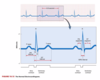
Explain the three principal deflections in an EKG
-
P wave:
- Produced when a signal from the SA node spreads through the atria and depolarizes them
- Atrial systole begins
- Time required for impulses to travel from the SA node to the AV node
- Atrial systole begins
- Produced when a signal from the SA node spreads through the atria and depolarizes them
-
QRS complex:
- small downward deflection (Q), a tall sharp peak (R), and a final downward deflection (S)
- Produced when the signal from the AV node spreads through the ventricular myocardium and depolarizes the muscles
-
T Wave:
- generated by ventricular repolarization immediately before diastole
- The ventricles take longer to repolarize than to depolarize; the t wave is therefore smaller and more spread out than the QRS comples, and it has a rounder peak

Explain the relationship of the ECG and electrical activity and contraction of the Myocardium

What is a deviation from the regular, SA node-driven sinus rhythem of the heartbeat called?
arrhythmia
Explain ventricular fibrillation arrhythmia
the hallmark of a heart attack (myocardial infarction)
- most in patients with a history of coronary artery disease
- In striking contrast to the steady sinus rhythm, the ECG shows weak, chaotic ventricular depolarization
- Electrical signals travle randomly about the mycardium and return to repeatedly restimulate the same area instead of dying out like a normal ventricular depolarization
What are the two main variables that govern flow?
Pressure, which impels a fluid to move
resistance, which opposes flow
What is the device to measure blood pressure?
sphygmomanometer
a calibrated mercury monometer with its open lower end attached to an inflatable pressure cuff wrapped around the arm
What is it called to listen to the sounds made by the body?
auscultation
(AWS-cul-TAY-shun)
Explain the heart sounds
- The first and second heart sounds, symbolized S1 and S2
- often described as a “lubb-dupp”
- S1 = louder and longer
- S2 = softer and sharper
- often described as a “lubb-dupp”
- In children and adolescents it is normal to hear a third heart sound (S3)
- in adults this is called triple rhythm or gallop, which may indicate an enlarged and failing heart
Explain the phases of the cardiac cycle
-
Ventricular filling.
- During Diastole, the ventricles expand and their pressure drops beloow that of the atria
- As a result, the AV valve open and blood flows into the ventricles (vent pressure rise)
-
Three phases:
- First one-third: rapid ventricular filling
- Second one third: diastasis, marked by slower filling. P wave of the EKG, depolarization of atria
- Third one third: atrial systole completes the filling process. right atrium contracts slightly before left
- At end of stage, each ventricle contains an end-diastolic volume (EDV) of 130 mL
-
Isovolumetric contraction
- atria repolarize, relax, and remain in diastole for the rest of the cardiac cycle
- Ventricles depolarize, generate the QRS complex, and begin to contract
- reverse the pressure gradient between atria and ventricles
- Heart sound S1 begins at this phase
- Called isovolumetric because even though the ventricles contract, they do not eject blood yet and there is no change in their volume (pressure in aorta and lungs too high)
-
Ventricular ejection
- Begins when ventricular pressure exceeds arterial pressure and forces the semilunar valves to open
- T wave then occurs late in this phase, beginning at the moment of peak ventricular pressure
- the amount ejected = stroke volume (SV) and precentage is called ejection fraction, end-systolic volume (ESV) is the amount after ejection
-
Isovolumetric relaxation
- This is early ventricular diastole, when the T wave ends and the ventricles begin to expand
- closing of the semilunar valves

what happens if the right ventricle pumps more blood into the lungs than the left can handle on return?
blood accumulates in the lungs, causes pulmonary hypertension, edema and a rist of drawing in one’s own body fluid
What is the amount ejected by each ventricle in 1 minute called?
How do you calculate it?
cardiac output (CO)
If HR is heart rate (beats/min) and SV is stroke volume (mL/beat),
CO = HR x SV
how is heart rate measured?
by taking a persons pulse at same point where an artery runs close to the body surface
What is tachycardia
is a persistent, resting adult heart rate above 100 bpm
can caused by stress, anxiety, stimulants, heart disease or fever
What is bradycardia?
is a persistent, resting adult heart rate below 60 bpm
it is common during sleep and in endurance-trained athletes
Endurance training enlarges the heart and increases its stroke volume, enabling it to maintain the same ouput with fewer beats
What are factors that raise heart rate called? and what are factors that lower heart rate called?
Positive chronotropic agents = raise HR
Negative chronotropic agents = lower HR
Where are there cardiac centers the autonomic nervous system?
medulla oblongata that contain the cardiac centers
initiate autonomic output to the heart
(cardiostimulatory effect and cardioinhibitory effect)
Explain the sympathetic postganglionic fiber interactions (adrenergic)
they release norepinephrine, which binds to beta-adrenergic fibers in the heart
- Activates the cyclic adenosine monophosphate (cAMP) second- messenger system in the cardiocytes and nodal cells
- cAMP activates an enzyme that opens a Ca2+ channel in the plasma membrane
- The Ca2+ inflow accelerates depolarization of the SA node and contraction of the cardiocytes, so it speeds up the heart
- In addition, cAMP accelerates the uptake of Ca2+ by the sarcoplasmic reticulum and thereby enables cardiocytes to relax more quickly
- By accelerating both contraction and relaxations, norepinephrine and cAMP increase the heart rate
Explain the parasympathetic vagus nerve effect on the autonomic nervous system and heart
have cholinergic, inhibitory effects on the SA and AV nodes
- Acetylcholine (ACh) binds to muscarinic receptors and opens K+ gates in the nodal cells
- As K+ exits the cells, they become hyperpolarized and fire less frequently, so the heart slows down
- The vagus nerves have a faster-acting effect on the heart than the sympathetic nerves becasue ACh acts directly on the ion channels in the plasma membrane; sympathetic effects are slower because of the time taken fort eh cAMP system to act on the ion channels
Explain the types and effects of other imputs received to the medulla from muscles, joints, arteries and brainstem
-
Proprioceptors:
- in muscles and joints provide information on changes in physical activity
-
Baroreceptors (pressoreceptors):
- pressure sensors in the aorta and internal carotid arteries
-
Chemoreceptors:
- occur in the aortic arch, carotid arteries and the medulla oblongata itself, and are sensitive to blood pH, CO2 and O2 levels
- They are more important in respiratory control than in cardiovascular control but they do influence the heart rate
What are the effects with an electrolyte with greatest chronotropic effects (potassium)
- In hyperkalemia, a potassium excess, K+ diffuses into the cardiocytes and keeps the memebrane voltage elevated, inhibiting cardiocyte repolarization
- The myocardium becomes less excitable, the heart rate becomes slow and irrgular and the heart may arrest in diastol
How does an excess of calcium affect the heart rate?
- calcium excess (hypercalcemia) causes a slow heartbeat, whereas a calcium deficiency (hypocalcemia) elevates the heart rate
- Such calcium imbalances are relatively rare, however, and when they do occure, they primary effect is on contraction strength
Name and explain the three variables of stroke volume
Increased preload or contractility increases stroke volume, whereas increased afterload opposes the emptying of the ventricles and reduces stroke volume
-
Preload
- amount of tension (stretch) in the ventricular myocardium immediately before it begins to contract
-
Contractility
- how hard the myocardium contracts for a given preload
- increase caused by factors that make the cardiocytes more responsive to stimulation
- Factors that affect contractility are called:
- positive inotropic agents - increase
- Calcium- increases strength
- increases excitation-contraction coupling of muscle
- Prolongs the plateau of the myocardial action potentials
- Calcium- increases strength
- Negative inotropic agents - decrease
- too much calcium (hyperkalemia) has a negative effect because it reduces the strength of myocardial action potentials and thus reduces the release of Ca2+ in the sarcoplasm
- positive inotropic agents - increase
- how hard the myocardium contracts for a given preload
-
Afterload
- is the sum of all forces a ventricle must overcome before it can eject blood
- The most significant contribution to afterload is the blood pressure in the aorta and pulmonary trunk immediately distal to the semilunar valve; it opposes the opening of these valves and thus limits stroke volume
Explain the vessel walls of arteries and veins
-
Tunica Interna:
- lines the inside of the vessel and is exposed to the blood
- Simple squamous epithelium called endothelium
- Selectively permeable barrier to materials entering or leaving bloodstream, secretes chemicals that stimulate dilation or constriction of vessels, repels blood cells and platelests so blood flow is not obstructed
-
Tunica Media:
- middle layer, usually the thickest
- consists of smooth muscle, collagen, and in some cases elastic tissue
- strengthens the vessel s and prevents blood pressure from repturing them, and it regulates the diameter of a blood vessel
-
Tunica Externa:
- outermost layer
- Consists of loose connective tissue that often merges with that of neighboring blood vessels, nerves, or other organs
- It anghors the vessel to adjacent tissues and provides passage for small nerves, lymphatic vessels and smaller blood vessels that supply the tissues of the larger vessels
Name the three classes of arteries
They are divided into three classes by size
-
Conducting (elestic or large) arteries
- are the largest, examples include aorta, common carotid and subclavian arteries, pulmonary trunk, and common iliac arteries
- They have a layer of elastic tissue called the internal elastic lamina at the border between the interna and media, but microscopically, it is incomplete and difficult to distinguish from the tunica media
- These areteries expand as they receive blood during ventricular systole and recoil during diastole
-
Distributing (muscular or medium) arteries:
- are smaller branches that distribute blood to specific organs
- Examples include brachial, femoral, renal, and splenic arteries
- they tipically have up to 40 layers of smooth muscle constituting about three-quarters of the wall thickness
-
Resistance (small) arteries
- usually too variable in number and location to be given individual names
- They exhibit up to 25 layers of smooth muscles and relatively little elastic tissue
- They have a thicker tunica media in proportion to the lumen
- aka arterioles

Name and describe some of the arterial sense organs
These receptors transmit information to the brainstem that serves to regulate the heartbeat, blood vessel diameters and respiration
-
Carotid sinuses
-
Baroreceptors (pressure sensors) that monitor blood pressure
- adjust blood pressure (baroreflex)
- Ascending the neck on each side is a common carotid artery
- They have a relatively thin tunica media and an abundance of glossopharyngeal nerve fibers
-
Baroreceptors (pressure sensors) that monitor blood pressure
-
Carotid bodies
- Located near the branch of the common carotid arteries, these are oval receptors about 3x5 mm in size, innervated by sensory fibers of the glossopharyngeal nerves
- They are chemoreceptors that monitor changes in blood composition
- primarily transmit signals to the brainstem respiratory centers, which adjust breaking to stabilize the blood pH and its CO2 and O2 level
-
Aortic bodies
- These are one to three chemoreceptors located in the aortic arch near the arteries to the head and arms
- same structure and function as carotid bodies, but transmit their signals to the brainstem via the vagus nerves
What are the arterioles, capillaries and venules
microvasculature (microcirculation)
What is the job of capillaries and what do they consist of?
exchange of materials from tissues into the blood stream
They consist of only an endothelium and basal lamina
Name and describe the types of capillaries
distinguished by the ease with which they allow substances to pass through their walls and by structural differeences that account for their greater or lesser permeability
-
Continuous capillaries
- occur in most tissues, such as skeletal muscles
- Their endothelial cells, held together by tight junctions, form a continuous tube
- Contain protein-carbohydrate layer = basal lamina
- endothelial cells separated by narrow intercellular clefts
- Some continuous capillaries exhibit cells called pericytes that lie external to the endothelium
-
Fenestrated capillaries
- Have endothelial cells riddled with patches of filtration pores (fenestrations)
- Allow for the rapid passage of small molecules, but still retain most proteins and larger particles in the blood stream
- Important in organs that engage in rapid absorption or filtration
-
Sinusois (discontinuous capillaries)
- Irregular blood-filled spaces in the liver, bone marrow, spleen, and some other organs
- Twisted, tortuous passageways that conform to the shape of the surrounding tissue
- proteins and blood cells can pass through pores
Name and explain the types of veins?
-
Postcapillary venules
- the smallest of the veins beginning with diameter of about 10 to 20 um. Receive blood from capillaries directly or by way of the distal ends of the thoroughfare cahnnels
- Exchange fluid with surrounding tissues
-
Muscular venules
- receive blood from teh postcapillary venules, up to 1 mm diameter
-
Medium veins
- up to 10 mm in diameter. Most veins with individual names are in this category (radial and ulnar veins for example)
- many contain venous valves to direct flow to heart
-
Venous sinuses
- veins with especially thin walls, large lumens and no smooth muscle (coronary sinus and dural sinuses of brain.
- not capable of vasoconstriction
-
Large veins
- Greater than 10 mm, they have smooth muscles in all three tunics.
- Have relatively thin tunica media
- Ex. Vena cavae, pulmonary veins, jugular veins and renal veins
What are portal systems
- Blood flows through two consecutive capillary networks before returning to the heart
- occurs in the kidneys; connecting the hypothalamus and anterior pituitary and connecting the intestines to the liver
What is an anastomosis
a point of convergence between two blood vessels other than capillaries
- In an arteriovenous anastomosis (shunt), blood flows from an artery directly into a vein and bypasses the capillaries
- helps to reduce heat loss in the extremities during cold weather
Name and describe how blood suppply to the tissue can be expressed
-
flow
- amount of blood flowing through the organ, tissue or blood vessel in a given time (mL/min)
-
Perfusion
- flow per given volume or mass of tissue (mL/min/g)
During the blood pressure reading, what is recording and what do they mean?
- Systolic pressure - peak arterial BP attained during ventricular contraction
- Diastolic pressure - is the minimum arterial BP occuring during the ventricular relaxationg between heartbeats
- The difference between systolic and iastolic pressure = pulse pressure
-
Mean arterial pressure (MAP) = mean pressure you would obtain if you took measurements at several intervals throughout the cardiac cycle
- diastolic + 1/3 (pulse pressure)
- What is arteriosclerosis?
- What is atherosclerosis?
- Hypertension
- Hypotension
-
arteriosclerosis =
- when there is an increase in stiffness of the arteries (hardening of the arteries)
-
atherosclerosis =
- growth of lipid deposits in the arterial walls = plaques
-
Hypertension =
- high BP = commonly considered to be a chronic resting blood pressure higher than 140/90
-
Hypotension =
- Cronic low resting BP
Explain the types of peripheral resistance?
the opposition to flow that the blood encounters in vessels away from the heart
-
Blood viscosity
- most significant is the erythrocyte count and albumin concentration to determine the level of viscosity of blood
-
Vessel length
- The longer th eliquid travels in the tube, the more cumulative friction it encounters, pressure and flow declines wiht distance
-
Vessel Radius
- The only significant way of controlling peripheral resistance from moment to moment are vasoconstrictions, the carrowing of a vessel and vasodilation, the widening of a vessel
Explain the types of regulation of blood pressure and flow
-
Local Control
- Autoregulation = ability of the tissues to regulate their own blood supply
- Platelets, endothelial cells and the perivascular tissues secrete a variety of vasoactive chemicals that stimulate vasodialation under such conditions as trauma, inflammation, and exercise
- If tissue’s blood supply is cut off from a time and then restored, it often exhibits reactive hyperemia - an increase above the normal level of flow
- Over a long time, a hypoxic tissue can increase its own perfusion by angiogenesis = the growth of new blood vessels
-
Neural Control
- In addition to local control, the blood vessels are under remote control by the central and autonomic nervous system
- Vasomotor center (medulla oblongata_ exerts sympathetic control over blood vessels throughout the body
- The vasomotor center is an integrating center for three autonomic reflexes - baroreflex (pressure regulation), chemoreflexes (pH, chemistry, O2 and CO2 concentrations), and medullary ischemic reflex (response to reduced perfusion of the brain, aka medulla oblongata monitors its own blood supply)
-
Hormonal Mechanisms
- Angiotensin II - vasoconstrictor that raises BP
- Aldosterone - salt-retaining hormone = water retension, increasing blood volume and pressure
- Natriuretic peptides - increase Na excretion by kidnesy, reducing blood volume and pressure
- Antidiuretic hormone - promotes water retentions, increases blood volume and pressure, also a vasoconstrictor
- Epinephrine and norepinephrine - stimulates vasoconstriction and raises BP. however, in coronary blood vessels they bind to beta-adrenergic receptors and cause vasodilation, increasing blood flow the th emyocardium during exercise
Name the chemicals that are taken up by the capillaries and the ones that are given off by the capillaries
- Taken up by the capillaries:
- CO2 and other wastes, and many of the sam esubstances as they give off: glucose and fatty acids released from storage in the liver and adipose tissue; calcium and other minerals released from bone; antibodies secreted by immune cells; and hormones secreted by the endocrine glands
What are three routes that chemicals pass through capillary walls
- the endothelial cell cytoplasm
- intercellular clefts between the endothelial cells
- filtration pores of the fenestrated capillaries
Name and describe the mechanisms of movement through the capillary wall
-
Diffusion
- glucose and oxygen, being more concentrated in the systemic blood than in the tissue fluid, diffuse out of blood
- Carbon dioxide and other wastes being more concentrated in the tissue fluid, diffuse into the blood (O2 and CO2 diffuse in opposite directions)
-
Transcytosis
- Process in which endothelial cells pick up material on one side of the plasma membrande by pinocytosis or receptor-mediated endocytosis, transport the vesicles across the cell, and discharge the material on the other side by exocytosis
- Fatty acids, albumin and some hormones such as insuline move this way
-
Filtration and Reabsorption
- typically, fluid filters out of the arterial end of a capillary and osmotically reenters it at the venous end
- This fluid delivers materials to the cells and removes their metabolic wastes
- Hydrostatic pressure is a physical force exerted by a liquid against a surface such as a capillary wall
- These forces are opposed by colloid osmotic pressure (COP), the portion of the osmotic pressure due to protein.
- The difference between the COP of blood and tissue fluid is called oncotic pressure
- dens to draw water into the capillary by osmosis, opposing hydrostatic pressure
- These opposing forces produce a net filtration pressure (NFP) of 13 mm Hg out
- Net reabsorption pressure - reabsorbing into capillarys

What is an edema?
the accumulation of excess fluid in a tissue
- often shows as swelling of the face, fingers, abdomen, or ankles but also occurs in internal organs where its effects are hidden from view
- Often occurs when fluid filters into a tissue faster than it is reabsorbed
What are three fundamental causes of edema? name and describe
-
Increased capillary filtration
- numerous conditions can increase the rate of capillary filtration and caccumulation of fluid in the tissues.
- Kidney failure, leads to water retention and hypertension. Histamine dilates arterioles and raises capillary pressure and makes the capillary wall more permeable
-
Reduced capillary reabsorption
- Capillary reabsorption depends on oncotic pressure, which is proportional to the concentration of blood albumin
- Deficiency of albumin (hypoproteinemia) produces edema by reducing the reabsorption of tissue fluid
- commonly seen in regions of famine due to dietary protein deficiency
-
Obstructed lymphatic drainage
- Lymphatic system, is a network of one-way vessels that collect fluid form the tissues and return it to the bloodstream. Obstructions of these vessels or the surgical removal of them can interfere with fluid drainage
Name and describe methods for venous return
-
The pressure gradient
- generated by the heart is the most important force in venous flow, even though it is substantially weaker in the veins than in the arteries
-
Gravity
- flow downhill
-
The skeletal muscle pump
- In the limbs, the veins are surrounded and messaged by the muscles. Contracting muscles squeese the blood out of the compressed parts of a vein, and the valves ensure that this blood can go only toward the heart
-
The thoracic (respiratory) pump
- Aids the flow of venous blood from the abdominal to the thoracic cavity. When you inhail your thoracic cavity expands and then the internal pressure drops
-
Cardiac suction
- During ventricular systole, the tendinous cords pull the AV valve cusps downward, slightly expanding the atrial space
- This creates a slight suction that draws blood into the atria from the venae cavae and pulmonary veins
What is it called when the body sits still and blood accumulates int he limbs?
venous pooling
What is circulatory shock? what are the categories?
is any state in which cardiac output is insufficient to meet the body’s metabolic need
There are two categories of circulatory shock:
-
Cardiogenic shock:
- caused by inadaquate pumping by the heart, usually as a result of myocardial infarction
-
Low venous return (LVR) shock:
- in which cardiac output is low because too little blood is returning to the heart
Name and describe the three principal forms of low venous return (LVR) shock
-
Hypovolemic shock
- most common form, is produced by a loss of blood volume as a result of hemorrhage, trauma, bleeding ulcurs, burning or dehydration
- Dehydration is the major cause of death from heat exposure
- most common form, is produced by a loss of blood volume as a result of hemorrhage, trauma, bleeding ulcurs, burning or dehydration
-
Obstructed venous return shock
- Occurs when any object, such as a growing tumor or aneurysm, compresses a vein and impedes its blood flow
-
Venous pooling (vascular) shock
- occurs when the body has a normal total blood volume, but too much of it accumulates in the lower body
- Neurogenic shock - is a form of venous pooling shock that results from a sudden loss of vasomotor tone, allowing the vessels to dialate
What is it clled when bacterial toxins trigger vasodilation and increased capillary permeability
septic shock
What is it called when exposure to an antigen to which a person is allergic, such as a bee venom
anaphylactic shock
What are the main functions of the lymphatic system?
-
Fluid recovery
- continually filters from blood capillaries into the tissue spaces.
- Reabsorbs excess and returns to the blood
-
Immunity
- picks up foreign cells and chemicals from the tissue. Fluid passes through lymph nodes, where immune cells stand guard against foreign matter
-
Lipid absorption
- In the small intestine, special lymphatic vessels called lacteals absorb dietary lipids that are not absorbed by the blood capillaries
What are the components of the lymphatic system:
- Lymph - the recovered fluid
- Lymphatic vessels - which transport the lymph
- Lymphatic tissue - composed of aggregates of lymphocytes and macrophages that populate many organs of the body
- Lymphatic organs - which these cells are especially concentrated and which are set off from surrounding organs by connective tissue capsules
Describe lymph
usually a clear, colorless fluid, similar to blood plasma but low in protein
- Originates as tissue fluid that has been taken up by the lymphatic vessels
Explain lymphatic vessels
- Lymph flows through a system of lymphatic vessels (lymphatics) similar to blood vessels.
- These begin with macroscopic lymphatic capillaries (terminal lymphatics), which penetrate nearly every tissue of the body but are absent from the central nervous system, cartilage, cornea, bone, and bone marrow
- closed at one end (ie. blind end)
- valves - based on pressure differences open and close
- lacteal
State the route from the tissue fluid back to the bloodstream for lymphatic system
lymphatic capillaries -> collecting vessels -> six lymphatic trunks -> two collecting ducts -> subclavian veins
Thus, there is a continual recycling of fluid from blood to tissue fluid to lymph and back to the blood

What are they lymphocytes that play various roles in defense and immunity (name and describe)
-
Natural killer (NK) cells
- large lymphocytes that attack and destroy bacteria, transplanted tissues, and host cells that have either become infected with viruses or turned cancerous
-
T lymphocytes (T cells)
- Lymphocytes that mature in the thymus and later depend on the thymic hormones; thymus-dependent
-
B lymphocytes (B cells)
- lymphocytes that differentiate into plasma cells - connective tissue cells that secrete antibodies.
- mature in the bone marrow of humans
-
Macrophages
- very large, avidly phagocytotic cells of the connective tiessues. They develop from monocytes that have emigrated from the bloodstream
- Is an antigen-presenting cell (APCs) = phagocytes
-
Dendritic cells
- branched, mobile APCs found in the epidermis, mucous membranes, and lymphatic organs
- Play an important role in alerting the immune system to pathogens that have breached the body surfaces
-
Reticular cells
- branched, stationary APCs that contribute to the connective tissue framework (stroma) of the lymphatic organs
What are the major lymphatic vessel ducts?
empty into venous system
- Thoracic duct - left side
- Right lymphatic duct - right side
What is it called when lymphocytes are made?
lymphopoeisis
Explain they types of lymphoid tissues
are aggregates of lymphocytes in the connective tissues of mucous membranes and various organs
- The simplest form is diffuse lymphatic tissue, in which the lymphocytes are scattered rather than densely clustered
-
Lymph Nodules: (follicles)
- Small and widespread
-
MALT (mucosa-associated)
- Internal epithelia - digestive track
-
Tonsils/adenoids
- Upper pharynx
Name and describe the lymphatic organs
-
Thymus
- Location: posterior to sternum
- Function: maturation of T cells, hormonal control of immunity (thymosins(
-
Lymph nodes
- Location: widely distributed, localized along spine and in axillary, cervical and inguinal regions
- Function: Filtration, stimulation of immune system
-
Spleen
- Location: located in upper left abdomen
- Function: damaged blood cell removal, Iron/blood storage, Immune response
- Red bone marrow
- Tonsils
Explain the function of the lymphatic system?
-
Fluid homeostais:
- Fluids returned from capillaries
- Flow provides fro lymphocytes circulation
- Fluids returned from capillaries
-
Lipid distribution:
- Lacteal
-
Defense:
- Lymphocyte control
- production
- maintenance
- distribution
- Lymphocyte control
What are viruses, bacteria, fungi and other microbes that cause disease called?
pathogens
Discribe the three lines of defense the body has against disease agents
-
The first line of defense:
- consists of external barriers, notably the skin and mucous membrane
-
The second line of defense:
- Consists of several nonspecific defense mechanisms against pathogens that break through the skin or mucous membrane
- These defenses include leukocytes and macrophages, antimicrobial proteins, natural killer cells, inflammation and fever
-
the third line of defense:
- mediated by the immune system, which not only defeats a pathogen but leaves the body with a “memory” of it, enabling one to defeat it so quickly in future encounters that the pathogen causes no illness

Explain non-specific defense mechanisms
-
Physical barriers
-
Skin
- Desmosomes = attachment ponts to skin that prevent penetration
- Keratin = protein of skin
- Hair
- Secretions
- sweat, lysozyme (enzymes that breakdown bacterial cell walls), mucus (grabs onto bacteria to get rid of them)
-
Skin
-
Macrophages
- Macrophages
- Fixed or free (wandering)
- Diapedesis
- Chemotaxis - move with chemical signals
- Target adhesion (stick to areas and distory invading bacteria)
- Macrophages
-
Immunological Surveillance
-
NK cells
- Recognize abnormal membrane proteins
- Virus-infected and cancerous cells
- Mechanism: perforins (perforate)
- NK cell activity
-
NK cells
-
Interferons
- Proteins produced by lymphocytes and macrophages
- Coordinate anti-viral and anti-tumor activities
-
Complement System
- Complement Activity
- Membrane destruction
- Inflammation (histamine release)
- Macrophage chemotaxis
- Opsonization (enhanced phagocytosis)
- Assist antibody activity
- Classical and alternative pathways
- Antibodies 1st or not
- Complement Activity
- Inflammatory Response
-
Fever
- Pyrogen-activated thermostat reset
What are the properities of the immune system?
- Specificity - attacks against specific antigens
- Versatility (diversity) - can respond to a whole bunch of different things
- Memory - cn create a response
- Tolerance for self - do not attack self cells
What are the forms of immunity?
Innate
- Genetically determined
Acquired
- Active (natural or induced)
- Passive (natural or induced)
What are the cellular participants of immune response (name and describe)
-
Cytotoxic T cells - (army rangers/ navy seals)
- Cell- mediated immunity
- Result: death of cells
-
B cells - (air force that pushes buttons from afar)
- Humoral immunity
- Result: production of antibodies
Explain immune activation
-
Antigen presentation
- ‘foreign’ (or not!) molecule ‘presented’ to lymphocytes
- MHC Proteins:
-
MHC Class I
- In all nucleated cells
- Self recognition
- Antigen presentation:
- Viral/bacterial infected cells
- “hot dog in a bun” (bun stays the same, but different type of hot dog (proteins)
- Binds to CD8 cells
-
MHC Class II
- Present only in antigen-presenting cells (APCs)
- eg. macrophages and lymphocytes
- Antigen presentation:
- Antigenic fragments (outside substance foreign)
-
Binds to CD4 cells (helper T cells)
- Molecular interactions
- T cell Summary
- Incredibly divers (> 3,000 alleles @ 3 loci)
- Present only in antigen-presenting cells (APCs)
-
MHC Class I
Name the cells involved in cell-mediated immunity
-
Cytotoxidc T cells (do the real work)
- The killers
-
Memory T cells
- The diary
-
Suppressor T cells
- The reins
Describe the antigen recognition for cell-mediated immunity
-
Cytotoxic and suppressor T cells
- CD8 markers -> MHC I
-
Costimulation
- Chemical signals confirming infection
Explain cell-mediated immunity activity: CD8 T cells
-
Cytotoxic T cells = death
- via proliferation (perference = molecular swards)
- Memory T cells
-
Suppressor T cells
- time release immune suppression
- after a certain amount of time they become active and shut down cell activity
- time release immune suppression

how can cytotoxic T cells kill? (methods)
-
Lymphotoxin
- bind to enzymes in the cells and disrupt cellular mechinery
-
Cytokins
- alter the structure of DNA, flip on a switch to cause self distruction (apoptosis)
-
Proforins
- destruction of cell membrane
What are the cells involved in humoral immunity
-
B cells
- Antibody producers
-
Helper T cells
- Stiumlate B cell activity

Explain the activation of the humoral immunity
- Sensitization: clonal selection
- Antigen binding to variable membrane antibodies
- Activation: Via activated TH interaction
- Antibody production
- Memory B cell production - become activated when they see the antigen
- Clonal selection - sensitization
- CD 4 Cell Activation
- MHC Class II -> TH cell activation, Memory TH cells
- B cell activation

Explain the structure of antibodies (Humoral Immunity)
- Y-Shaped proteins
- Constant and variable regions
- Antigen binding sites
- Antigenic determinant (binding) sites
Name and describe the classes of antibodies
-
IgM:
- good agglutinator, first antibody secreted
-
IgG:
- Most common, cross placenta
-
IgA:
- Found in secretions, colostrum (first milk from mom)
-
IgD:
- Found on surface of B cells
-
IgE:
- Causes cells to release histamine(basophil and mast cells)
Explain the activity of antibody
- Neutralization of viral/bacterial binding
- Agglutination/precipitation - clump up
- Complement activity
- Opsonization - some are very attracted to macrophages
- Stimulation of inflammation
Explain the details on the primary and secondary response with antibody/immune activity
-
Primary Response
- First exposure
- Antibody titer - test to see if exposed
- Peaks 1-2 weeks after exposure
- Plasma cells die quickly
- Suppressor T cells take over
- Antibody titer - test to see if exposed
- First exposure
-
Secondary Response
- Memory B cells -> Plasma cells
- Large scale production of antibodies
- Pathogen stopped
- Memory B cells -> Plasma cells
- Comparing:
- Secondary response = much more rapid response, does not show specificity, variety, tolerance for self
- *** does show memory
- Secondary response = much more rapid response, does not show specificity, variety, tolerance for self

Name and describe types of Homeostatic Imbalance
-
Immunodeficiency
- Genetic Abnormalities
- eg. interleukin receptor malformation
- or CD8 protein malformation
- eg. interleukin receptor malformation
- Acquired
- Hodgkin’s disease (lymph node cancer)
- AIDS (TH cell infection)
- AIDS progression
- Genetic Abnormalities
-
Autoimmune Diseases
- Failure to recognize self
-
Hypersensitivity
- Allergies
- Over-reaction to non-harmful antigens
- Localized degranulation (histamine release)
- Eg hayfever
- Allergies

Explain Anaphylaxis
widespread degranulation, vasodilation
your immuse response to something
What are the cardiac muscle structures?
Intercalated discs
Desmosomes
Gap junctions
Explain cardiac muscle metabolism
AEROBIC!!!
Tons o’ mitochondria
Mostly fueled by fatty acids
Resistant to fatigue
Explain the two types of cardiac muscle cells that run the physiology of the organ
-
Conducting cells (“officers”)
- Autorhythmicity
- Cannot maintain stable resting potential
- Spontaneous depolarization
- Prepotential
- “funny current”
- Autorhythmicity
-
Contractile cells (“grunts”)
- Most cardiac muscle cells
- Similar to skeletal muscles cells
- Stimulated by conducting system
- Contractile cell physiology
- Three stages
- Rapid depolarization
- Plateau
- Repolarization
- Three stages
- Refractory period (s)
- Ca2+ levels critical!
- Prolonged depolarization
What are the components of Cardiac Electrophysiology
-
SA Node
- “pacemaker” of the heart
- In right atrium
- Fastest beating (80-100 bpm) autorhythmic unit
-
AV Node
- Delys ventricular contraction
- Maximum rate: 230 bpm
-
AV bundle, Bundle branches and Purkinje Fibers
- Conduction to apex of heart and ventricles
Explain the methods for detection and measurement of cardiac activity
- Electrocardiogram (ECG)
- Electrodes (leads)
- Placement
- ECG readings
- P wave (atrial depolarization)
- QRS complex (Ventricular depolarization)
- T wave (ventricular repolarization)
- Diagnostics:
- Amplitude
- Intervals
Explain the cardiac cygles (overview)
-
Systole
- Contraction and expulsion
-
Diastole
- Relaxation and filling
- Blood moves from high to low pressure areas
Explain the phases the heart goes through during Pumping
-
Ventricular filling
- A/V valves open
- Venous return (passive)
- Atrial contraction
-
Isovolumetric contraction
- All valves closed (Why??)
-
Ventricular Ejection
- Semilunar valves open
- Maximum ventricular pressure
-
Isovolumetric Relaxation
- Early diastole, all valves closed
- Very low chamber pressure

Explain the heart sounds
-
S1 = “lub”
- Closing of the A/V valves
-
S2 = “dup”
- Closing of the semilunar valves

What is cardiac output and how is it calculated
Cardiac output = heart rate x stroke volume
Cardiac output directly proportional to HR and SV

Explain heart rate modification overview (chronotropic effects)
- ANS Control
- Hormonal control
Explain ANS heart rate modification/control
-
Dual innervation
- Autonomic tone
-
Cardioacceleratory centers (via SNS)
- Increases heart rate
-
Cardioinhibitory centers (via PSNS)
- Decreases heart rate
- Mechanisms:
-
Sympathetic stimulation
- Increase in Ca2+ permeability ->depolarization
-
Parasympathetic stimulation
- Increase in K+ permeability -> hyperpolarization
-
Sympathetic stimulation
Explain hormonal control of heart rate control/modification
- epinephrine and norepinephrine (increase HR)
- Bind to beta receptors
- Increases cAMP inside cells
- Increases SA node frequency
- Increases Myocardial conduction
Explain stroke volume terminology
- EDV: End diastolic volume (maximum)
- ESV: End systolic volume (minimum)
- Stroke volume (SV) = EDV - ESV
- Ejection fraction = SV/EDV x 100
- Cardiac output (CO) = SV x HR

Explain the stages of heart rate/stroke volume
-
Preload
- Caused by venous return and atrial contraction
- Increase EDV = Increased preload = increased SV
- Starling’s law
- increased blood = increased stretching = increased optimum length-tension relationship
-
Ventricular Contractility
- Changes in strength of ventricular contraction
- ANS: sympathetic stimulation increases contractility
- Hormones: epinephrine, norepinephrine, thyroid hormones increase contractility
-
Afterload
- Based on aortic (and vascular) pressure
- Needs to be overcome to eject blood
- Increased afterload = decreased stroke volum
What is the cardiac reserve?
difference between resting and maximal cardiac output
pumping potential
Explain the large vessel histologyical layering of blood vessels
- Tunica intima: endothelium + CT
- Tunica media: smooth muscle + CT
- Tunica externa: CT only
Explain veins (compared to arteries)
- Less muscular
- Thinner walled
- Less eleastic (higher compliance)
- Often contain valves
Explain the characteristics of arteries
- More elastic
- Contractile (regulatory function)
- vasoconstriction/ vasodilation
Name and describe the types of arteries
-
Elastic arteries
- The largest vessels
- Aortae, pulmonary vessels, initial aortic branches
- Function:
- Bulk flow of blood
- Absorb pressure changes
- Pressure reservoirs: Store energy
- The largest vessels
-
Muscular arteries
- Smaller diameter
- ex. brachial, femoral, renal, hepatic, gastric
- Function: local distribution of blood (perfusion)
- Smaller diameter
-
Arterioles
- Local control of blood flow into capillary beds
- Influence peripheral resistance of sasulature
Name and discribe the atrial sense organs
-
Carotid and aortic sinuses
- Baroreceptors (BP)
-
Carotid and aortic bodies
- Chemoreceptors (pH and CO2/O2)
Explain the characteristics of capillaries
- Site of material exchange
- Endothelial tube, one cell thick
- Form networks called capillary beds in tissue
- Anastomosis - lots of areas of redundances (multiple ways to get from point A to B)
- Flow regulated by precapillary sphincter
What are the types of capillaries (describe)
-
Continuous
- Most common
- Allow diffusion of water, solutes, lipid-soluble substances
-
Fenestrated
- Perforated walls
- allow passage of larger materials: hormones, nutrients, wastes
-
Sinusoids
- Found in liver, spleen, marrow
- Allow for material exchange
Explain the characteristics of veins
- Highly distensible (compliant)
- volume reserviors
- Valves (low venous pressure)
- Blood flow aided by skeletal muscle contraction
- Venoconstriction
- Prevents blood loss
Explain the hemodynamics of cardiovascular system
**Main function**
Targeted perfusion of tissues to match demand
- Tools: (aka effectors!!)
- Heart: change in cardiac output
- Vessels: change in peripheral resistance
What is the equation definition for flow?
change in pressure / resistance
Explain the flow through veins and arteries with size
Where are baroreceptors (mechanoreceptors) located? and what do they control?
Location:
- Carotid sinuses, aortic sinuses, right atrium
Control:
- Neural message!!!
Explain the feedback loops for both Low BP and high BP

Explain the endocrine response to low blood pressure
- Location: adrenal glands, kidneys and hypothalamus
- Messages:
- NE and E (sympathetic activation)
- Activation of RAAS
- Erythropoeitin (EPO)
- Antidiuretic Hormone (ADH)
- Response:
- Sympathetic activation
- increase CO, peripheral vasoconstriction
- ADH, aldosterone, hypothalamus, EPO
- increase blood volume
- Sympathetic activation
Explain the endocrine response to high blood pressure
- Receptor location: heart
- Message: ANP, BNP
- Response:
- Increase water and sodiume loss at kidneys
- decrease thirst (hypothalamus)
- Inhibition of ADH, aldosterone, NE and E
- Peripheral vasodilation
Explain the response to chemical composition irregularity
- Stimuli: increase CO2, decrease O2, decrease pH
- Receptor location:
- carotid and aortic bodies (chemoreceptors)
- Responses:
- Cardioacceleratory centers stimulated
- Cardioinhibitory centers inhibited
- Vasomotor centers stimulated
- Respiratory centers stimulated
Explain the response to excercise
- light
- vasodilation to skeletal muscle
- Increased venous return (respiratory pump)
- increased CO
- Heavy
- greatly increased CO
- Shunting blood from ‘non-essential’ organs
- Long term adjustments:
- increases SV, BP maximums
- Increased cardiac health
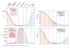
What happens if hydrostatic pressure increases above osmotic pressure?
fluid move out of capillaries
= filtration
ie. at the arterial end
What happens if the hydrostatic pressure is less than the osmotic pressure
fluid move IN to capillaries
= REABSORPTION
ie. at the venous end
Explain the forces at work in capillaries
-
Hydrostatic
- Capillary hydrostatic pressure (CHP)
- Interstitial hydrostatic pressure (IHP) = negligible
-
Osmotic
- Capillary (blood colloid osmotic pressure -BCOP)
- proteins are the key
- Interstitial fluid (ICOP) = negligible
- Capillary (blood colloid osmotic pressure -BCOP)
-
Net filtration pressure (NFP)
- net hydrostatic pressure - net osmotic pressure
- values changes from arterial end to venous end
What is high hydrostatic pressure (CHP) due to?
MAP
What is high osmotic pressure (CHP) due to?
dissolved plasma proteins
In the middle of capillary what does it look like?
- fluid leaving, due to low venous pressure
- solutes and fluids leaving together
- NFP = no net flow out of capillary
