Lecture 2: Motility of the GI tract Flashcards
How do the circular and longitudinal muscles of the GI tract differ in function?
Inner circular: contraction decreases diameter of the segment
Outer longitudinal: contraction decreases length of the segment

What are slow waves and how do they relate to AP’s?
- Oscilating depolarization and repolarizations of the membrane potential, but are themselves NOT AP’s!
- AP’s occur when depolarization moves the membrane potential to or above the threshold

What are Phasic contractions; where do they occur in the GI tract?
- Periodic contractions followed by relaxation
- Esophagus, stomach (antrum), SI, and all tissues involved in mixing and propulsion

What are tonic contractions; where do they occur?
- Maintain a basal level of contraction w/o regular period of relaxation
- Stomach (orad), lower esophageal, ileocecal, and internal anal sphincters

How are slow waves related to tonic contractions?
- Even sub-threshold depolarization produces weak, basal contractions
- Even w/o occurence of AP’s, the smooth muscle is not completely relaxed and is exhibiting tonic contractions
How is the number of AP’s on top of a slow wave related to contraction?
Greater # of AP’s on a slow wave, the larger the phasic contractions

What 3 factors cause an increase in the amplitude of slow waves and the number of AP’s?
- Stretch
- ACh
- Parasympathetics
What 2 factors cause a decrease in the amplitude of slow waves and number of AP’s?
- NE
- Sympathetics
What are the main things that the submucosal and myenteric plexus control in the GI?
Submucosal: GI secretions and local blood flow
Myenteric: GI movements
What generates the spontaneous slow wave activity?
Pacemaker regions in the myenteric and submucosal plexuses
What kind of cells are the pacemaker for GI smooth muscle; how do they function?
- Interstitial cells of Cajal (ICC)
- Slow waves occur spontaneously in the ICC and spread rapidly to smooth muscle via gap junctions
- Electric activity in the ICC drives the frequency of contractions
Smooth muscle cells respond to slow wave depolarization by increasing?
Ca2+ channel open probability = more likely to depolarize and generate an AP
Most of the muscles of mastification are innervated by the motor branch of which nerve; what control and causes mastification?
- CN V = Trigeminal N.
- Controlled by nuclei in the brain stem
- Caused by chewing reflex
What are the 3 phases of swallowing; which are voluntary vs. involuntary?
- Oral phase (voluntary) - initiates the swallowing process
- Pharyngeal phase (involuntary)
- Esophageal phase (involuntary)
What are the order of events during the pharyngeal phase of swallowing?
1) Soft palate pulled upward
2) Epiglottis moves
3) UES relaxes
4) Peristaltic wave of contractions is initiated in pharynx
5) Food is propelled through open UES
What controls the esophageal phase of swalloing and what is seen during this phase?
- Controlled by swallowing reflex and the ENS
- 1° Persistaltic wave
- 2° Peristaltic wave - only if necessary!
How is the involuntary swalowing reflex controlled?
Sensory receptors in pharynx sense food –> afferent sensory input via vagus/glossopharyngeal N. –> swallowing center (medulla) –> brain stem nuclei –> efferent input to pharynx
What is a 1° peristaltic wave; controlled by; affect of vagotomy?
- Continuation of pharyngeal peristalsis
- Controlled by medulla
- Cannot occur after vagotomy
What is a 2° peristaltic wave; controlled by; affect of vagotomy?
- Occurs if 1° wave fails to empty the esophagus or if gastric contents reflux into the esophagus
- Medulla and ENS are involved
- Can occur in absence of oral and pharyngeal phases
- Occurs even after a vagotomy
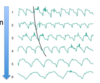
How does swallowing change the intraluminal pressure along the esophagus?
- Immediately after swallowing pressure drops in UES, but then quickly increases so that sphincter closes and food does not come back up.
- This pattern continues segmentally as the food bolus moves through the esophagus and toward the LES/Fundus

Is the pressure high or low in the UES, LES, and Fundus prior to swallowing?
Pressure is high, because these sphincter should be closed
What is the receptive relaxation phenomenon?
The LES and Fundus relax much earlier than would be expected, this is because they are preparing to receive the food bolus

The intrathoracic location of the esophagus poses the challenge of keeping air out of the esophagus at the upper end, and acidic gastric contents out of the lower end. How are these problems solved?
UES and LES are closed, except when food bolus is passing from pharynx to esophagus or from esophagus to stomach
Gastroesophageal reflux occurs when; often seen in what type of patients?
- Intra-abdominal pressure is increased
- Pregnant and morbidly obese
How is the physiology of the esophagus affected by achalasia; what are results of this disease?
- Impaired peristalsis
- Incomplete LES relaxation during swallowing
- Backflow of food (regurgitation), difficulty swallowing both liquids and solid (dysphagia), chest pain
What leads to achalasia?
- Lack of VIP or enteric NS has been knocked out
- Damage to nerves in esophagus, preventing it from squeezing foof into the stomach
What occurs anatomically during GERD; why does it happen; leads to what complications?
- Changes in the barrier between the esophagus and stomach (i.e., LES relaxes abnormally or weakens)
- Motor abnormalities that result in abnormally low pressure in the LES; if intragastric pressure increases, as may occur following a large meal, during heavy lifting, or pregnancy
- Backward wash of acid, peside and bile into esophagus = heartburn and acid regurgitation
- Can lead to complications such as irritation of the lining of the esophagus (esophagitis), scar tissue in esophagus (stricture of esophagus), and Barret’s esophagus
A particularity of the stomach are the 3 layers of muscle, what are they?
- Circular
- Longitudinal
- Oblique
What region of the stomach is affected by receptive relaxation and what occurs to the pressure and volume; what type of reflex is this?
- Decreased pressure and increased volume in orad region
- Vagovagal reflex!
What is the contractile activity of the orad region of stomach; controlled by what hormone?
- Minimile contractile activity, so very little mixing of food occurs here
- CCK decreases contractility and there is increased gastric distensibility
What is the primary contractile event in the stomach, what region does it occur in?
- Peristaltic contraction (mid stomach –> pylorus)

How do contractions change as they move further through stomach and what is the max frequency?
- Contractions increase in force and velocity as they approach the pylous
- Max frequency is ~ 3 – 5 waves/min

Explain retropulsion and why this is occuring?
- The pylorus opens/relaxes for a very short period of time so only a small portion of the food bolus is moved into the duodenum. Most of the gastric contents are propelled back into the stomach for further mixing and further reduction in particle size.
- This occurs due to segmental contraction

What increases AP and force of gastric contractions?
Parasympathetic stimulation, gastrin and motilin
What decreases AP and force of gastric contractions?
Sympathetic stimulation, secretin, and GIP
What 4 factors increase the rate of gastric emptying; how long does gastric emptying take?
- Decreased distensibility of the orad (means it is very distended)
- Increased force of peristaltic contractions of the caudad stomach
- Decreased tone of the pylorus
- Increased diamter and inhibition of segmental contractions of the proximal duodenum
- Takes ~ 3 hrs
What 4 factors lead to inhibition of gastric emptying?
- Relaxation of the orad
- Decreased force of peristaltic contractions
- Increased tone of pyloric sphincter
- Segmentation contractions in the intestine
What is the Entero-Gastric reflex?
Negative feedback from duodenum will slow down the rate of gastic emptyting
How do acid, fats, and hypertonicity of the duodenum contribute to the Entero-gastric reflex?
- Acid in duodenum –> Stimulates secretin release –> Inhibits stomach motility via gastrin inhibition
- Fats in duodenum –> Stimulate CCK and GIP –> Inhibit stomach motility
- Hypertonicity in duodenum –> (unknown hormone) –> Inhibit gastric empyting

Symptoms, causes, and treatmet for slow gastric emptying?
Sx: fullness, loss of apetite, nausea, sometimes vomiting
Causes: gastric ulcer (scar tissue), cancer (physical obstruction), eating disorder (anorexia, bulimia, obesity), vagotomy
Tx: pyloroplasty, ballon dilation
What is Gastroparesis, causes, commonly seen with what disease, and symptoms?
- Slow empyting of stomach/paralysis of stomach
- ~ 20% Type I DM and some Type II affected
- Cause: damage to the vagus nerve, idiopathc dx, high blood glucose (diabetic gastroparesis)
Sx: nausea, vomiting, an early feeling of fullness when eating, weight loss, abdominal bloating, and abdominal discomfort
What are migrating myoelectric complexes (MMC), how often do they occur, and what are they mediated by?
- Periodic, bursting peristaltic contractions
- Occur at 90 min. intervals, during fasting
- Motilin-mediated
- Inhibited during feeding
*Help prevent bacterial overgrowth and/or to move large particles along*

What does the motility of the small intestine aim to do?
- Mix the chyme w/ digestive enzymes and pancreatic secretions
- Expose the nutrients to the intestinal mucoas for absorption
- Propel the unabsorbed chyme along the small intestine to the large intestine
What is the purpose of segmental contractions in the small intestine; what occurs?
- Mix the chyme and expose it to pancreatic enzymes and secretions, but produces no forward, propulsive movement along the SI
- Section of SI contracts, splitting the chyme and sending it in both orad and caudad directions
- Section now relaxes, allowing split bolus of chyme to merge back together

What is the purpose of peristaltic contractions and what occurs?
- Propel the chyme toward the large intestine
- Contraction occurs orad to (behind) bolus; simutaneously the section caudad to bolus will relax
- Chyme is propelled down the SI, as this sequence repeats itself

How do circular and longitudinal cells work together to accomplish the propulsive movements in the SI?
- Circular and longitudinal muscles work in opposition to complement each other’s action - reciprocal innervation
- When the circular muscle of a segment contracts, the longitudinal muscle simultaneously relaxes and vice versa
How do slow waves operate in the small intestine?
- Always present whether contractins are occuring or not
- DO NOT intiate contractions like they do in the stomach
- Spike potential (AP) are necessary for muscle contraction to occur
- Slow wave frequency sets the maximum frequency of contraction
What is the slow wave frequency gradient of the small intestine from duodenum to ileum?
Duodenum = 12 cycles/min
Jejunum = 10 cycles/min
Ileum = 8 cycles/min

What is the function of the Myenteric plexus and Submucosal plexus in the small intestine?
Myenteric - mainly regulates the relaxation and contraction
Submucosal - sense the lumen enviornment
Describe what occurs in the small intestine when the food bolus is sensed by ECL cells?
- ECL cells in intestinal mucosa will release serotonin (5-HT)
- Serotonin binds to receptors on IPANs that, when activated inititate the peristaltic reflex in that segment
What occurs behind the bolus after the peristaltic reflex has been initiated by ECL cells?
- ACh, Substance P, and Neuropeptide Y (excitatory NT’s) are released in circular muscle, while these pathways are simultaneously inhibited in the longitudinal muscle
- This segment will then narrow and lengthen (contraction)
What occurs in front of the bolus after the ECL cells have initiated the peristalsis reflex?
- Inhibitory pathways (i.e., VIP, NO) are activated in circular muscle, while excitatory pathways are activated in longitudinal muscle
- This segment will widen and shorten (relax)
How does neural input control the contractions of the small intestine (ENS and ANS)?
ENS: mediates the peristaltic reflex
ANS: the PNS stimulates and SNS inhibits contractions
Which hormones function to stimulate contractions in the small intestine?
Serotonin, prostaglandins, gastrin, CCK, motilin, and insulin
Which hormones functions to inhibit contractions in the small intestine?
Epinephrine, secretin and glucagon
What coordinates the vomiting reflex and how do these signals get there?
- Coordinated by the medulla
- Nerve impulses transmitted by vagus and sympathetics to multiple brain stem nuclei

What kind of medications can stimulate the chemical trigger zone and lead to vomiting reflex?
Opioids - apomorphine and morphine
What are the events that occur during the vomiting reflex?
- Reverse peristalsis in small intestine
- Stomach and pylorus relaxation
- Forced inspiration to increase abdominal pressure
- Movement of the larynx
- LES relaxation
- Glottis closes
- Forceful expulsion of gastric contents
How is the flow of contents from the small intestine to large intestine regulated at the ileocecal junction; what reflex is this?
- Distention of ileum causes relaxation of the sphincter (allows contents to flow from ileum to colon)
- Distention of colon causes contraction of the sphincter (prevents passage of contents from colon to the ileum

How does innervation of the internal and external anal sphincter’s differ?
Internal - innervated by Pelvic splanchnics (PNS)
External - innervated by Pudenal n.
What are the 4 types of innervation of the large intestine; what does each innervate?
1. ENS (myenteric plexus) - beneath teniae coli, innervates muscle layers
2. PNS
- Vagus: Cecum, ascending and transverse colon
- Pelvic (S2-S4): Descending and sigmoid colon, rectum
3. SNS (T10-L2)
- SMG: Proximal regions
- IMG: Distal regions
- Hypogastric plexus: Distal rectum and anal canal
4. Somatic pudendal nerves: external anal sphincter
What are mass movements in the large intestine and how often do they occur?
- Occur over large distances, 1-3x/day
- Stimulate defecation reflex
- Final mass movement propels the fecal content into the rectum

How does slow motility and excess motility in the large intestine affect fecal composition/absorption?
Slow (constipation) - greater absorption of water and vitamin + hard feces in transverse colon
Fast (diarrhea) - less absorption and loose feces

How does the rectum fill intermittently and what will occur?
- Mass movement and segmentation contractions
- As it fills with feces, SM walls of the rectum contracts and internal anal sphincter relaxed (rectosphincteric reflex)
- External anal sphincter is tonically closed (voluntary control)

How are the the rectosphincteric reflex and defecation controlled?
- Under neural control: partially by ENS and reflex is reinforced by acitivity of neurons within the spinal cord
- Sensation of rectal distention and voluntary control of the external anal sphincter are mediated by pathways within the spinal cord that lead to the cerebral cortex

What is Hirschsprung disease (AKA mega colon) caused by, results in, symptoms, and treatment?
Cause: ganglion cells absent from distal colon
Result: VIP levels low –> SM constriction/loss of coordinated movement –> colon contents accumulate (“colon equivalent of achalasia”)
Sx: Failure to pass meconium, poor feeding, jaundice, vomiting (newborns). Constipation, swollen belly, malnutrition (older children)
Tx: surgical resection of colon segment lacking ganglia
What is the intestino-intestinal reflex?
- Short reflex
- Generally inhibitory
- Involved only the ENS, completely independent of the ANS (Atopine has no affect on it!)
- Ileocecal sphincter (SM) acts by intestino-intestinal reflexes
What is the gastoileal reflex (gastroenteric)?
Gastric distention relaxes ileocecal sphincter
What is the gastro- and duodeno-colic reflexes?
- Distention of stomach/duodenum initiated mass movements
- Transmitted by way of the ANS