Lecture 1 COPY Flashcards
What does the brain float in?
cerebral spinal fluid
How much cerebral spinal fluid is made in a day?
500cc
How much CSF is there in total?
150cc
What makes CSF?
Choroid plexus
Where is excess CSF reabsorbed?
arachnoid granulations
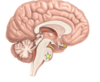
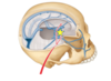
Describe the flow of CSF.
-Made in the choroid plexus -through foramen of monroe -into the third ventricle -through the cerebral aqueduct -into the fourth ventricle -through 3 foramen (2 lateral: foramen of lueschka; 1 medial: foramen of magendie) -now in the cerebellar cistern -through foramen magnum -down spinal cord and back up to be reabsorbed
How does CSF get into the venous system?
Arachnoid granulations
What does the dura split in two to form?
superior sagittal sinus
What PADs the brain?
The meninges: Pia mater (inner most) Arachnoid mater (middle) (spider webs) Dura mater (outside)
If you break a bridging vein what could happen?
A subdural hematoma
What does stenosis mean? What would happen if you had stenosis in a foramen for the CSF?
stenosis is narrowing of something if you had narrowing in the foramen then it would cause a backup of CSF and built up pressure
What part of the dura mater separates the two hemispheres?
cerebral falx
What part of the dura separates the cerebellum?
the cerebellar tentorium
What is the Pterion?
The place were the 4 bones meet on side of head, weak spot.
What are the four lobes of the brain?
Occipital Parietal Temporal Frontal
What is the central sulcus?
between frontal and parietal lobes
What lobe is the primary motor cortex in?
Frontal
What lobe is the primary somato-sensory cortex in?
Parietal
T/F the right side of your brain controls the left side of your body
True
What is the cavernous sinus?
lateral to the sella turcica, houses passage for CN3,4,6,V1,V2
Label Veins
PPT 1 Slide 28
What is the danger triangle?
corner of lips to bridge of nose. do not get an infection here bc it can travel to the brain
What is Broca’s area?
LEFT SIDE broken language, form words/sentences
What is wernicke’s area?
LEFT SIDE understanding language. can speak words but will be wordy
Pyramidal or Lateral Corticospinal tract
motor tract
Posterior Columns or Dorsal Columns tract
Vibration and position
Anterolateral or Spinothalamic tract
pain and temperature and CRUDE touch
What is the decussation point for the lateral corticospinal tract?
pyrimidal decussation in the medulla
What is the decussation point for the posterior column tract?
internal arcuate fibers in the medulla
What is the decussation point for the anterolateral tract?
Anterior commissure (spinal cord)
Where does the lateral corticalspinal tract nerves synapse?
anterior horn (2 neurons, upper and lower)
Where does the posterior column tract nerves synapse?
primary and secondary synapse at the gracile nucleus secondary and 3rd neuron synapse at the thalamus: VPL
What is the VPL?
Ventral posterior lateral nucleus
Where does the anterior lateral tract nerves synapse?
the primary neuron and secondary neuron synapse at the posterior horn the secondary and 3rd neuron synapse at the thalamus VPL
Label the blood supply vessels to the brain
PPT 1 Slide 58
Label the blood supply to the hemispheres of the brain
PPT 1 Slide 63
What is a homunculus? What does it look like?
The two monkeys haning upside down. What parts of the brain correspond to the sensory or motor cortex
Where is the primary auditory cortex?
in the sylvian fissure
Identify the structure on the diagram with the stars

cerebellum
Identify the structure with the star on the diagram

pons
Identify the structure on the diagram

midbrain
identify the structure on the brain

cerebrum
what three parts make up the brainstem?
- midbrain
- pons
- medulla oblongata
Identify the lobe on the diagram

parietal lobe
Identify the lobe on the diagram

frontal lobe
Identify the lobe on the diagram

temporal lobe
Identify the lobe on the diagram

occipital
Identify the structure with the star

central sulcus
Identify the structure with the star

cavernous sinus
Identify the structure with the star

superior sagittal sinus
Identify the structure in the diagram

inferior petrosal sinus
Identify the structure with the blue arrow.

Ophthalmic nerve (CN V1)
Identify the structure with the arrow.

oculomotor nerve (CN III)
Identify the structure with the arrow.

trochlear nerve (CN IV)