Lecture 1 - Abdominal Anterolateral wall and posterior wall Flashcards
(49 cards)
What kinds of fascia are in the abdominal anterolateral wall?
It has superficial fascia that is connective tissue. There are two kinds:
1.) Above the umbilicus : single sheet of connective tissue that is continous with the superficial fascia in other regions of the body.
2.) Below the umbilicus: There are two layers that are the fatty superficial layer called camper’s fascia and the membranous deep layer called Scarpa’s fascia. Superficial vessels and nerves run between these layers of fascia.
What kind of muscles are in the anterolateral abdominal wall?
Two main groups:
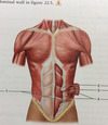
1.) flat muscles - three of them that are situated laterally on either side of the wall.
2.) vertical muscles: two of them that are situated near the mid line of the body.
How are the flat muscles oriented and how does this effect the wall?
The flat muscles are stacked up on one another and their fibers run in different direcetions but they all meet and cross each other. This strenghtens the wall and reduces the risk of herniation. Also, each flat muscle forms an aponeurosis which covers the vertical abdominis muscle. These become intertwined in th emiddle forming the linea alba.

- ) External Oblique
- ) Rectus Abdominus
- ) Transverse Abdominus
- ) Internal Oblique
Where is the pyrimidalis muscle?
At the bottom of the abdominal wall, in the medial side. Small muscle
What is the arcuate line?
It is behind the rectus muscle, 1/3 the distance between the pubic symphysis and the umbilicus. The point at which the aponeurosis of the three lateral abdominal muscles pass anterior to the rectus abdominis.
What is in direct contact with the internal aspect of the rectus abdominis muscle?
transversalis fascia
What is the vasculature in terms of arteries in the anterior abdominal wall and where do these arteries come from?
- ) sueprior epigastric artery that comes from the internal thoracic artery.It travels deep to the rectus abdominis.
- ) Inferior epigastric artery that branches from the external iliac artery and also travels deep to the rectus abdominis.
- ). Superficial epigastric artery which brances from the femoral artery and travels superficial to the rectus sheath.
How is the anterior abdominal wall innervated?
- Innervated by the anterior rami of spinal nerves T7 to L1. These intercostal nerves travel between the neurovascualr plane between the internal and innermost intercostal muscles.
- When they pass the costal margin they become thoracoabdominal nerves, these travel betweent the neurovascular plane between the internal oblique and the transversus abdominis.
Thoracoabdominal nerves - T7-T11 - how they travel?
Enter abdominal wall at the costal margin.
Thoracoabdominal nerves - T12 - how it travel?
Called the subcostal nerve and it runs under the 12th rib
Thoracoabdominal nerves - L1 - what it divide into?
- iliohypogastric nerve
- ilioinguinal nerve
what are the abdominal quadrants and describe how each one is cut?
There are 4 and they are cut by 2 lines. The transumbilical plane which cuts horizontally creating and upper and lower quadrant. Then there is the median plane which cuts vertically creating a left and right quadrant.
- right upper
- right lower
- left upper
- left lower
Umbilicus is often used as a key landmark, what is its dermatome, vertebral level, and where is it?
Dermatome: T10
Vertebral level: L3/L4
At the midline
What is mcBurney’s point ?
It is 1/3 point on a line that is from the right anterior superior iliac spine to the umbilicus.

What are the abdominal Regions?
There are 9 different abdominal regions:
- ) Right hypochondriac
- ) Epigastric
- ) Left Hypochondriac
- ) Right lumbar
- ) Umbilical
- ) Left lumbar
- ) Right inguinal/iliac
- ) Hypogastric
- ) Left inguinal/iliac

What does the gubernaculum become?
In men: becomes the scrotal ligament
In women: upper: suspensory ligament of teh ovary, lower: round ligament of the uterus.
What does the process vaginalis become?
tunica vaginalis
What is the tunica vaginalis?
serous sheath of the testis and epididymis
How does scrotum come out?
The testicles go down and through what was the anterior abdominal wall to make a pouch called the scrotum.
What is the inguinal ligament and where is it?
It is a fibrous, thickened and folded margin of the external oblique aponeurosis. It froms the floor of the inguinal canal and it extens from the anterior superior iliac spine and the pubic tubercle.

What is the Lacunar Ligament ?
The deeper fibres of the external oblique aponeurosis pass posteriorly to attach lateral to the pubic tubercle to form an arch.

Pectineal Ligament ?
Most lateral lacunar ligament fibers continue to run along pecten pubis. It is medial to the femoral canal. Forms the posterior border of the femoral canal.

Reflected inguinal ligament?
Superior fibers of external oblique aponeurosis and lacunar ligament fan upwards crossing the linea alba instead of inserting onto the pubic tubercle.


