Lecture 05. Nervous System Flashcards
Nervous System
The master controlling and communication system of the body.
Functions of Nervous System
- Sensory input
- Motor output
THREE basic steps:
- Through sensory nerve endings, it receives information about changes in the body and external environment and it transmits coded messages to the spinal cord and brain.
- The spinal cord and brain process this information, relate it to past experience, and determine what response, if any, is appropriate to the circumstances.
- The spinal cord and brain issue commands primarily to muscle and gland cells to carry out such responses.
Subdivisions of Nervous System
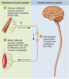
- Central Nervous System (CNS): Consists of the brain and spinal cord, which are enclosed and protected by the cranium and vertebral column.
- Peripheral Nervous System (PNS): Consists of all the nervous system except the brain and spinal cord. It is composed of nerves and ganglia.
- *Nerve:** Bundle of nerve fibers (axons) wrapped in fibrous connective tissue. Nerves emerge from the CNS through foramina of the skull and vertebral column and carry signals to and from other organs of the body.
- *Ganglion:** A knot-like swelling in a nerve where the cell bodies of neurons are concentrated.

Subdivisions of Peripheral Nervous System
Sensory (afferent) Division: Carries sensory signals from various receptors (sense organs and simple sensory nerve endings) to the CNS. This is the pathway that informs the CNS of stimuli within and around the body.
Somatic Sensory Division: Carries signals from receptors in the skin, muscles, bones, and joints.
Visceral Sensory Division: Carries signals mainly from the viscera of the thoracic and abdominal cavities, such as the heart, lungs, stomach, and urinary bladder.
Motor (efferent) Division: Carries signals from the CNS to gland and muscle cells that carry out the body’s responses. Cells and organs that respond to commands from the nervous system are called effectors.
Somatic Motor Division: Carries signals to the skeletal muscles. This output produces muscular contractions that are under voluntary control, as well as involuntary muscle contractions called somatic reflexes.
Visceral Motor Division (autonomic nervous system, ANS): Carries signals to glands, cardiac muscle, and smooth muscle. We usually have no voluntary control over these effectors, and this system operates at an unconscious level. The responses of this system and its effectors are visceral reflexes.
The autonomic nervous system has two further divisions:
Sympathetic Division: Tends to arouse the body for action (accelerating the heartbeat and increasing respiratory airflow).
Parasympathetic Division: Adapts the body for energy intake and conservation. (It stimulates digestion but slows down the heartbeat and reduces respiratory airflow).
Histology of Nervous Tissue
Highly Cellular (very little extracellular component)
Made up of TWO pricipal types of cells:
- Neurglia: Support cells
- Neurons: Electrically conductive cells (much less numerous)

Properties of Neurons
Nerve Cell: The functional unit of the nervous system.
THREE fundamental physiological properties:
- Excitability (irritability): All cells possess excitability, or responsiveness to environmental changes called stimuli. Neurons have developed this property to the highest degree.
- Conductivity: Neurons respond to stimuli by producing traveling electrical signals that quickly reach other cells at distant locations.
- Secretion: When the electrical signal reaches the end of a nerve fiber, the neuron usually secretes a chemical called a neurotransmitter that crosses a small gap between cells and stimulates the next cell.
Functional Classes of Neurons
THREE general classes of neurons:
- Sensory (afferent) Neurons: Specialized to detect stimuli such as light, heat, pressure, and chemicals and to transmit information about them to the CNS. These neurons can begin in almost any organ of the body but always end in the brain or spinal cord; the word afferent refers to signal conduction toward the CNS. Some sensory receptors, such as pain and smell receptors, are themselves neurons. In other cases, such as taste and hearing, the receptor is a separate cell that communicates directly with a sensory neuron.
- Interneurons (association neurons): Lie entirely within the CNS. They receive signals from many other neurons and carry out the integrative function of the nervous system— they process, store, and retrieve information and “make decisions” about how the body responds to stimuli. About 90% of human neurons are interneurons. The word interneuron refers to the fact that they lie between, and interconnect, the incoming sensory pathways and the outgoing motor pathways of the CNS.
- Motor (efferent) Neurons: Send signals predominantly to muscle and gland cells, the effectors that carry out the body’s responses to stimuli. They are called motor neurons because most of them lead to muscle cells, and efferent neurons to signify that they conduct signals away from the CNS.

Structure of Neuron
- Neurosoma/Soma (cell body): Control center of neuron.
- Dendrites: Tree branch structure. Receives signals from other neurons. The more dendrites = the more information neuron is able to receive.
- Trigger Zone: Where neuron first generates action potential.
- Axon: Specialized for rapid condction of nerve signals to points remote from the soma.
- Myelin Sheath: An insulating layer around a nerve fiber, (like the rubber insulation on a wire). It is formed by oligodendrocytes in the central nervous system and Schwann cells in the peripheral nervous system.
- Synaptic Knob: A little swelling that forms a junction (synapse) with a muscle cell, gland cell, or another neuron.

Multipolar Neurons
Have one axon and two or more (usually many) dendrites.
Examples:
- Includes most neurons of the brain and spinal cord.

Bipolar Neurons
Have one axon and one dendrite.
Examples:
- Olfactory cells of the nasal cavity
- Some neurons of the retina
- Sensory neurons of the inner ear

Unipolar Neurons
Have only a single process leading away from the soma. They are represented by the neurons that carry sensory signals to the spinal cord. Also called pseudounipolar because they start out as bipolar neurons in the embryo, but their two processes fuse into one as the neuron matures. A short distance away from the soma, the process branches like a T, with a peripheral fiber carrying signals from the source of sensation and a central fiber continuing into the spinal cord. In most other neurons, a dendrite carries signals toward the soma, and an axon carries them away. In unipolar neurons, however, there is one long fiber that bypasses the soma and carries nerve signals directly to the spinal cord. The dendrites are the branching receptive endings in the skin or other place of origin, while the rest of the fiber is considered to be the axon (defined in these neurons by the presence of myelin and the ability to generate action potentials).

Anaxonic Neurons
Have multiple dendrites but no axon.
They communicate over short distances through their dendrites and do not produce action potentials.
Examples:
- Brain
- Retina (help in visual processes such as the perception of contrast)
- Adrenal Medulla

Types of Neuroglia
**Oligodendrocytes (CNS): **Form myelin in brain and spinal cord (lipid-based substance) acts as insulation, increasing the conduction velocity of the nerve fiber.
Ependymal cells (CNS): Line cavities of brain and spinal cord; secrete and circulate cerebrospinal fluid
**Microglia (CNS): **Phagocytize and destroy microorganisms, foreign matter, and dead nervous tissue
**Astrocytes (CNS): **Cover brain surface and nonsynaptic regions of neurons; form supportive framework for the CNS; stimulate formation of blood– brain barrier; nourish neurons and secrete growth stimulants; influence synaptic signaling between neurons; help to regulate composition of the extracellular fluid in the CNS; form scar tissue to replace damaged nervous tissue
**Schwann cells (PNS): **Form neurilemma around all PNS nerve fibers and myelin around most of them; aid in regeneration of damaged nerve fibers
Satellite cells (PNS): Surround somas of neurons in the ganglia; insulate them and regulate their chemical environment.
Charges
Electrically forces produced by ions. Force increases as charge increases as well as with decreasing distance.
Within the cell there is a thin membrane of negatively charged ions inside of the cell and a thin membrane of positively charged ions on the outside of the cell - this determines resting membrane potential. (-70mmu)
Membrane Channels
Voltage Gated:
Ligand Gated:


