Lecture 02. Histology Flashcards
Histology
(Microscopic Anatomy)
The study of tissues and how they are arranged into organs.
Tissue
(Four Primary Types)
Tissue: A mass of similar cells and cell products that forms a discrete region of an organ and performs a specific function. Composed of cells and matrix, matrix composed of fibers and ground substance.
**FOUR **primary types of tissue:
- Epithelial
- Connective
- Nervous
- Muscular
Tissues differ from each other in the types and functions of their cells, the characteristics of the matrix (extracellular material) that surrounds the cells, and the relative amount of space occupied by cells versus matrix.
Matrix: Composed of **Fibrous Proteins and Ground Substance (Extracellular Fluid**, Interstitual Fluid, or Tissue Fluid) - in cartilage and bone the matrix is rubbery or stony.
Epithelial Tissue
Tissue composed of layers of closely spaced cells that cover organ surfaces or form glands; serves for protection, secretion, and absorption.
Classified by number of layers:
- Simple Epithelium: Single layer of cells.
- Stratified Epithelium: More than one layer of cells.
Classified by shape:
- Squamus: Flattened, longer than they are tall.
- Cuboidal: As tall as they are wide.
- Columnar: Taller than they are wide.
Uique forms:
- **Transitional: **Streching capabilities.
- **Pseudo-stratified: **Cells appear as though they occur in a few layers, but all of the cells rest on the basement membrane.
Locations:
- Epidermis
- Inner lining of digestive tract
- Liver and other glands

Simple Squamous Epithelium
- *Microscopic appearance:** Single layer of thin cells, shaped like fried eggs with bulge where nucleus is located; nucleus flattened in the plane of the cell, like an egg yolk; cytoplasm may be so thin it is hard to see in tissue sections; in surface view, cells have angular contours and nuclei appear round
- *Representative locations:**
- Air sacs (alveoli) of lungs
- Inner lining (endothelium) of heart and blood vessels
- Serous membranes of stomach, intestines, and some other viscera
- Surface mesothelium of pleurae, pericardium, peritoneum, and mesenteries
Functions:
- Allows rapid diffusion or transport of substances through membranes
- Secretes lubricating serous fluid

Simple Cuboidal Epithelium
Microscopic appearance: Single layer of square or round cells; in glands, cells often pyramidal and arranged like segments of an orange around a central space; spherical, centrally placed nuclei; often with a brush border of microvilli in some kidney tubules; ciliated in bronchioles of lung Representative locations:
- Kidney tubules
- Liver
Functions:
- Absorption and secretion

Simple Columnar Epithelium
- *Microscopic appearance:** Single layer of tall, narrow cells; oval or sausage-shaped nuclei, vertically oriented, usually in basal half of cell; often shows a brush border of microvilli; ciliated in some organs; may possess goblet cells (helps create mucus).
- *Representative locations:**
- Inner lining of stomach, intestines, gallbladder, uterus, and uterine tubes
Functions:
- Absorption and secretion of mucus and other products
- Movement of egg and embryo in uterine tube

Pseudostratified Columnar (Cilia) Epithelium
- *Microscopic appearance:** Looks multilayered; some cells do not reach free surface but all cells reach basement membrane; nuclei at several levels in deeper half of epithelium; often with goblet cells; often ciliated
- *Representative locations:**
- Respiratory tract from nasal cavity to bronchi
Functions:
- Secretes and propels mucus
- Traps dust particles and moves them away from lungs

Stratified Squamous Epithelium
KERATINIZED
Microscopic appearance: Multiple cell layers with cells becoming increasingly flat and scaly toward surface; surface covered with a layer of compact dead cells without nuclei; basal cells may be cuboidal to columnar
Representative locations:
- Epidermis; palms and soles are especially heavily keratinized
Functions:
- Resists abrasion
- Prevents water loss through skin
- Resists penetration by pathogenic organisms
- *NONKARATINIZED**
- *Microscopic appearance:** Same as keratinized epithelium but without the surface layer of dead cells
- *Representative locations:**
- Tongue
- Esophagus
- Anal canal
- Vagina
Functions:
- Resists abrasion and penetration by pathogenic organisms

Stratified Cuboidal Epithelium
- *Microscopic appearance:** Two or more layers of cells; surface cells roughly square or round
- *Representative locations:**
- Sweat gland ducts
- Egg-producing vesicles (follicles) of ovaries
- Sperm-producing ducts (seminiferous tubules) of testes
Functions:
- Contributes to sweat secretion
- Secretes ovarian hormones
- Produces sperm

Transitional Epithelium
- *Microscopic appearance:** Somewhat resembles stratified squamous epithelium, but surface cells are rounded, not flattened, and often bulge above surface; typically five or six cells thick when relaxed, two or three cells thick when stretched; cells may be flatter and thinner when epithelium is stretched (as in a distended bladder); some cells have two nuclei
- *Representative locations:**
- Urinary tract—part of kidney, ureter, bladder, part of urethra
Function:
- Stretches to allow filling of urinary tract
Connective Tissue
Tissue with usually more matrix than cell volume; often specialized to support, bind, and protect organs.
Functions of Connective Tissue:
- Binding of organs: Tendons bind muscle to bone, ligaments bind bone to bone, fat holds the kidneys and eyes in place, and fibrous tissue binds the skin to underlying muscle.
- Support: Bones support the body, and cartilage supports the ears, nose, trachea, and bronchi.
- **Physical Protection: **The cranium, ribs, and sternum protect delicate organs such as the brain, lungs, and heart; fatty cushions around the kidneys and eyes protect these organs.
- Immune Protection: Connective tissue cells attack foreign invaders, and connective tissue fiber forms a “battlefield” under the skin and mucous membranes where immune cells can be quickly mobilized against disease agents.
- Movement: Bones provide the lever system for body movement, cartilages are involved in movement of the vocal cords, and cartilages on bone surfaces ease joint movements.
- Storage: Fat is the body’s major energy reserve; bone is a reservoir of calcium and phosphorus.
- Heat Production: Metabolism of brown fat generates heat in infants and children.
- Transport: Blood transports gases, nutrients, wastes, hormones, and blood cells.
Locations:
- Tendons and ligaments
- Cartilage and bone
- **Blood and lymph **
Fibrous Connective Tissue
(Also called fibroconnective tissue or connective tissue proper)
Classified together because the fibers are so conspicuous, also includes cells and ground substance.
Cells that make fibrous tissue:
- Fibroblasts: Large, fusiform cells that often show slender, wispy branches. They produce the fibers and ground substance that form the matrix of the tissue.
- Macrophages: Large phagocytic cells that wander through the connective tissues. They phagocytize and destroy bacteria, other foreign matter, and dead or dying cells of our own body. They also activate the immune system when they sense foreign matter called antigens. They arise from certain white blood cells called monocytes or from the stem cells that produce monocytes.
- Leukocytes (white blood cells - WBCs): WBCs travel briefly in the bloodstream, then crawl out through the capillary walls and spend most of their time in the connective tissues. The two most common types are neutrophils, which wander about in search of bacteria, and lymphocytes, which react against bacteria, toxins, and other foreign agents. Lymphocytes often form dense patches in the mucous membranes.
- Plasmacells: Certain lymphocytes turn into plasma cells when they detect foreign agents. The plasma cells then synthesize disease-fighting proteins called antibodies. Plasma cells are rarely seen except in inflamed tissue and the walls of the intestines.
- **Mast cells: **These cells, found especially alongside blood vessels, secrete a chemical called heparin that inhibits blood clotting, and one called histamine that increases blood flow by dilating blood vessels.
- Adipocytes (fat cells): These appear in small clusters in some fibrous connective tissues. When they dominate an area, the tissue is called adipose tissue.
**THREE **types of protein fibers found in fibrous connective tissues:
- Collagenous Fibers: These fibers, made of collagen, are tough and flexible and resist stretching. Collagen is the body’s most abundant protein, constituting about 25% of the total. It is the base of such animal products as gelatin, leather, and glue. In fresh tissue, collagenous fibers have a glistening white appearance, as seen in tendons and some cuts of meat (often called white fibers). In tissue sections, collagen forms coarse, wavy bundles. Tendons, ligaments, and the deep layer of the skin (the dermis) are made mainly of collagen. Less visibly, collagen pervades the matrix of cartilage and bone.
- Reticular Fibers: These are thin collagen fibers coated with glycoprotein. They form a spongelike framework for such organs as the spleen and lymph nodes.
- Elastic Fibers: These are thinner than collagenous fibers, and they branch and rejoin each other along their course. They are made of a protein called elastin, whose coiled structure allows it to stretch and recoil like a rubber band. Elastic fibers account for the ability of the skin, lungs, and arteries to spring back after they are stretched. (Elasticity is not the ability to stretch, but the tendency to recoil when tension is released.)
Ground Substance: Amid the cells and fibers in some connective tissue sections, there appears to be a lot of empty space, this space is occupied by the featureless ground substance. Ground substance usually has a gelatinous consistency resulting from three classes of large molecules composed of protein and carbohydrate: glycosaminoglycans (GAGs), proteoglycans, and adhesive glycoproteins. The ground substance absorbs compressive forces and protects the more delicate cells from mechanical injury. GAGs also form a very slippery lubricant in the joints and constitute much of the jellylike vitreous body of the eyeball. In connective tissue, such molecules form a gel that slows down the spread of bacteria and other pathogens (disease-causing agents). Adhesive glycoproteins bind plasma membrane proteins to collagen and proteoglycans outside the cell. They bind all the components of a tissue together and mark pathways that guide migrating embryonic cells to their destinations in a tissue.
Types of fibrous Connective tissue:
- Loose Connective Tissue: Much of the space is occupied by ground substance. (Areolar and Reticular)
- Dense Connective Tissue Fiber occupies more space than the cells and ground substance, and appears closely packed in tissue sections. (Dense Regular and Dense Irregular)

Areolar Connective Tissue
- *Microscopic appearance:** Loose arrangement of collagenous and elastic fibers; scattered cells of various types; abundant ground substance; numerous blood vessels
- *Representative locations:**
- Underlying nearly all epithelia
- Surrounding blood vessels, nerves, esophagus, and trachea
- Fasciae (fibrous tissue) between muscles
- Mesenteries (attaches intestine to interior wall of abdomen)
- Visceral layers of pericardium (covers heart) and pleura (covers lungs)
Functions:
- Loosely binds epithelia to deeper tissues
- Allows passage of nerves and blood vessels through other tissues
- Provides an arena for immune defense
- Provides nutrients and waste removal for overlying epithelia

Reticular Connective Tissue
- *Microscopic appearance:** Loose network of reticular fibers and cells, infiltrated with numerous lymphocytes and other blood cells
- *Representative locations:**
- Lymph Nodes
- Spleen
- Thymus
Functions:
- Framework for soft organs

Dense Regular Connective Tissue
- *Microscopic appearance:** Densely packed, parallel, often wavy collagen fibers; slender fibroblast nuclei compressed between collagen bundles; scanty open space (ground substance); scarcity of blood vessels
- *Representative locations:**
- Tendons and ligaments
Functions:
- Ligaments tightly bind bones together and resist stress
- Tendons attach muscle to bone and move the bones when the muscles contract

Dense Irregular Connective Tissue
- *Microscopic appearance:** Densely packed collagen fibers running in random directions; scanty open space (ground substance); few visible cells; long fibers in the tissue appear as short, chopped-up pieces in thin histological sections, as in photo
- *Representative locations:**
- Deeper portion of dermis
- Sheaths around cartilage and bone
Functions:
- Provides strength
- Resists tearing

Adipose Tissue
- *Microscopic appearance:** Dominated by adipocytes (large, empty- looking cells with thin margins and nucleus pressed against inside of plasma membrane). Cells usually shriveled by histological fixative. Tissue sections often pale because of scarcity of stained cytoplasm. Blood vessels often visible.
- *Representative locations:**
- Subcutaneous fat beneath skin; Breast, Heart Surface, Mesenteries
- Surrounding organs; Kidneys and Eyes
Functions:
- Energy storage
- Thermal insulation (heat production by brown fat)
- Protective cushion for some organs
- Filling space
- Shaping body
Cartilage
A connective tissue consisting of a dense matrix of collagen fibres and elastic fibres embedded in a rubbery ground substance. The matrix is produced by cells called chondroblasts, which become embedded in the matrix as chondrocytes. They occur, either singly or in groups, within spaces called lacunae (sing. lacuna) in the matrix.
The surface of most of the cartilage in the body is surrounded by a membrane of dense irregular connective tissue called perichondrium. Cartilage contains no blood vessels or nerves - except in the perichondrium.
THREE different types:
- Hyaline Cartilage
- Fibrocartilage
- Elastic Cartilage

Hyaline Cartilage
- *Microscopic appearance:** Clear, glassy matrix, often stained light blue, violet, or pink in tissue sections; fine, dispersed collagen fibers, not usually visible; chondrocytes often in small clusters of three or four cells (cell
nests) , enclosed in lacunae; usually covered by perichondrium - *Representative locations:**
- Forms a thin articular cartilage, lacking perichondrium, over the ends of bones at movable joints
- Forms supportive rings and plates around trachea and bronchi
- Forms a boxlike enclosure around the larynx
- A costal cartilage attaches the end of a rib to the breastbone
- Forms much of the fetal skeleton
Functions:
- Eases joint movements
- Holds airway open during respiration
- Moves vocal cords during speech
- A precursor of bone in the fetal skeleton and forms the growth zones of long bones of children

Elastic Cartilage
- *Microscopic appearance:** Elastic fibers form weblike mesh amid lacunae; always covered by perichondrium
- *Representative locations:**
- External ear
- Epiglottis
Functions:
- Provides flexible, elastic support

Fibrocartilage
- *Microscopic appearance:** Parallel collagen fibers similar to those of tendon; rows of chondrocytes in lacunae between collagen fibers; never has a perichondrium
- *Representative locations:**
- Pubic symphysis (anterior joint between two halves of pelvic girdle)
- Intervertebral discs that separate bones of spinal column
- Menisci, or pads of shock- absorbing cartilage, in knee joint
- At points where tendons insert on bones near articular hyaline cartilage
Functions:
- Resists compression and absorbs shock in some joints
- Often a transitional tissue between dense connective tissue and hyaline cartilage (for example, at some tendon–bone junctions)

Bone
- *Microscopic appearance** (compact bone, c.s.): Calcified matrix arranged in concentric lamellae around central canals; osteocytes occupy lacunae between adjacent lamellae; lacunae interconnected by delicate canaliculi
- *Representative location:**
- Skeleton
Functions:
- Physical support of body
- Leverage for muscle action
- Protective enclosure of viscera
- Reservoir of calcium and phosphorus

Blood
- *Microscopic appearance:** RBCs appear as pale pink discs with light centers and no nuclei; WBCs are slightly larger, are much fewer, and have variously shaped nuclei, which usually stain violet; platelets are cell fragments with no nuclei, about one-quarter the diameter of erythrocytes
- *Representative locations:**
- Heart and blood vessels
Functions:
- Transports gases, nutrients, wastes, chemical signals, and heat throughout body
- Provides defensive WBCs
- Contains clotting agents to minimize bleeding
- Platelets secrete growth factors that promote tissue maintenance and repair

Nervous Tissue
Tissue containing excitable cells specialized for rapid transmission of information to other cells.
- *Microscopic appearance:** Most sections show a few large neurons, usually with rounded or stellate cell bodies (neurosomas) and fibrous processes (axon and dendrites) extending from the neurosomas; neurons are surrounded by a greater number of much smaller glial cells, which lack dendrites and axons
- *Representative locations:**
- Brain
- Spinal cord
- Nerves
- Ganglia
Function:
- Internal communication
Muscular Tissue
Muscular tissue is specialized to contract when it is stimulated.
THREE types of muscular tissue:
- Skeletal
- Cardiac
- Smooth (walls of viscera)
Skeletal Muscle
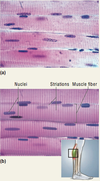
- *Microscopic appearance:** Long, threadlike, unbranched cells (fibers), relatively parallel in longitudinal tissue sections; striations; multiple nuclei per cell, near plasma membrane. Voluntary control.
- *Representative locations:**
- Skeletal muscles, mostly attached to bones but also including voluntary sphincters of the eyelids, urethra, and anus
- Diaphragm
- Tongue
- Some muscles of esophagus
Functions: VOLUNTARY CONTRACTIONS.
- Body movements
- Facial expression
- Posture
- Breathing
- Speech
- Swallowing
- Control of urination and defecation
- Childbirth

Cardiac Muscle Tissue
- *Microscopic appearance:** Short branched cells (myocytes); less parallel appearance than other muscle types in tissue sections; striations; intercalated discs; one nucleus per cell, centrally located and often surrounded by a light zone. Involuntary control.
- *Representative location:**
- Heart
Functions:
- Pumping of blood

Smooth Muscle Tissue
- *Microscopic appearance:** Short fusiform cells overlapping each other; nonstriated; one nucleus per cell, centrally located. Involuntary control.
- *Representative locations:**
- Usually found as sheets of tissue in walls of viscera and blood vessels
- In iris and associated with hair follicles
- Involuntary sphincters of urethra and anus
Functions:
- Swallowing
- Contractions of stomach and intestines
- Expulsion of feces and urine
- Labor contractions
- Control of blood pressure and flow
- Control of respiratory airflow
- Control of pupillary diameter
- Erection of hairs
